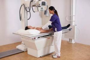
A chest x-ray is a non-invasive diagnostic method that uses x-rays. It is one of the most widespread and accessible to a wide range of patients and allows you to study internal organs and bone structures, identify their pathologies and developmental anomalies. This procedure is prescribed most often. Moreover: teachers of schools, colleges and universities, as well as persons who work with food products, are required to undergo it every year.
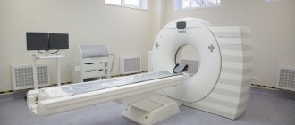
Don't know where to get a chest x-ray, but want to be sure that the pictures will be clear? Contact the diagnostic center of the multidisciplinary clinic CELT. Our radiologists have over 15 years of experience, and they have perfectly mastered the technique of performing the procedure. They have at their disposal modern stationary and mobile devices made in Germany and Japan, which allow them to carry out diagnostics in accordance with international standards.
X-ray of the chest organs - 1,000 rubles.
X-ray of the chest organs (survey) - RUB 2,500.
15 - 20 minutes
(duration of procedure)
Indications
The list of indications for a chest X-ray examination is very wide. We invite you to familiarize yourself with the main ones.
Contraindications
Contraindications are relative, so the procedure can be performed as an exception if there is a danger to the patient’s life. They are due to the negative impact that even minimal doses of radiation can have on a patient who is in a certain condition:
- Pregnancy and breastfeeding in women;
- The patient's age is up to 15 years;
- General serious condition of the patient.
| Groups of diseases/conditions | Suspicion of diseases | Symptoms indicating them |
| Lung diseases |
|
|
| Diseases of the heart and blood vessels |
| X-rays are carried out along with other diagnostic methods (for example, ECG). Symptoms:
|
| Diseases of the spinal column |
|
|
| Injuries |
| In the picture you can see:
|
When is radiography performed?
The study should be carried out for preventive purposes and to confirm or refute the suspected diagnosis. Every year, during the next medical examination, patients undergo radiography for early diagnosis of cancer and tuberculosis.
When the first symptoms appear, therapy is much more difficult and longer. This poses a great danger to the patient. A favorable prognosis for such serious diseases depends on timely diagnosis and proper treatment.
Radiography has limitations on the number of times it can be performed. For preventive purposes, the examination should be carried out once a year. This amount is quite enough. If it is necessary to monitor the progress of treatment, this figure can increase within reasonable limits and according to strict indications.
A chest X-ray is prescribed by doctors if there are suspicions of the following pathologies:
- lung abscess;
- pneumonia;
- pleurisy;
- tuberculosis;
- diseases of the lymphatic system;
- any neoplasms;
- heart disease;
- silicosis;
- the presence of foreign bodies in the lungs;
- congenital pathologies of the development of internal organs and skeleton.
In addition to suspected diseases, fluorography is performed if one or more of the following symptoms are present:
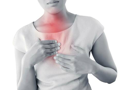
- the appearance of discomfort in the limbs and thoracic region;
- spinal injuries;
- infectious diseases;
- curvature of the spinal column;
- pathology of bone development, as a result of which a person experiences severe pain;
- breathing problems;
- cough that does not respond to treatment for a long time.
Indications and contraindications for undergoing fluorography
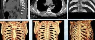
Depending on the indications and the expected diagnosis, patients are prescribed a local or general X-ray. The first method allows you to visualize the required organ or its separate part. Thanks to high-quality images, doctors can detect the exact location of the pathology. This greatly increases the diagnostic value of radiography.
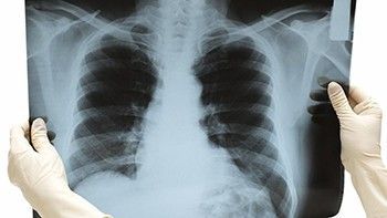
In the overview image you can see all the organs of the chest. However, the quality of such an image will be worse. The choice of a specific method is carried out separately for each patient. Based on what the diagnostics shows, additional local and overview images can be taken.

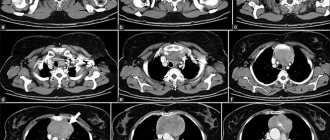
An x-ray is obtained by passing ionizing radiation through the body of the subject. Therefore, diagnostics has one big drawback - radiation.
The principle of obtaining images comes down to the passage of rays from soft tissues and reflection from dense ones. The resulting images can be used to visualize images of organs and tissues.
In recent years, all clinics have used only high-precision equipment. Fluorography is considered a safe method for the person being examined. However, doctors agree that it is impossible to undergo diagnostics, even with significant indications, more than 6 times a year.
It is permissible to perform chest X-rays on pregnant women only for health reasons.
Sometimes the study may be prescribed to patients with a contrast enhancer. In such a situation, an allergy test is performed. If the results reveal that the patient is intolerant to contrast, this will be a strict contraindication.
Preparing for a chest x-ray
X-ray of the sternum and OGK in general does not require any preliminary preparation. As during any X-ray examination, the patient should not wear metal objects; glasses and jewelry should be removed; women with long hair will be asked to raise them. You will also need to expose the chest area so that clothing does not degrade the image quality. The procedure will not take more than 15 minutes. Although direct contact of the patient’s body and the X-ray tube on modern equipment is no more than 0.02 seconds.
Performing an emergency x-ray
Typically, a chest x-ray is not a life-saving procedure. When an ambulance arrives to the patient, specialists provide first aid. For example, there is a real threat to the health and life of the patient. If we are talking about pneumonia, the patient needs antibiotic therapy. If a patient has a heart attack, resuscitation measures are often necessary. After first aid is provided, the person will be taken to a medical facility.
The urgency of an x-ray is determined by the severity of his condition. All necessary tests are carried out at the hospital. The list also includes a chest x-ray. This diagnostic method is not urgent. Only in extreme cases, the study is performed urgently.
How are pelvic bones x-rayed?
The patient is asked to remove metal objects from the body and report whether there are grafts or other foreign objects in the body. Pictures are taken in frontal and lateral projection:
- The direct projection requires the patient to assume a supine position with his legs straightened and feet turned inward. A special cushion is placed under the knees. If this position causes pain, the patient is placed on his stomach, raising the pelvis from the healthy side;
- The lateral projection requires the patient to lie on their side with the lower limb flexed at the hip. If pain symptoms are present, it is allowed to bend both legs.
Taking X-rays at home
During the diagnosis, the person needs to take a vertical position. Due to a number of diseases, some patients cannot fulfill this condition. In this case, x-rays are performed in the ward or at home. Such patients may include:
- people with critical body temperature;
- patients with severe infectious pathologies;
- seriously ill;
- patients with fractures;
- cancer patients;
- children, old people, disabled people.
When carried out correctly, the resulting images do not differ from classical radiography.
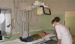
How the research is carried out
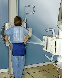
Chest X-rays are always performed in a room specially designed for this purpose. Medical staff explains how to prepare and helps you take the correct position in front of the scanning machine. Before taking a photo, you will need to take a deep breath and hold your breath. You cannot strain too much at this moment. This may cause distortion of the images, which will complicate diagnostic measures.
The procedure takes about 1 minute. As soon as all the manipulations come to an end, the doctor will begin deciphering the resulting images.
results
Based on the data received, a conclusion will be issued. The specialist always describes the location of the heart and the full characteristics of the tissues. A description of blood vessels, lymph nodes and bronchi is required. Sometimes a radiologist detects foreign bodies and neoplasms in the images. Without fail, everything that the photographs show will be recorded in the conclusion.
Based on the results of a chest x-ray, the presence of fluid in the pleural cavity, diffuse changes, pneumothorax, reticular and linear changes are revealed. Inflammatory diseases of the lungs and pleura are visualized by significant darkening. If the darkening has a heterogeneous structure, this is a sign of pulmonary overhydration. Regardless of what the diagnosis showed, the patient must seek advice from the doctor who issued the referral.
What does an x-ray of the pelvic bones show?
Deciphering the results requires not only clear images, but also relevant knowledge from the doctor. This is due to the fact that changes of the same type have different interpretations. That is why in the process it is necessary to take into account the patient’s history and complaints: each disease or anomaly has certain clinical manifestations. So:
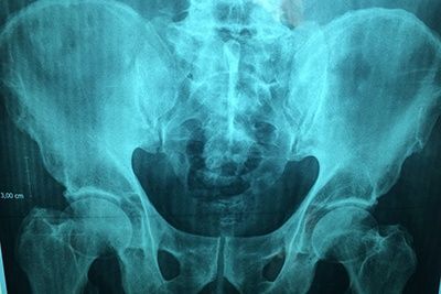
- Displacements in the hip joint noticeable in the image indicate its dislocation;
- Anomalies in the structure of the femoral head and acetabulum are signs of dysplasia;
- Narrowing of the joint space and the presence of osteophytes indicate a degenerative disease such as osteoarthritis;
- Darkening is a sign of malignant neoplasms, shadows are metastatic foci;
- Bone fragments confirm the suspicion of a fracture;
- Clearly visible regeneration of bone structures is a symptom of aseptic necrosis.
You can make an appointment for an x-ray of the pelvic bones at the CELT clinic right now through our website or by phone.
Where to take the study

Patients are often prescribed a chest x-ray. The test can be taken at a public clinic at the place of registration or registration for free. To do this, you must provide a compulsory medical insurance policy, a medical card, and a referral from a doctor. Only the majority of those in need of emergency diagnostics are not always able to undergo the procedure quickly. Due to the large number of patients, the queue at the X-ray room is quite long.
Therefore, private medical centers are becoming increasingly popular. They allow you to get examined without long waits.
Our website presents a huge selection of medical centers, in each of which you can undergo a study without a long wait and at a low cost. When making an appointment by phone, we offer an additional discount to everyone who applies.
Our website provides a convenient search and filtering system for all parameters. Thanks to well-thought-out functionality, you don’t have to waste time manually filtering clinics. It is enough to enter the necessary data in the parameters and the system will automatically select medical centers for x-rays.
Our distinctive feature is the provision of free advice to patients. If difficulties arise with the choice, everyone has the opportunity to contact the operator to find answers to all questions. The specialist makes an advance appointment for radiography at the selected clinic.
Grodno Regional Children's Clinical Hospital
Details Published November 24, 2017
*X-ray examinations should be carried out only in X-ray rooms, the design and maintenance of which strictly comply with sanitary and technical requirements;
*When protecting patients from ionizing radiation, using personal protective equipment to shield the patient’s body and thyroid gland;
*For justified medical reasons;
*When ensuring continuity of the results of X-ray examinations at all stages of medical care for patients;
• qualified medical personnel with special training;
• Using optimal techniques that ensure the highest achievable diagnostic information content and radiation safety of patients.
• Recommendations for limiting and reducing radiation exposure to patients during X-ray examinations
• Only a doctor can order a medical X-ray examination.
• Diagnostic X-ray examination is carried out according to clinical indications, only as prescribed by a doctor and with the consent of the patient.
• When referring for an x-ray examination, the doctor is obliged to justify the indications for it and indicate a preliminary diagnosis, without substituting vague wording for the type of examination. Otherwise, the referral should be regarded as unfounded and examinations in such areas are PROHIBITED.
• In emergency conditions, an X-ray examination is carried out regardless of the timing of the previous examination.
• X-ray examinations of pregnant women must be carried out using all protective equipment.
• Studies should be carried out, if possible, in the second half of pregnancy.
• Research should be carried out with minimal exposure, current strength and irradiation field size, with the obligatory use of a tube.
• Persons involved in restraining patients or children during X-ray examinations must be protected by the necessary means (protective aprons, gloves, etc.).
• When conducting X-ray examinations, the presence of more than one patient and unauthorized persons in the procedure room is STRICTLY PROHIBITED.
• Conduct research only on working X-ray equipment equipped with all technical means of protecting patients and personnel.
• Special precautions must be taken to protect patients awaiting testing.
Recommendations for limiting and reducing radiation exposure to
personnel during X-ray examinations
• The staff of X-ray rooms is obliged to constantly improve their knowledge in mastering X-ray techniques, striving to minimize the time of that part of the study that takes place with the X-ray tube turned on.
• It is mandatory to use personal protective equipment against radiation if personnel are in the treatment room during X-ray examinations. The number of protective equipment must be at least two sets.
• Protective aprons, leaded glass, and other protective equipment must be stamped or marked indicating their lead and the date of inspection.
• During radiography, the laboratory assistant is at the control panel of the device, observing the patient.
The office staff is obliged
• Know the principles of operation and operating conditions of X-ray diagnostic equipment in the office.
• Do not allow the presence in the cabin of persons not related to the X-ray examination. Know first aid techniques.
• Know the coordinates of the organization and persons to whom the accident is reported.
• Monitor the frequency of checking the technical condition of X-ray equipment, grounding, ventilation, darkroom lighting, chemical reagents.
• Know accurately the purpose of the controls of X-ray equipment.
• Be in the office when carrying out preventive and repair work with X-ray equipment.
• Be prepared to provide first aid to the victim in the event of an accident.
• Wear a dosimeter at all times. Correctly follow the rules for its operation.
• Protect the patient as much as possible by using lead aprons and collars to protect the thyroid gland. Protective equipment made from leaded rubber must be placed in covers made of film materials.
• Conduct X-ray examinations only as prescribed by a doctor. Enter the received dose into the outpatient card in a special insert.
• For pregnant women, X-ray examinations are carried out according to strict indications in the 2nd half of pregnancy.
• Children are more susceptible to ionizing radiation than adults. The reason is that the child’s body is in the process of development, and X-rays are dangerous primarily for actively dividing cells. Short stature causes exposure to more than required body surface area. For this reason, safety issues when performing an x-ray on a child are significant and relevant.
• Preventative testing is strictly prohibited for patients under 17 years of age. Diagnostic x-rays are prescribed only for justified indications. At the same time, of all diagnostic procedures, preference is given to those that are accompanied by the lowest dose of radiation.
• Radiation doses in X-ray diagnostics are low. However, you should not neglect safety measures. X-rays with unreasonably frequent and prolonged exposure to the human body can lead to undesirable health consequences.
Strict adherence to recommendations regarding X-ray diagnostics significantly reduces the radiation dose to the patient.
• Eliminating or limiting the influence of other sources of radiation, especially natural ones, is not easy. But medical exposure is an area that can be impacted. This means that by reducing the radiation exposure during diagnostic studies, it is possible to reduce the total radiation dose resulting from the influence of natural and man-made factors.