Magnetic resonance diagnostics is considered a popular method for identifying diseases. Screening helps doctors, without injuring the patient or radiation exposure, examine the condition of internal organs, tissues and systems, as well as identify signs of abnormalities in the initial stages of progression. Electromagnetic diagnostics are used in all medical fields.
Tomography is considered a safe procedure for the body, but it has a list of contraindications, one of which is the presence of metal implants and electronic devices in the body. The latter is due to the interaction of ferromagnets with a magnetic field, which can lead to the movement of loose metal parts. Such a process can distort images and reduce the reliability of the study.
When people first encounter an MRI, they wonder about safety. Doubts regarding tomography with tattoos are associated with the use of pigments with metallic elements when applying designs. The risk of patient discomfort during scanning and distortion of results exists in cases where the coloring agent contains particles of iron, cadmium, lead and other ferromagnetic substances. Pigments without such compounds do not affect the quality of the picture in any way and do not interact with the induction field of the device.
Why are tattoos considered a limitation for MRI?
Advanced tomographic scanners have powerful sources that generate a magnetic field: electromagnets or permanent magnets. MRI allows you to monitor the functioning of all vital systems and identify emerging disorders without invasion. The nuclear resonance technique involves studying the concentration of protons in the body. Each of them has its own magnetic abilities, because it is surrounded by particles with different properties.
Need to know! The magnetic field of tomography equipment is so powerful that any metal object can begin to move. Such a process can negatively affect a person and even lead to death.
Three decades ago, tattoos were created with metal needles of different diameters, using blue, red and black inks. To ensure the durability of the painting, chemicals were added to the composition, which contained a large amount of metal. This meant that microscopic metal particles remained in the epidermis. Therefore, it was previously difficult to predict how metal particles would react to exposure to a magnetic field in tattooed patients.
SO WHAT DOES THIS HAVE TO DO WITH TATTOOS?
More than 20 years ago, tattoo ink sometimes included small pieces of metal. Recently, such inks are practically not used. Some patients with tattoos that were done a long time ago have reported slight discomfort or even severe pain during an MRI. It is believed that the reason for this was that magnetic forces pulled the metal fragments of the ink so strongly that it caused a burning sensation at the tattoo site. However, even if there is no pain at all, these metal fragments can cause artifacts that distort the MRI scan image.
If the tattoo was done within the last 20 years, then it is certain that it will not be a problem for an MRI. Even if a tattoo is over 20 years old, this does not mean that the ink absolutely contains metal. And even if they contain metal, this does not mean that there will definitely be problems with MRI.
However, it makes sense to tell your doctor about any tattoos you have. Remember that there is always an alternative to MRI. For example, people with pacemakers and metal implants must find other diagnostic methods.
MRI and tattoos
What will happen to the “tattoo” after the MRI?
Over the course of several years, the metal elements located in the epidermis gradually begin to be eliminated. What to do if research on electromagnetic equipment is necessary for health reasons, but you have a tattoo? In such situations, diagnosticians recommend checking the place where the pattern is applied using appropriate equipment - a device for determining the amount of metal. You can use a metal detector or any of its equivalents.
Note! You don’t need to immediately go to the store to buy a metal detector; it can detect iron molecules present in the skin. Contact your doctor; perhaps he will suggest an easier way to search for ferromagnetic particles or prescribe another type of diagnosis.
Medical staff began to take a more careful approach to examining patients on MRI more than 15 years ago. Previously, people with tattoos could undergo screening without hindrance and did not ask doctors whether they could have a tomography with a “tattoo.” However, during diagnosis, patients informed doctors about the presence of such manifestations in the area where the tattoo was present:
- tingling;
- burning skin;
- nagging pain.
After completion of the session, the tattooed area of the skin takes on a reddened appearance or small spots of different locations were noted on it.
MRI: research features
It is not always possible to make an appointment for an MRI “today for tomorrow,” even for a fee. Although in some centers there are no long queues, and one of the private Minsk clinics does this test around the clock. There are many people interested, and people are not stopped by the high cost of research.
Radiation diagnostics doctor at the Minsk City Clinical Oncology Center and the national representative of Belarus at the section of young scientists of the European Society of Radiology (ESR) and the European Congress of Radiology (ECR) Konstantin Kenigsberg told when it is worth doing an MRI, whether it is possible to go for the study with a tattoo, piercing or braces and what to do if you have claustrophobia and are afraid of lying in an MRI machine.
1. What is the essence of the MRI method?
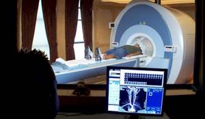
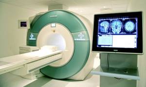
— Magnetic resonance imaging is one of the most complex medical devices. To put it simply, under the plastic shell of the tomograph there is a large magnet in the form of a cylinder, which creates a very powerful magnetic field in the room. As a result, with the help of additional parts of the device, it is possible to receive a signal from hydrogen protons in our body.
Hydrogen in the human body is found in various molecules, and they have different concentrations in different tissues. We apply energy to hydrogen protons and monitor how quickly the protons restore their original properties and emit energy back.
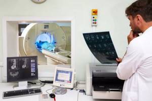
The device captures this energy and produces diagnostic images. Since the properties of protons differ depending on the molecules and tissues in which they are located, these tissues in the images differ in color, and, very importantly, by changing the program settings, we ourselves can influence this. For example, make the liquid bright and the fatty tissue dark. This is one of the main advantages of the method - high soft tissue contrast.
2. How does MRI differ from computed tomography and x-rays?
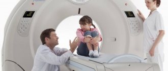
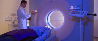
— Externally, computed tomography (CT) and MRI machines are very similar to each other. There is the device itself, which has a tunnel, and a patient table that moves inside. However, the operating principle of the technology is completely different.
The main advantages of the MRI method over all x-rays, including CT, are the absence of ionizing radiation and amazing soft tissue contrast.
The CT method, in contrast to classical radiography, allows you to obtain not one image with summary information about all the structures of the body within the frame, but many images of any area in high resolution with a certain slice thickness and step. But here it is important to understand the specific task of the study, because the more images and the better their quality, the greater the dose the patient will receive.
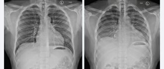
X-ray, fluoroscopy, X-ray computed tomography are based on the fact that on one side of the patient there is an X-ray tube emitting bremsstrahlung X-ray radiation, and on the other side there is some kind of signal receiver - a screen, cassette or detector. Different tissues of the body slow down the rays passing through them at different speeds. That’s why, for example, on X-ray photographs the lungs are black (the rays fly through them almost without stopping, because there is mostly air there), and the bones are bright (it’s not so easy to fly through dense and strong bone).
3. When should you get an MRI?
— If we are not talking about traumatic brain injury or fractures, then MRI is several times more effective as a diagnostic method. We use additional programs that allow us to analyze biochemical and physiological processes in healthy and altered tissues and carry out accurate diagnosis of stroke even in the first hours, when other methods are useless, tumors, infectious (encephalitis, meningitis) and inflammatory (for example, multiple sclerosis) diseases, developmental anomalies. With the help of MRI, the doctor can see the blood vessels of the brain even without the use of contrast agents.
According to statistics, MRI of the spine is the most popular MRI examination in our country, and this is not without reason. In addition to the fact that there is nothing better for diagnosing the spinal cord and nerves, vertebrae, herniated discs, spinal cord and nerves, and cerebrospinal fluid have different colors on MR images. This makes the diagnosis clear.
MRI is the only method for studying the bone marrow and all its pathologies: benign and malignant vertebral tumors, inflammatory and degenerative changes.
There is a misconception that if there is a problem with the bones, then a CT scan should be done. Actually this is not true. Bone consists of periosteum and bone marrow. So, for bone fractures and bone tumors, you should definitely start with X-ray or CT, and for everything else, do an MRI.
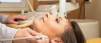
In addition to bones, joints contain ligaments, tendons, menisci, cartilage... Something can be examined with good information using ultrasound or radiography, but to get maximum information about all structures of the joint, it is better to undergo an MRI.
In terms of diagnosing diseases of the abdominal organs, there is its own specificity. In this case, ultrasound is a cheaper and harmless method, CT with contrast is more expensive, but much more informative, and MRI with or without contrast is expensive, time-consuming, but harmless and very informative. The choice is largely determined by the specific task, and the decision which method to choose should be made by the attending physician. As a rule, they start with an ultrasound, and for a detailed assessment they resort to CT with contrast or MRI.
If we are talking about a tumor, it is better to immediately do an MRI. MRI is the gold standard in diagnosing tumors of the cervix and uterine body, vagina, prostate and rectum. Most studies are carried out precisely for this purpose. If you have a good apparatus and experienced doctors, the stage of the tumor process is determined in conclusion.
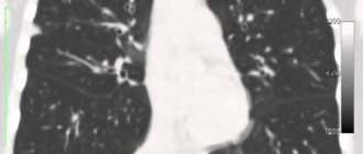
On the other hand, MRI is a frankly weak method in diagnosing the lungs, at least because it is technically very difficult to obtain good images in a short time. X-ray methods work better here. The second weak point of MRI is the periosteum. It has a black signal because there are simply few hydrogen protons, which means that to diagnose fractures, you again need to use radiography or CT.
4. How many times a year can an MRI be done?
- There are no restrictions. The method is considered harmless in the absence of contraindications.
5. Is it possible to do an MRI on the part of the body and head where there is a tattoo?
— It’s possible, but no one can guarantee that the research will be described reliably, since the picture may be distorted. After all, the higher the magnetic field, the greater the image distortion may be due to the presence of metal in the tattoo on the examined area. You also need to monitor your condition during the study and, if something happens, call a laboratory assistant in time.
Many people watched the series “House M.D.,” where in one of the episodes the machine “sucked” a tattoo out of a patient’s skin using a strong magnetic field. None of my colleagues, of course, have encountered this in their practice, because the metal content in tattoo ink is not that high.
It is worth making a reservation: the devices differ in many characteristics. The main one is the magnetic field strength, measured in Tesla. In our country there are low-field (0.3−0.4 Tesla), high-field (1.5 Tesla) and ultra-high-field (3 Tesla) machines.
Metal in the magnetic field of a tomograph has three properties that are unpleasant for us: it is attracted, heats up, and distorts the image. And although when undergoing an MRI at 1.5 Tesla you will not see the “sucking” of the tattoo from the skin, theoretically in isolated cases in the field of 1.5-3 Tesla you can feel a slight heating of the skin in the tattoo area. You should not be afraid of this, because throughout the entire study the patient has a special signaling device in his hands - a pear. When you click on it, a laboratory assistant will immediately come to the patient, who will ask what happened and, if necessary, complete the procedure.
6. Is it possible to do an MRI of the brain if there is permanent makeup on the face?
— It is possible, but you should not expect that the study area will be described in detail. If you want to explore your eyeballs, come without makeup. If you came with makeup, you felt a burning sensation in the eyelid area, squeeze the bulb that is given to you during the examination, wait for the laboratory assistant and get out of the machine. There is no need to be patient.
The literature describes cases of small eyelid burns during MRI on a 3-Teslav machine, but the patients in those situations were in serious condition and unconscious, so they could not inform the staff about their condition in a timely manner.
If you are going to see a doctor and especially an MRI about vision problems, then come without makeup. This way you will get more information. I can’t resist and ask you not to use too much perfume. It is in your interests that the staff focus on your health problem, and not think about ventilating the room.
7. Is it possible to do an MRI if you have braces?
— You can do this, but it is better to choose a device with a field strength of 1.5 Tesla or less. The device will not tear out the braces, but they may vibrate a little during the examination. What's worse is that on diagnostic images nothing will be visible within a radius of a few centimeters from the braces.
But this information on braces is relevant for studying, for example, the paranasal sinuses, but not for diagnosing the lumbar spine.
8. Is it possible to do an MRI if you have a piercing?
- It is forbidden. The piercing must be removed before the examination.
9. Do I need to remove rings, earrings, chains, bracelets before an MRI?
- They definitely need to be removed. If you are doing an MRI and forgot to take off your watch, put away your phone or bank card, the watch will stop, the phone will fly away and stop working, and the plastic card will stop issuing money.
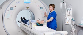
Before the examination, the patient is invited to the locker room, where he completely empties his pockets and removes all jewelry and hairpins. When examining the spine, women must remove their bra. When examining the pelvis and lower back, you need to remove your jeans.
The operating temperature in the tomograph room is 17−22 degrees. Research takes from 10 minutes to an hour. It is better to take a T-shirt and lounge pants made of cotton fabric without metal inserts. You cannot do research in woolen clothes.
10. What are the contraindications for MRI?
— The strength of the tomograph’s magnetic field is more than 10 thousand times stronger than the Earth’s magnetic field. Therefore, when signing up for a study, pay special attention to whether you have in your body:
- pacemaker, neurostimulator, implanted hearing aid and other devices;
- clips on vessels. If there was an arterial aneurysm and it was operated on with a metal clip, MRI is strictly contraindicated;
- metal fragments, especially in the orbital area. There is a risk that the fragments may dislodge and injure surrounding tissue.
In the cases described above, MRI is not performed in principle, since this procedure is very dangerous for the patient.
As for joint prostheses, it is important to know what metals and alloys they are made of. Each prosthesis has its own name and passport; if it does not indicate whether it is possible to undergo an MRI, there is an excellent online database where you can look. We, professionals, also use this site.
If you don’t know the name of the prosthesis, check with the place where it was installed and take the corresponding extract. They may ask for it in the MRI room.
All modern stents (metal meshes installed in the lumen of blood vessels to widen them) are made of non-magnetic metals. They are usually an alloy of nickel and titanium and are not a contraindication for MRI.
11. Is it possible to do an MRI if you have claustrophobia?
- If a person is truly claustrophobic, he simply will not be able to withstand the study inside the apparatus for 10-30 minutes. He will press the bulb in the first minute and ask you to remove it from the machine.
In this case, you should either turn to other methods (ultrasound, radiography, CT) or undergo an MRI on a low-field machine. There will be a magnet at the top and bottom, and open space on the sides. But these devices are much less powerful compared to 1.5 Tesla, and the study will be much less informative.
12. Is it possible to do MRI during pregnancy?
— Previously, MRI was not performed on patients in the first trimester of pregnancy just in case. Simply because a teratogenic effect on the fetus has not been proven. But in 2021, a large scientific work was published by Canadian researchers who tracked more than 15 thousand pregnant women and found no signs of harmful effects of MRI either on pregnant women or on subsequently born children.
In general, there are not many situations when pregnant women really need an MRI. We are talking here about very serious diseases, such as stroke, fetal developmental anomalies (but most of these issues are resolved by ultrasound), fetal cerebral ischemia (here MRI has no alternative) and tumors in pregnant women themselves.
13. How to prepare for an MRI?
- Firstly, you need to make sure that you have no contraindications.
Secondly, if you have been prescribed an MRI of the abdominal cavity, pelvis or the whole body, you need to start preparing 24 hours in advance. It is necessary to exclude gas-forming foods from the diet, and if possible, come for the study on an empty stomach. If the study is in the afternoon, do not eat for six to eight hours. An adult should take antispasiolytics 30-40 minutes before the test, if the person tolerates them well. It is best to clarify the dosage and which drug to take when signing up for the study. Immediately before the MRI, you need to go to the toilet and empty your rectum. Gas in the rectum creates artifacts and makes examination of the prostate, uterus or pelvis uninformative.
When examining the brain, spine, soft tissues of the neck and joints, special preparation is not needed.
When choosing a place to conduct a study, be guided by the profile of the institution: with a shoulder injury, it is better to go to clinics with a traumatological focus, if you suspect a tumor, to oncology centers. Private centers usually have specialists in various fields, but if in doubt, check these points when making an appointment. The most important indicator of the study is the qualifications and experience of the doctor.
The second important point is the choice of device. If you just have a back pain, and the doctor has not found any signs of a serious illness, but you still want to see what the reason is, then you can choose any device. If the task is serious, I would recommend only 1.5 and 3 Tesla machines. Yes, 3-T diagnostics are more expensive, but when studying the nervous and musculoskeletal systems, small tumors, small regions (pituitary gland, orbits, prostate), the information content of the study increases. You can draw a parallel with the diagonal of the screen - you can watch a movie on your phone, but you won’t be able to examine the model of the main character’s watch in detail.
Among the 1.5-T machines there are entry-level, intermediate and expert-level devices. This can also be clarified when registering for the study.
It is best to come to an MRI with the results of laboratory tests, conclusions of specialized specialists and data from previous studies on disks. In international practice, MRI is a method of expert diagnostics. This test is ordered to answer a specific question, such as ruling out a herniated disc, confirming a stroke, or assessing the effect of chemotherapy for liver metastases. The specificity is that the MRI doctor practically does not communicate with patients and must have all the necessary information along with the images. Then his work will be effective. If this does not happen, the doctor will not receive an answer to his question.
Website tut.by
Is it possible to do an MRI with tattoos on the body?
How do doctors explain the discomfort during examination that is felt on the skin where the drawing was made? Under the influence of powerful magnetic forces, metal elements are attracted to each other.
Due to the force of friction, the patient develops hyperthermia. Molecules of metal particles move in tissues, heading towards the magnetic emitter. At this time, the person being examined feels pain and/or burning on the skin. After receiving numerous complaints about unpleasant symptoms during MRI, the list of restrictions was expanded to include tattoos and other objects with a metal connection.
Important information! The coloring elements in modern tattoos do not have metal particles. Dermatology has determined that iron molecules can trigger the development of melanoma and other malignant tumors in humans. If there is a possibility of the presence of a titanium alloy, then you should consult a doctor for advice.
Permission to perform tomographic diagnostics is given by the attending physician. Most likely, he will not risk your health and clarify whether the tattoo contains metal or not. An alternative will be offered: ultrasound testing or computer screening.
MRI compared to other diagnostic methods
When choosing the optimal diagnostic method, the orthopedic traumatologist focuses on information content and expediency.
MRI has a number of undoubted advantages over its “brothers” in the diagnostic workshop.
Thus, magnetic resonance imaging, in contrast to radiography and CT, traditional for traumatology and orthopedics, is indispensable for determining the condition of soft tissues.
The method allows you to visualize structures such as menisci, intra-articular ligaments, synovial folds, and identify types of damage such as edema, foci of infiltration, and ligament ruptures. X-rays and CT scans better reflect the condition of bone structures.
In addition, MRI provides visualization of organs in different planes and sections. The images taken in different projections can then be converted into a three-dimensional model. Often, it is this feature of MRI that allows one to see pathology that remains invisible when undergoing other diagnostic methods.
The choice of research technique always depends on the indications and individual characteristics of the patient. In particularly complex diagnostic cases, all possible examinations are prescribed to compare data and obtain the most complete clinical picture.
Is it dangerous or not to do an MRI with tattoos?
In 2021, diagnostics using an electromagnetic scanner are permitted under the following conditions:
- Using modern dyes to create a tattoo without metal particles.
- The presence of an old tattoo, since there are not necessarily metal molecules left in it.
- Even if the “tattoo” contains ferromagnetic elements, this is not always considered a limitation for MRI. Diagnosis is carried out if its results exceed the potential risk of further development of the disease.
In any case, consult a specialist to avoid negative consequences.
Contrast agent for MRI: contraindications
MRI with contrast is prescribed to study the lumen of blood vessels, blood flow characteristics, and also to more clearly determine the boundaries of the tumor. In this case, during the procedure, patients are injected intravenously with gadolinium-based dyes. Such substances are most often absolutely harmless, however, they also have a number of contraindications. The main contraindications to MRI with contrast include:
- Individual intolerance to a substance that causes an allergic reaction: observed in a very small number of patients;
- Pregnancy or lactation: gadolinium can cause intrauterine growth retardation and can also negatively affect the baby during breastfeeding if it enters the body with milk;
- Cardiovascular failure.
Is it safe to do an MRI with a tattoo on the body: the opinion of scientists
German researchers at the Max Planck Institute tested more than 300 volunteers to determine the safety of tomography for patients. They found that diagnosis using an MRI scanner does not pose a big risk for people with drawings on their bodies. The article was published in the New England Journal of Medicine, and a brief description of the experiment can be found on the institute's official website.
Many patients with tattoos assume that MRI is dangerous for them. They are afraid of getting burns and other injuries, as well as possible cancer. This is due to the use of various chemicals in ink. Some of them contain iron. A powerful magnetic field can actively interact with such molecules. Will this process cause damage to the skin? Scientists have not found an answer to this question.
German researchers, together with scientists from University College London, tested the diagnosis of 330 volunteers and the diagnosis of 932 tattoos. They collected data on the size of the pictures, information about in which countries and under what conditions the patterns were created, and also what colors were used.
The head of the research team, Nikolaus Weiskopf, says that most of the participants did not notice any side effects. Among all, there was only one case where researchers discovered the consequences of tomography in a patient. They manifested themselves as a burning sensation on the skin. All sensations went away on their own a day after the session. The participant in the experiment did not require medical assistance.
In an MRI of the brain, a high-frequency magnetic field is directed at the head. however, it can also affect the upper body, where some patients have tattoos. Even the weakest readings of the device are tens of thousands of times stronger than the planet’s magnetic field. But Weiskopf says the impact of the scanner (three types of tomographs were used in testing) was minimal. The results indicate a negligible risk for tattooed patients.
What is MRI?
To understand how MRI and tattoos are related in general, you need to understand what this examination is. Magnetic resonance imaging is a method of obtaining images of the internal areas of the human body, which are subsequently used to diagnose various diseases of the internal organs.
The tomograph allows radiofrequency pulses and a powerful magnetic field to make “slices” (as images of internal areas of the body are called). The device sends signals to the human body, and after a certain period of time, receives them already reflected from the tissues. In turn, the computer, having received such modified signals, converts them into 3D images.
The magnetic force of the device is so great that metal objects located in the room during the examination will heat up. This may result in personal injury and equipment damage. Even if they are small metal particles, the resulting image may be distorted.
Make an appointment now!
Where to get an MRI with tattoos in Moscow?
Use to explore dozens of current offers from local diagnostic centers. On the service you will find out the current prices for MRI, reliable addresses of clinics and their ratings. Our team has provided a convenient search system for medical centers by district and metro, a sorting panel based on various criteria, as well as an online map of the city. To make an appointment for an MRI, call the hotline at 8 (495) 363-40-76. You can consult with the company operator, find out which clinics are currently running promotions and book a place for examination with a bonus of up to 1,000 rubles.