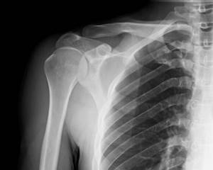
As you know, medicine is developing rapidly - new diagnostic methods are appearing, innovative models of treatment and rehabilitation are being introduced, and various new pharmaceutical products are entering the market. But at the same time, there are still some fundamental ways of working as a medical specialist, which over the years have proven their benefit and importance in treating patients. For example, x-ray. This diagnostic method is known to everyone and is successfully used in many healthcare institutions, including veterinary specialists who have positive experience in the practice of x-rays.

Why is this technique so popular? In fact, everything is quite simple. The fact is that during a routine examination, the doctor relies on the patient’s complaints and testimony, as well as his own observations and practical experience. And most often, these components are quite enough to make a correct diagnosis and draw up a course of treatment. But there are more serious cases that are simply impossible to detect on your own. In other words, an experienced doctor can detect massive diseases during an external examination, but to identify a tumor it is necessary to resort to the help of special equipment - in this case, an x-ray. Thus, we can conclude that this diagnostic method is necessary if serious diseases are suspected:
- Oncological diseases;
- Purulent-necrotic processes in bone tissue;
- Diseases affecting the lungs (tuberculosis, for example);
- Polyps;
- Cyst.
And, of course, X-rays are necessary when there is a possibility of fractures or cracks in bone tissue. Thus, the scope of application of the X-ray apparatus is quite diverse and includes a large number of medical areas:
- Dentistry;
- Traumatology;
- Pulmonology;
- Surgery.
But still, we should not forget that this diagnostic method is based on radiation, which has its contraindications. Today, the use of this type of diagnosis is contraindicated in the following cases:
- Bleeding;
- Pathologies of the thyroid gland;
- Hypersensitivity to iodine;
- Open pneumothorax;
- Severe form of kidney and liver diseases.
In addition, pregnancy is a contraindication for X-rays, and they try not to prescribe such diagnostics to children under 16 years of age. But first things first.
What is an X-ray?

X-rays are invisible beams of ionizing radiation that pass through the body and are altered by different tissues to create pictures.
The result is a two-dimensional picture showing bones, lungs and many other organs.
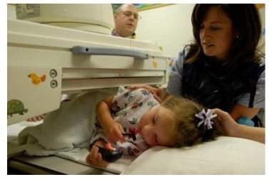
The scan itself is completely painless, but requires your child to remain still while it is performed. Sometimes parental involvement is necessary to help the child remain calm in the treatment room. For the safety of other organs, lead blankets are used.
What is computed tomography?
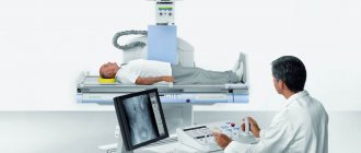
Computed tomography, or CT, uses an X-ray source that rotates around the patient's body to create a 3-dimensional image of the body.
The tomograph produces many “slices” that the computer reconstructs into images. These images provide more detailed information about internal organs than a single x-ray.
Despite the fact that CT can sometimes provide more relevant information for the treatment of a child, this technique has a higher level of radiation compared to a single x-ray.
The CT scanner looks like a big donut. The child should lie still while the table is advanced through the donut (CT scanner). Although a CT scan is a relatively quick and painless test without touching the child, sometimes children can be frightened by a large machine. The presence of parents during the examination helps the child feel calmer. Sometimes anesthesia is used for young children to help them remain still.
To examine organs and blood vessels where intravenous contrast agent is used, an intravenous catheter is required. Contrast agents are usually safe. Because the contrast agent is eliminated from the body through the kidneys, it may not be suitable for children with severe kidney problems. In rare cases, allergic reactions to the contrast agent may occur. Therefore, before the IV drug is given, you will be asked if your child has any unusual allergies or kidney problems. It is very important to discuss all of these issues with your doctor before undergoing the procedure.
Indications for radiography in children
X-rays can reveal a huge number of different pathologies of internal organs, soft tissues and the osteoarticular system. However, there are several diseases in which the question of at what age a child can be x-rayed recedes into the background, and the specialist prescribes an examination without hesitation. These are injuries to the spine, limbs or skull, foreign bodies in the gastrointestinal tract and respiratory system, pleurisy or pneumonia, heart disease or other anomalies in the development of internal organs.
X-rays are often required when inflammatory processes develop. A child cannot always describe symptoms (especially at an early age), so sometimes a doctor prescribes an examination to make a diagnosis and treatment.
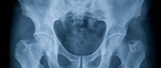
With hip dysplasia, the question of at what age can children be x-rayed fades into the background. Without proper treatment, this congenital pathology can lead to impaired leg function, leaving the child disabled for life. To diagnose a dangerous pathology, a specialist may prescribe an x-ray at its first signs. Even at the age of 4-5 months.
What is radioisotope research?
Radiation medicine uses small amounts of radioisotope drugs, which are concentrated in various organs and tissues. They emit so-called gamma rays. Special
The camera converts these rays into electronic signals that create an image. The radiation dose depends on the type of examination. As a rule, the drug is administered intravenously. The tests are painless, but your child must lie still during the procedure. You are advised to stay close to your child. Sometimes anesthesia is used for young children to help them remain still.
About the radiation doses in these studies
We are all exposed to low doses of radiation every day, which comes from soil, stones, building materials, air, water, as well as cosmic radiation. It's called natural or natural
radiation background. The radiation dose depends on where we live. For example, people living in the high mountains of Colorado are exposed to more cosmic radiation than people living at sea level. The radiation dose can be measured in different ways, and the radiation dose to the whole body and individual organs can also be determined. Radiation diagnostic parameters are individualized taking into account the size and shape of each patient. Determining doses even for typical studies can be difficult, much less comparing doses across studies. One solution to this problem is the use of millisieverts (mSv, mSv) as a unit of effective radiation dose for various types of radiation diagnostics.mSv

The radiation used in X-rays and CT scans is usually compared to the background radiation that we are exposed to every day. Comparing whole body dose to single organ dose can be misleading. However, this comparison may be useful in understanding the relative radiation dose to patients.
How dangerous is radiation diagnostics?
There is no reliable evidence that radiotherapy causes cancer. However, some studies of large populations exposed to radiation have shown a small increase in cancer risk even at low levels of radiation exposure, especially in children. Leading national and international organizations involved in radiation risk assessment agree that even minimal levels of radiation can increase the risk of cancer. In the interests of safety, even small doses of radiation must be considered dangerous.
The risk of cancer resulting from radiation diagnostics must be compared with the average statistical risk of developing cancer. The lifetime risk of dying from cancer is approximately 20-25%. For every 1000 children who have never undergone radiation diagnostics, 200-250 will die from cancer. The proportion of increase in a person's lifetime cancer risk after a single computed tomography (CT) scan remains controversial and is approximately 0.03-0.05%. This risk is for the general population and does not pose a direct risk to a specific child. Another way to assess the relative risk of CT is to compare the theoretical risk of abdominal CT with other risk groups. It is estimated that the risk of one CT scan of the abdomen is comparable to the risk of getting into an accident with:
• travel 12,000 km by car
• traveling 1600 km on a motorcycle
(estimates based on US traffic accident statistics)
These data show that, despite errors in estimating radiation dose, the risk of cancer after a single CT examination is very small. However, available research suggests that some risk exists and can accumulate.
Which parts of the body are most exposed to radiation?
Practical radiological studies show that in children, the lion's share of images are images of the musculoskeletal system - this area is most often irradiated. In children, injuries and bruises such as sprains, bruises and fractures are common. When a child enters the clinic with complaints of blows or obvious signs of pathology, it is recommended to take an x-ray.
Congenital pathologies of the musculoskeletal system are also added to the statistics when dislocations and subluxations in large joints are found in children, which requires x-ray diagnosis and further monitoring.
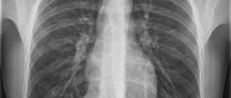
In second place in popularity are the chest organs, since an x-ray is required for a positive Mantoux reaction. A positive Mantoux test result may be a manifestation of pathology or simple irritation and scratching of the area being examined, but a diagnosis is necessary in any case.
How can I reduce my child's risk of radiation exposure?
There are various ways to reduce your child's radiation dose.
The Friendly X-Ray campaign promotes the following strategies to optimize radiology for children:
• Only perform tests that have a clear diagnostic benefit.
• Use the lowest possible radiation dose for adequate imaging based on the size of the child.
• Scan only the desired area of the body
• Avoid multiple scans
• Use alternative diagnostic tests (eg, ultrasound or MRI) whenever possible.
If my child's doctor thinks it is necessary to perform a CT scan, should I allow it?
As with any medical procedure, the benefit should always outweigh the risk. CT is a very valuable diagnostic test that can improve treatment and diagnose a number of diseases that other diagnostic methods do not detect. CT can provide the best course of treatment, avoid other tests or surgeries, and improve treatment outcomes. It is important to know that if your child has a serious illness that requires a CT scan, it is necessary to overcome the fear of radiation. In many cases, the merits and benefits of the test clearly outweigh the risk of radiation exposure to your child. If CT is the best test in your case, ask the staff if they are using proper techniques to minimize radiation dose.
How do I know that my diagnostic center is using proper techniques to minimize radiation dose?
Some centers that perform CT scans on adults do not use dose minimization techniques for pediatric patients. You will never know unless you ask about it. You have the right to this information. Your diagnostic center must provide you with information about how they reduce radiation doses. It is also necessary to find out whether the center has the appropriate certification for performing CT scans, for example, in the USA - accreditation from the American College of Radiology.
Alternative diagnostic methods
A CT scan may be the best way to obtain the diagnostic information needed to make clinical decisions in your child's treatment. In some cases, the doctor may decide that
It is safer to place the child for observation instead of performing a CT scan. Waiting while being monitored may be difficult for you and your family, but can provide the same end result without exposing your child to an unnecessary dose of radiation. Ultrasound (ultrasound) and magnetic resonance imaging (MRI) are diagnostic methods that do not use radiation. They can sometimes provide similar diagnostic information and can be a useful alternative. MRI is a lengthy procedure and sometimes requires anesthesia, which carries its own risks. A CT scan may be the only way to obtain the information needed to make clinical decisions about the best treatment for your child. You should ask your doctor and radiologist if there are alternative testing options for your child.
Stages of joint dysplasia
Depending on how susceptible the articular apparatus and its parts are to pathology, the following stages of dysplasia are distinguished:
- preluxation: the mildest degree, when the head of the femoral bone can move inside the acetabulum, and the joint itself is characterized by instability;
- subluxation: the stage when anatomical and morphological changes begin to form, the head of the femur moves away from the glenoid cavity, but remains within the limbus (a cartilaginous plate that prevents the head from moving upward);
- dislocation: in this case, the head of the femur completely comes out of the socket of the joint, while the limbus moves upward, and the ligaments of the joint are stretched.