Call the clinic +7 928 828 4001
Prices for services
| Service | Price |
| Phlebologist consultation | 1350* ք |
| Ultrasound (duplex) of the veins of the lower (upper) extremities | 1900 ք |
| Doppler ultrasound of the vessels of the lower (upper) extremities | 3800 ք |
| Ultrasound (duplex) of the arteries of the lower extremities | 1900 ք |
| Ultrasound (duplex) of extracranial brachiocephalic arteries | 1900 ք |
| Ultrasound (duplex) of the arteries of the upper extremities | 1900 ք |
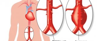
Using this method, blood supply disorders in the brain and neck vessels are determined. The bracheocephalic arteries consist of vessels responsible for supplying blood to the head, brain, shoulders and arms. The aorta is the largest vessel in the body. Large arteries branch off from it. On the right is the brachiocephalic or bracheocephalic trunk. It divides into the carotid and subclavian arteries. And they are already divided into small vessels. On the left are the carotid and subclavian arteries. They extend directly from the aorta and form the brachiocephalic trunk. Next, they divide into small vessels. The internal carotid and basilar arteries, as well as their branches, supply blood to the brain. They are arranged in a circle so that in case of obstruction in some areas, blood flows from other vessels in this circle.
What are the advantages of ultrasound scanning of the brachiocephalic vessels in the neck:
Ultrasound scanning of the brachiocephalic arteries and vessels is not expensive, it does not cause pain and it goes away very quickly. It provides a lot of necessary information that will help the doctor make the correct diagnosis.
Using BCA ultrasound it is possible to identify:
- Initial vascular damage;
- The very condition of the vessels;
- The presence of atherosclerosis and stenosis;
- The nature of blood flow and its speed;
- Vein thrombosis;
- Vascular injuries;
- Varicose veins.
And the same anatomical deviations in the neck vessels themselves, for example, the entwining of vessels around the neck.
A fascinating “walk” through the veins and arteries
I bring to your attention an article about a medical diagnostic procedure - duplex scanning of blood vessels.
Duplex scanning of blood vessels, otherwise known as vascular Doppler ultrasound (USDG), is essentially an ordinary, familiar, well-known ultrasound scan.
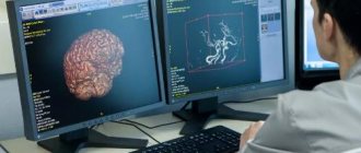
Many patients undergoing medical examination are prescribed an ultrasound examination of the brachiocephalic vessels. I will try to talk about the essence of this study, what information it gives to your attending physician, and why this study was included in the list of diagnostic measures for medical examination of the adult population.
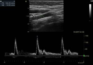
So, as a “tour guide,” I invite you to a short and, I hope, educational journey through the veins and arteries. The study is called duplex because it combines two modes - a two-dimensional image and a Doppler mode - a study based on measuring blood flow velocity. The 2D image looks like this.
This is a familiar black and white image, or rather gray, in which the manufacturers of modern ultrasound scanners promise not just 50, but as many as 256 shades! Doppler is a mode for studying blood flows, their speed and direction. It is the inclusion of the Doppler that is accompanied by the appearance of somewhat unusual sounds during the examination, sometimes slightly frightening to patients. These sounds are a kind of interpretation of the characteristics of blood flow. Each artery has its own “melody”. The external sleepy one is harsh and rough, the internal sleepy one is softer and more pleasant. And the veins generally “sound” like a light breeze.
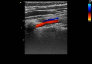
This is what duplex scanning looks like on the monitor.
There is a color doppler that adds bright colors to a boring black and white picture. Patients, having seen the image, are usually interested in what is “red” and “blue” there?
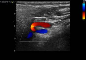
Both “red” and “blue” are blood. The color depends only on the direction of blood flow relative to the sensor. What moves towards the sensor is colored red, what moves away from the sensor is colored blue.
This is no longer a duplex, but a triplex study. Actually, any modern ultrasound examination is triplex, but the name “duplex” has been preserved as more familiar.
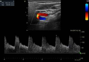
Vascular examination is harmless to the patient, non-invasive (no penetration into the body) and does not require prior preparation. And now a little about the study of the brachiocephalic arteries (BCA), the same study that is included in the list of studies under the clinical examination program.
Brachiocephalic arteries is a definition that includes the carotid and vertebral arteries. The carotid arteries have nothing to do with the quality of sleep. They owe their unusual name to a small anatomical formation on the internal carotid artery. This formation, or rather the expansion of the artery, is called the carotid or carotid sinus. If you squeeze the neck in the area of this sinus, the number of heart contractions decreases, the person seems to fall asleep and may even lose consciousness. This is where the name carotid arteries comes from.
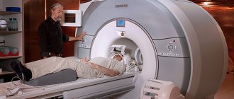
The examination is performed with the patient lying on his back, sometimes with a cushion under the neck for better vision of the arteries with the sensor.
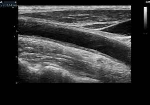
This is what the common carotid artery looks like on an ultrasound scanner.
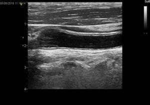
The carotid arteries can tell a lot of interesting things about a person. First of all, we are interested in the diameter and shape of the arteries, as well as the presence of atherosclerotic plaques that are dangerous to human health. The presence of atherosclerotic plaques or the predisposition of blood vessels to their appearance is indicated by such an indicator as the thickness of the “intima-media” complex or, in other words, the thickness of the inner layer of the vascular wall.
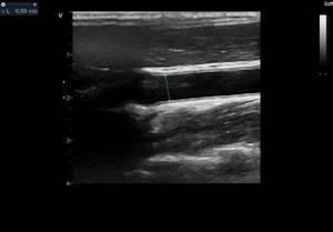
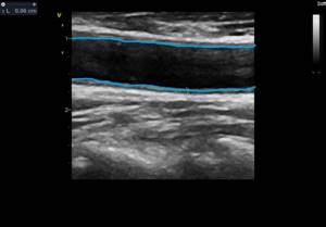
IMM thickness is an indicator of the severity of the atherosclerotic process in the body. Normally, it should be no more than 1.0 mm. The intima is a thin white stripe, the media is a thin black stripe following the intima, and the dark inside is the lumen of the artery.
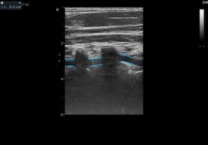
The following image shows a vessel that is not affected by atherosclerosis. The thickness of the intima-media complex is 0.6 mm. The blue line is drawn along the inner layer of the vascular wall - the intima.
But in the next image, the intima-media complex, in other words, the vessel wall, is already thickened (1.4 mm) - these are already manifestations of atherosclerosis.
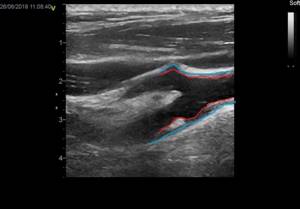
As atherosclerosis progresses, the intima-media complex not only thickens, but atherosclerotic plaques form, which cause narrowing (stenosis) of the vessel. The degree of stenosis is expressed as a percentage. Atherosclerotic plaques look something like this. This is the site of bifurcation (branching) of the common carotid artery (CCA) into its two branches - the internal carotid artery (ICA) and the external carotid artery (ECA). Arterial branching sites are favorite sites for the appearance of atherosclerotic plaques. I outlined the contours of the vessel itself in blue, and the atherosclerotic plaque in red. The degree of vasoconstriction in this patient is about 30%.
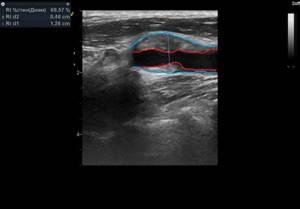
And in the next two pictures the process is already more advanced, the degree of narrowing of the vessel is 68%. The blue outline shows the vessel itself, and the red outline shows what the lumen of the vessel has become, changed due to atherosclerosis.
Atherosclerotic plaques can be extremely dangerous. Increasing in size, the plaque impairs blood flow in the vessel, up to its complete blockage. In addition, sometimes a fragment of atherosclerotic plaque can break off and block a smaller blood vessel, thus causing a stroke or heart attack.
If complicated plaques are detected and with a pronounced degree of narrowing of the vessel, a consultation with a vascular surgeon is indicated to decide on surgical treatment. If the process is moderate, the doctor prescribes treatment with statins - drugs that strengthen the surface of the plaque and prevent its destruction. It is also necessary to control your blood cholesterol levels, correct your diet and get rid of bad habits.

It is generally accepted that if atherosclerotic plaques are detected in the carotid arteries, then in the coronary arteries - those that feed the heart, there is almost one hundred percent probability that they are also present. We cannot easily and simply look into the arteries of the heart. Coronary angiography, the study of the blood vessels of the heart, is a complex study performed in a hospital setting. Therefore, ultrasound examination of brachiocephalic vessels (neck vessels) is a simple and informative method for diagnosing atherosclerosis. It is believed that atherosclerosis develops if a person has elevated levels of “bad” cholesterol - low-density lipoproteins.
To paraphrase popular wisdom, we can say that by the age of 50 every man should plant a tree, raise a son, build a house and... check his cholesterol level. It is advisable for a man who smokes to pay attention to his cholesterol by the age of forty. The same recommendations apply to representatives of the fair sex. In addition to the carotid arteries, vertebral arteries are also available for study. At the level of the sixth cervical vertebra, the vertebral arteries enter the bony canal formed by the transverse processes of the cervical vertebrae. Sometimes the artery enters the bone canal higher, at the level of the fourth or even third cervical vertebra.
The vertebral artery with a diameter of 3 mm is shown in the following image. Its walls are highlighted with blue lines. Dark vertical formations that interrupt the course of the artery are transverse processes of the cervical vertebrae. Ultrasound does not penetrate bone tissue, so the artery wall is not visible in these areas.
The vertebral arteries (VA) are smaller in diameter than the carotid arteries. The normal size is considered to be a diameter of 3 mm, if less than 3 but more than 2, this is called a “small” diameter, and less than 2 mm is already hypoplasia (underdevelopment) of the artery. This is a congenital condition, and it is impossible to influence the diameter of the arteries. When hypoplasia of the vertebral artery is detected on one side, the vertebral artery on the other side is usually increased in diameter. This ensures balance and ensures adequate blood supply to the brain. In addition to the diameter, we evaluate the shape of the arteries and the presence of tortuosity. The photo below shows an artery with a deformed course. It can be seen that the artery is curved, and this arc is painted in different colors.

Sometimes there are simply arteries curled into a ring! How can you avoid getting sick or dizzy with these? The vertebral vein runs parallel to the vertebral artery and above it in the bone canal. Blood in veins and arteries moves in different directions, so the vessels are painted in different colors.
Typically, the vertebral vein does not appear so bright and deep blue on an ultrasound. As a rule, it is slightly stained. Such brightness and expressiveness is already a bit of a pathology, which in conclusion is reflected in the phrase “difficulty of venous outflow.” This is not a disease yet, but rather a feature of the patient’s venous tone. But this condition already requires treatment, since it often manifests itself as tedious headaches and heaviness in the head. Remember what the doctor said from the movie “Formula of Love”? “The head is a dark object and cannot be examined.” A hundred years ago, maybe it was like that. Well, now it is very possible, and ultrasound examination of the brachiocephalic vessels (neck vessels) is an informative and safe diagnostic method that allows one to obtain important information about the blood supply to the brain.
It is enough just to do the research during medical examination. An ultrasound examination of the carotid and vertebral arteries at the second stage of clinical examination is prescribed to everyone who has elevated cholesterol levels or has complaints as prescribed by a neurologist.
Davidenko Yulia Gennadievna, head of the functional diagnostics office of the polyclinic of OKB No. 3.
Indications and contraindications for ultrasonography of brachiocephalic arteries and vessels:
Mostly people suffering from obesity, diabetes mellitus, hypertension, coronary heart disease, and cervical osteochondrosis suffer. And also if you have previously suffered a stroke, heart attack and people over forty years old. If such diseases are present, then an ultrasound examination must be performed at least once a year.
Also, people suffering from frequent headaches, fainting, sleep disturbances, tinnitus, spots in the eyes, and visual disturbances should also undergo ultrasound examination of the bracheocephalic trunk. And also if there is weakness in the legs and arms and memory becomes worse. There is only one contraindication, and that is the presence of wounds on the neck.