Diaphanoscopy of the scrotum is one of the instrumental diagnostic methods widely used in urological practice. It is highly informative, easy to carry out, has no contraindications and is affordable. Allows you to establish and differentiate testicular diseases, as well as detect pathological neoplasms.
This method is based on the ability of various fabrics to transmit light. For example, dense tumors completely absorb the rays. The degree of permeability of liquids depends on their composition: serous - almost transparent, purulent - cloudy, hemorrhagic - thick and dark. Knowing these features, a urologist-andrologist can make accurate diagnoses based on the results of the study.
Features of diaphanoscopy of the scrotum
The term "diaphanoscopy" is derived from two Greek words - "diaphanes" and "skopeo" - which translate as "transparent" and "to look".
This perfectly describes the essence of testicular diaphanoscopy. The procedure is aimed at illuminating pericutaneous formations using a light trigger and is based on the ability of tissues to transmit light differently, depending on their density. For example, bone structures do not even transmit x-rays, while liquid, on the contrary, transmits their light very well. This property determines diaphanoscopy as a diagnostic technique: the structure of the subcutaneous tissue assumes the presence of fluid, therefore, if there are purulent accumulations and blood impurities in it, they can be easily recognized by transillumination.
A diaphanoscope is a device whose design includes an element such as a tube. It can be straight or curved and have a light source. Diagnostics with its use in urology makes it possible to identify the cause of the pathological increase in the size of the scrotum and distinguish it from other pathological conditions.
Despite the fact that diaphanoscopy is inferior to ultrasound diagnostics in its information content, the ease of its implementation and the absence of the need for expensive equipment allow it to be widely used.
Diaphanoscope
The study is carried out using a device - a diaphanoscope. It is a powerful light bulb placed in a closed housing. Inside it there is also a lens system that collects light rays (a condenser) and a fan.
At the end of the diaphanoscope there is a tip - a conical-shaped light guide with a curved end. This design of the nozzle makes it possible to illuminate the eyeball from the opposite end of the equator.
The light guide is designed so that the rays are reflected several times on their way to the exit. By concentrating, they form a narrow beam of light. Often the kit includes tips with varying degrees of curvature. Light guides with a flexible end, made from a fiber bundle, are also produced.
The light of the diaphanoscope is cold, which eliminates the possibility of overheating the eye.
Indications and contraindications for testicular diaphanoscopy
Being absolutely painless and requiring no more than five minutes to complete, the procedure has no contraindications. It can be performed even on small children, which has been successfully practiced by urologists for decades.
As for the indications, they are quite extensive. Thus, it is used during the initial examination if the patient has specific complaints that suggest swelling or dropsy of the scrotum. Other indications are as follows:
- suspicion of a seminal cyst, which is a cavity neoplasm of the testicle or its epididymis, which contains seminal fluid;
- suspicion of an inguinal hernia in male patients under three years of age;
- diagnostic studies of benign or malignant neoplasms located on the testicles or spermatic cords;
- with testicular torsion, which is characterized by compression of blood vessels and nerves and the development of hydrocele of the testicular membranes;
- the need to determine the area of blood accumulation and its quantity if the patient has internal bleeding;
- during the postoperative period to assess the effectiveness of surgical intervention;
- to determine the localization of foreign bodies in traumatic injuries of the groin area.
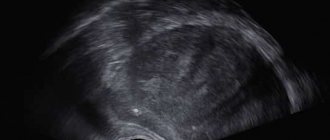
Hydrocele
Hydrocele of testicular membranes
Hydrocele of the testicular membranes (dropsy of the testicle, hydrocele - from the ancient Greek ὕδωρ - water and κήλη - swelling) is an accumulation of serous fluid between the proper membranes of the testicle, during which it increases in size.
Often found in children. Occurs in 1% of men. In most cases, a hydrocele is not very bothersome. Classification
In adults, the disease is often acquired; in children, it is usually congenital. Hydrocele of the testicular membranes is classified according to its genesis or severity of the disease.
By genesis: congenital and acquired. The congenital form can be communicating or non-communicating. Acquired form: primary (idiopathic) or secondary (symptomatic). According to the severity of the disease: acute or chronic form. Diagnostics
Inspection and palpation of the genitals. It should be distinguished from inguinal and inguinal-scrotal hernia, cysts, hydrocele of the spermatic cord, testicular tumors and spermatocele. The disease is quite common and is observed in both children and adults.
Congenital hydrocele
Normally, the laying of a testicle occurs in the abdominal cavity, then the formed testicle migrates to the scrotum under the influence of male hormones with the participation of the “Gunter’s” cord surrounded by the processus vaginalis of the peritoneum. The lumen of this process should be completely closed by the time the child is born. In the case when a violation of the obliteration (closure) processes occurs, a communicating hydrocele of the testicle or an inguinal hernia occurs, depending on the diameter of the process.
In this case, fluid from the abdominal cavity freely enters the testicular membranes through the duct. Sometimes only partial closure of the vaginal process occurs. In this case, the processus vaginalis of the peritoneum is obliterated at different levels of the inguinal canal and testicle. This leads to diseases such as isolated hydrocele of the testicle (testicular cyst), isolated hydrocele of the spermatic cord (spermatic cord cyst) and isolated hydrocele of the spermatic cord and testicle (spermatic cord and testicular cyst).
The tunica vaginalis of the testicle produces a fluid that serves as a lubricant for the testicle and promotes its free movement inside the scrotum. Normally, a balance is maintained between the production of this fluid and its reabsorption.
The so-called physiological dropsy is associated with a violation of this mechanism. It occurs in approximately 10% of newborns and in more than half of the cases disappears on its own by the end of the child’s first year of life. The reason for its development lies in the imperfection of the lymphatic system of the groin area in newborns and infants, which leads to slow resorption (absorption) of the resulting serous fluid between the membranes of the testicle. As the child grows, obliteration of the vaginal process and an increase in the absorption properties of its membranes are possible, which in a significant proportion of children leads to independent healing of dropsy.
Acquired hydrocele
Acquired dropsy occurs with acute or chronic inflammation of the testicle, with testicular trauma, with cardiovascular failure, with neoplasm of the scrotal organs. Surgery on the genitals can also lead to hydrocele. This is the so-called reactive “symptomatic” dropsy, which goes away as the underlying disease is treated.
The development mechanism is associated with compaction of the testicular membranes, which disrupts lymph flow with inhibition of microcirculation (impaired blood circulation). As a result, fluid accumulates between the shells. Hydrocele of the testicular membranes develops without pain and without any disorders. The accumulation of fluid occurs slowly and imperceptibly, sometimes spasmodically. The enlargement of the scrotum may be small, but sometimes it reaches the size of a goose egg and even the head of a child. With dropsy of the testicular membranes of very large sizes, difficulties arise during urination and sexual intercourse. Hydrocele has a smooth surface and a dense elastic consistency, painless on palpation, fluctuation is determined. The skin of the scrotum is loosely folded. The testicle usually cannot be felt, and only with slight dropsy can it be determined at the bottom of the swelling. During diaphanoscopy, transillumination of the entire formation is noted. The symptom of transillumination is negative only in cases where the testicular membranes are sharply thickened, there is a hematocele or pyocele (blood or pus in the testicular membranes), or a testicular tumor. Hematocele is a hemorrhage into the hydrocele cavity of the testicular membranes, which can occur as a result of injury, with hemorrhagic diathesis, after unsuccessful puncture of the hydrocele. Purulent hydrocele of the testicle occurs more often with orchitis and epididymitis as a result of infection during an abscess of the testicle or epididymis.
Diaphanoscopy and ultrasound examination
The diagnosis of hydrocele is established using ultrasound examination of the testicles and diaphanoscopy. Ultrasound examination reveals the accumulation of fluid between the membranes of the testicles and the unchanged testicle, and evaluates the volume of fluid and its liquor structure. Diaphanoscopy (from ancient Greek διαφανής “transparent” and σκοπέω “I observe”) is a method based on transillumination. With a hydrocele, the entire formation is evenly translucent. However, the method is not always informative. After suffering inflammation of the testicular membranes or hematocele, transillumination may be uneven. Diaphanoscopy also allows for differential diagnosis with hernias (intestine, omentum strand), when a violation of the passage of light through the swelling is detected.
Pathogenesis
The causes of congenital hydrocele are as follows. During the prenatal period, the testicle descends into the scrotum through the inguinal canal; along with the testicle, part of the peritoneum moves, which is called the processus vaginalis of the peritoneum. Then the lumen of the vaginal process of the peritoneum becomes overgrown. If this lumen does not close, fluid collects in it from the abdominal cavity. In addition, the cells of the inner lining of the peritoneum, which covers the process of the peritoneum from the inside, are themselves capable of producing fluid. The processus vaginalis may communicate with the peritoneum or be blind. If it communicates with the peritoneum, fluid can sometimes circulate from the hydrocele into the abdominal cavity. If a hydrocele is detected in a newborn child, treatment is not started. Such hydrocele of the testicle can disappear on its own when the process of the peritoneum heals and the fluid from the hydrocele cavity is absorbed.
Acquired hydrocele of the testicle occurs with inflammatory diseases of the scrotal organs, injuries of the scrotum and perineum, and impaired lymphatic drainage from the scrotum. Sometimes hydrocele of the testicular membranes can be reactive due to inflammatory processes in the testicles or epididymis or testicular torsion. This type of hydrocele disappears when the underlying disease disappears.
Treatment
Treatment of testicular hydrocele in inflammatory diseases of the testicle and its epididymis consists of treating the underlying disease: prescribing antibacterial therapy, rest and wearing a suspensor. To remove fluid from the hydrocele cavity, a puncture of the hydrocele is performed. The liquid is removed, and sclerosing drugs are injected into the cavity. This treatment method may have complications. If the puncture is unsuccessful, the testicular membranes may be damaged, hemorrhage occurs and blood accumulates in the hydrocele cavity. Sometimes an infection can enter the hydrocele cavity and an inflammatory process occurs. Surgery is considered a radical treatment method. For hydrocele of the testicular membranes, three types of surgical interventions are performed.
Winkelmann operation. During this surgical intervention, one of the layers of the testicular membrane is cut along the anterior surface, turned inside out and sutured behind the testicle. In this case, fluid accumulation no longer occurs.
Bergman's operation. Part of the inner layer of the testicular membrane is removed, the remaining part is stitched. In the postoperative period, antibacterial drugs are prescribed and a suspension is worn for some time.
Operation Lord. During this operation, the membranes of the testicle are dissected, the hydrocele is released, and the so-called corrugation of the vaginal membrane around the testicle is performed. In this case, the testicle itself is not freed from the surrounding tissues and does not dislocate into the wound. This allows you to reduce trauma to adjacent tissues and feeding vessels of the testicle.
However, there is no fundamental difference between the proposed operations (Winkelmann, Bergmann or Lord). So, in most cases, the surgeon determines the type of plastic surgery of the testicular membranes already during the operation. So, for example, it is irrational to perform a Winkelmann or Lord operation for large-sized dropsy, when there is an excess of membranes. Lord's operation is also not suitable for chronic dropsy, when the membranes become hard and their corrugation will lead to a poor result from an aesthetic point of view.
According to the course, acute and chronic hydrocele of the testicle are distinguished. Usually, dropsy of the testicular membranes is not accompanied by pain. Fluid in the membranes of the testicle can accumulate very slowly, in some cases the accumulation of fluid can occur intermittently. With hydrocele of the testicular membranes in the scrotum, you can palpate a pear-shaped formation, the base of which is at the bottom, and the narrowed apex is directed towards the inguinal canal. Sometimes fluid can get into the inguinal canal. Then the hydrocele of the testicle may have the appearance of an hourglass with a constriction in the area of the external inguinal ring. The size of a hydrocele can vary: from small increases in the size of the scrotum to a spherical formation the size of a football. Most often there is no pain when palpating. The skin on the scrotum remains unchanged and is easily displaced. Hydrocele of the testicle can be felt as a dense elastic formation. If the hydrocele is large, it can become an obstacle to sexual intercourse, and sometimes difficulty urinating.
Features of testicular diaphanoscopy
The procedure is very simple and does not require any preparation from the patient other than hygiene procedures. It is carried out by a urologist in his office, having previously darkened it (which will allow for the best X-ray results). The patient is asked to assume a supine position on the couch, exposing the area to be examined.
The diagnostician turns on the device and presses it firmly against the skin of the area being examined, palpating certain areas if necessary. During the procedure, he first examines the back surface of the scrotum, after which he slowly moves the device throughout the organ, determining the presence of foreign bodies or inclusions.
Normally, passing through soft tissue, the light acquires a red tint, clearly visualizing the shadow of the testicle. If the patient has certain pathologies, the picture will be different. So:
- exudate is more transparent;
- swollen areas and tumors transmit light less well;
- the stones are clearly visualized against the background of clear liquid.
It is important to understand that the diagnosis after this procedure is not final and requires an ultrasound examination.
How to increase research accuracy
Differences in the ability to transmit light may not be caused solely by inflammation. For example, in some patients, due to anatomical features, the walls of the sinuses have different thicknesses, or these formations themselves are located slightly asymmetrically. To reduce the inaccuracy of the study, the doctor can use the following methods:
- gradual increase in the intensity of transillumination using a rheostat, with the healthy sinus first appearing, and then the affected one;
- use of a two-lamp diaphanoscope for simultaneous examination of both frontal sinuses;
- combination of diaphanoscopy with photography.
Diaphanoscopy remains a method of initial examination, giving only an approximate idea of the presence of pathology and its nature. However, if changes are detected, the doctor more confidently refers the patient for additional studies - radiography, tomography, etc. This is especially important in pediatric practice to reduce the radiation exposure to the child.
Who is recommended for diaphanoscopy?

A patient with a persistent dry cough that is refractory to therapy may also be recommended for this test.
It is recommended to contact an ENT doctor and undergo an examination, including diaphanoscopy, for the following complaints:
- a feeling of pressure and pain in the area under the eye, near the cheekbone, often in combination with toothache, aggravated by chewing;
- difficulty breathing through the nose, discharge from it, usually greenish in color;
- deterioration in the evenings, headache;
- weakness, lack of appetite, fever;
- swelling of the infraorbital region or the area above the bridge of the nose;
- constant swelling of the eyelids in the morning, lacrimation, redness of the conjunctiva of the eye;
- prolonged dry cough that cannot be treated;
- pain in the depths of the orbit;
- deterioration of smell.
Often such signs accompany acute or chronic sinusitis, frontal sinusitis, ethmoiditis or neoplasms in the sinuses.
Rating: (votes - 1 , average: 5.00 out of 5)