Learn more about diseases starting with the letter “A”: Abdominal migraine, Absence, Brain abscess, Abuse headache, Agnosia, Pituitary adenoma, Adrenoleukodystrophy, Acalculia, Akathisia, Alternating syndromes, Amaurotic idiocy, Werdnig-Hoffmann amyotrophy, Kugelberg-Welander amyotrophy, Amnesia, Angioneurosis, Cerebral aneurysms, Anomalies of brain development, Chiari anomaly, Kimerli anomaly, Apallic syndrome.
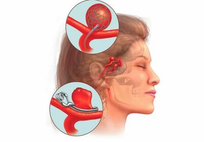
Cerebral aneurysm is a protrusion of the walls of the arteries of the brain. Divided into:
- Apoplexy, which is characterized by signs of intracerebral or subarachnoid hemorrhage that occurred after its sudden rupture;
- A tumor-like course, in which there is a symptomatic picture, including damage to the trigeminal, optic and oculomotor nerves.
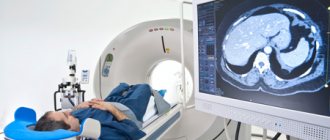
In order to diagnose this pathology, it will be necessary not only to take an anamnesis and a superficial neurological examination, but also to examine cerebrospinal fluid, skull radiography, magnetic resonance imaging, computed tomography and MRA. Successful treatment of an intracranial aneurysm often requires surgical treatment: clipping or endovascular occlusion.
An aneurysm is caused by degenerative changes, damage or underdevelopment of any of the layers of the vessel wall. In the normal state, there are three such layers: the outer layer is the adventitia, the middle layer is the muscular layer, and the inner layer is the intima. As a result of thinning and loss of elasticity under the influence of blood flow, protrusion of the vascular wall begins in the affected area. Most often, cerebral aneurysm appears in places where arteries branch, where the blood flow pressure is highest.
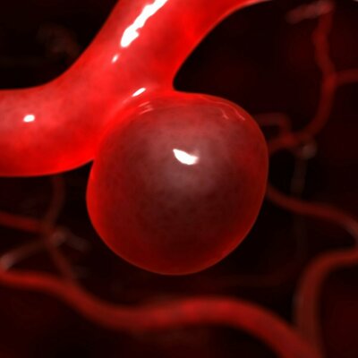
A cerebral aneurysm consists of three parts: the dome, the neck and the walls. The neck, like the vessel, has 3 layers, while the dome is formed only by the intima. It is this that is the thinnest place where the gap occurs.
According to approximate statistics, 5% of the world's population has cerebral aneurysms, which can be asymptomatic for a long time. As aneurysmal expansion increases, the vascular wall begins to thin, resulting in a rupture in the dome area, which results in a hemorrhagic stroke. In addition, most often this pathology is the cause of non-traumatic subarachnoid hemorrhages. Up to 85% of all cases of SAH fall into this category. The risk group for rupture of an intracranial aneurysm includes people aged 30-50 years.
Causes
With hyalinosis and atherosclerosis, after a TBI, against the background of hypertension, the structure of the walls of blood vessels is disrupted. Which leads to the formation of an acquired aneurysm. In rare cases, its appearance can be triggered by infectious emboli that have entered the arteries of the brain. Such a cerebral aneurysm is called mycotic. Pathology occurs if arterial hypertension and uneven blood flow are present.
With developmental anomalies leading to disruption of the normal anatomical structure of the vascular walls, a person may be born with a congenital protrusion. This pathology is most often combined with other malformations: coarctation of the aorta, polycystic kidney disease, arteriovenous malformation of the brain, connective tissue dysplasia and others.
How dangerous is the disease?
Firstly, this disease can develop in the human body for a long time and not manifest itself in any way. Secondly, the disease progresses rapidly and, filling with blood, the protrusion of the vessel wall begins to put pressure on the brain tissue and nerve endings, leading to a deterioration in cerebral circulation. Without proper treatment, there is a risk of aneurysm rupture, followed by cerebral hemorrhage and neurological disorders.
In order to promptly detect this disease and begin effective treatment, it is necessary to become familiar with its symptoms and causes.
Classification of the disease
>The disease can be classified according to its location. Aneurysms occur:
- Internal carotid artery;
- Anterior cerebral artery;
- Vertebro-basilar system;
- Middle cerebral artery;
- Multiple aneurysms affecting several vessels at the same time.
The latter type of disorder occurs in approximately 13% of patients.
In addition, aneurysms are distinguished by shape:
- Fusiform;
- Saccular, occurs fifty times more often. It can be multi-chamber or single-chamber.
By size they are distinguished:
- Giant - more than 25 millimeters;
- Large – 16-25 millimeters;
- Medium – 11-15 millimeters;
- Small – up to 10 millimeters;
- Miliary - up to 3 millimeters.
Types of disease
The classification of pathology is based on its form:
- Saccular aneurysm.
It is most common among cases of aneurysm in the brain area. - Dissecting aneurysm.
The formation has an elongated shape and is located between the layers of the vessel wall. Often found on the aorta due to the appearance of an intimal defect. Blood begins to gradually penetrate, stratifying the walls and causing the formation of a cavity. In the vessels of the brain, pathology is much less common than in the aorta. - Vertebral aneurysm.
It is also rare in the blood vessels of the brain and develops mainly on the wall of the aorta. The formation is cylindrical in shape, the vascular wall expands evenly.
At the initial stage of formation, the aneurysm does not manifest itself in any way. As the pathology develops, the symptoms become pronounced. This is due to the pressure of the formation on surrounding tissues or rupture and blood entering the brain tissue.
Aneurysms are divided into small (up to 11 mm), medium (up to 25 mm) and large (more than 25 mm) aneurysms by size. Also illness
can be congenital and acquired, with multiple and single formations.
Characteristic symptoms
Depending on whether a tumor-like or apoplectic aneurysm is observed in the patient, the symptomatic picture changes. During apoplexy, headaches in the fronto-orbital region are occasionally observed before rupture. But most often the breakup occurs suddenly.
The first symptom of this is sharp, intense cephalalgia. Initially, it manifests itself locally at the location of the aneurysm rupture, gradually becoming diffuse. The headache is accompanied by repeated vomiting and nausea. Meningeal symptoms are observed:
- Stiff neck;
- Hyperesthesia;
- Kernig and Brudzinski syndromes;
- Signs of mental disorder or epileptiform seizures appear, which can include mild confusion and acute psychosis.
- The patient may faint.
Subarachnoid hemorrhage, which occurs when an aneurysmal extension ruptures, is accompanied by a prolonged spasm of the arteries located in the area of the aneurysm. Moreover, in 65% of cases, such vascular spasm can cause ischemic stroke with damage to the brain substance.
Rupture of a cerebral aneurysm may also be accompanied by hemorrhage into the ventricles or substance. In 22% of cases, an intracerebral hematoma is observed, accompanied by focal symptoms, depending on the location of the rupture and increasing over time. The most severe variant of the development of pathology occurs when blood enters the ventricles of the brain. It occurs in approximately 14% and is almost always fatal.
It is worth noting that depending on the location of the aneurysm, focal symptoms are very diverse:
- Violation of the anterior cerebral artery is accompanied by mental disorders and paresis of the lower extremities;
- An aneurysm in the area of the bifurcation of the carotid artery causes visual impairment;
- With a rupture in the middle cerebral artery, speech impairment and hemiparesis of the opposite region are noted;
- In the vertebrobasilar system, the characteristic signs are: dysarthria, dysgraphia, ataxia, nystagmus, alternating syndrome, facial nerve paresis and damage to the trigeminal nerve.
Since the protrusion of the cerebral vessels in the cavernous sinus extends beyond the dura mater, when it ruptures there is no hemorrhage into the cranial cavity.
A tumor-like aneurysm progresses over time and, reaching a significant size, compresses nearby parts of the brain, which leads to the appearance of characteristic symptoms of an intracranial tumor, manifested in the cavernous sinus or in the area of the optic chiasm, that is, the chiasma. The anomaly is accompanied by:
- Reduced acuity and narrowing of visual fields.
- Optic nerve atrophy;
- A combination of paresis of the III, IV and VI pairs of the cranial nerve with damage to the branches of the trigeminal nerve (typical of a cerebral aneurysm in the cavernous sinus);
- Oculomotor disorders with the development of strabismus or weakening/impossibility of convergence that accompany lesions of the III, IV and VI pairs;
- Trigeminal neuralgia;
- In the absence of timely treatment, a cerebral aneurysm can cause destruction of the skull bones that can be diagnosed by radiography.
Symptoms
There are several characteristic signs of an intracranial aneurysm:
- Violation of the tone of the facial muscles on one side.
- Headaches that come on suddenly.
- Dilated pupils.
- Pain in the eyes, blurred vision.
- Numbness, loss of sensitivity.
- Vomiting and nausea.
- Double vision, loss of consciousness.
- Photophobia.
- Speech impairment.
At the very beginning of the development of the disease, there are practically no manifestations. Noticeable signs begin to appear as the pathology develops, its size increases, or the cavity ruptures. Symptoms depend on the amount of blood shed and the location of the process.
Get an aneurysm diagnosis at Clinic No. 1
- Neurologist appointment
- Appointment with a phlebologist
- MRI of the head
- CT head
For one-time payment for services - 20% discount
Call
Diagnostic methods
Often, cerebral aneurysm is asymptomatic and is detected by chance. If clinical symptoms are present, to make a diagnosis, a neurologist needs:
- Taking anamnesis;
- Neurological examination, which reveals focal and meningeal symptoms. Based on them, it is possible to establish the localization of the aneurysm and make a topical diagnosis;
- Magnetic resonance or computed tomography allows you to clarify the diagnosis;
- X-ray examination shows the presence of bone destruction and reveals petrified aneurysms;
- Cerebrospinal fluid examination is performed if it is impossible to use more accurate diagnostic methods. If blood is found in the cerebrospinal fluid during a lumbar puncture, this may indicate subarachnoid hemorrhage;
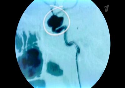
- Angiography is considered the most informative method, allowing one to clarify the shape, size and location of a cerebral aneurysm. In this case, magnetic resonance angiography does not require the introduction of a contrast agent and can be performed even in the acute period when an aneurysm ruptures. Using MRA, you can obtain a three-dimensional volumetric image of vessels or their two-dimensional cross-sectional projection.
Diagnosis is complicated by the need to distinguish an apoplectic aneurysm from a transient ischemic attack, an epileptic attack and meningitis, and a tumor-like aneurysm from a cyst, tumor and abscess of the brain with similar symptoms.
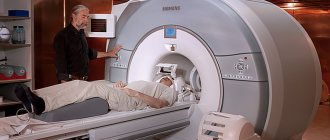
Is a cerebral aneurysm visible on an MRI?
Magnetic resonance imaging involves layer-by-layer scanning of the anatomical zone under consideration to obtain images in increments of 1 mm. The method is safe and approved for use in children and pregnant women. MRI is based on the phenomenon of nuclear magnetic resonance, characteristic of tissues saturated with fluid.
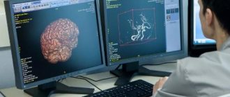
A conventional head scan produces detailed images of brain structures. For a more thorough examination of the arteries, angiography is used. The combination of standard MRI and vascular mode allows you to obtain maximum information about the state of the brain and is informative regarding aneurysms with a diameter of 3 mm or more.
The angio mode is based on the effect of fluid flow. In constantly moving blood, nuclear magnetic resonance does not occur, so the signal from the vessels differs from that of magnetized tissues. The computer program essentially eliminates the arteries from the image. This allows you to assess the topography, size and permeability of the latter.
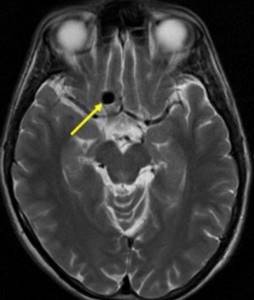
Aneurysmal dilatation of cerebral vessels on the M-scan (indicated by an arrow)
MRI scans show vessels of different diameters. A brain aneurysm will look like a thickening along the artery. The method makes it possible to identify blood clots inside the aneurysmal sac and give more accurate predictions regarding the patient’s condition.
The question of whether an MRI shows a cerebral aneurysm often arises when diagnosing small protrusions. Dilatation of small diameter or unusual location may indeed go unnoticed. If vascular pathology is suspected, radiologists recommend repeating the study with contrast. Gadolinium preparations are used as an enhancer. These are secure connections. Gadolinium-based products do not interact with the tissues of the human body and are excreted unchanged by the kidneys within 6-12 hours. Contrast allows you to more clearly visualize large arteries and study small-diameter vessels.
The use of the amplifier has virtually no restrictions. Contrast is not used in children, pregnant women, or those with end-stage liver and kidney diseases. When using an amplifier, the scanning time increases by 2 times, since essentially two examinations are performed - native and after intravenous administration of gadolinium.
If a person is at risk for an aneurysm and has neurological symptoms, MRI of the head is often supplemented with X-ray methods for studying blood vessels.
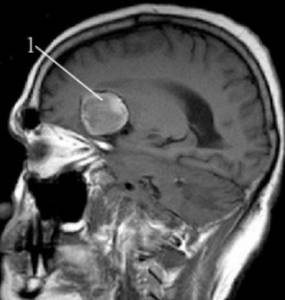
Giant aneurysm (indicated by number 1) of the middle cerebral artery on an MR image
Diagnosis depends on the power of the equipment, experience and skills of the doctor who interprets the images. To obtain accurate results, it is better to examine cerebral vessels in specialized centers that have high-field tomographs with a magnetic field strength of 1.5 Tesla.
Therapy for cerebral aneurysm
If there is a small or miliary aneurysm, the patient must be regularly monitored by a neurologist or neurosurgeon to monitor the size and course of the pathology. It involves conservative therapy to avoid an increase in the size of the aneurysm. It includes normalization of heart rate and blood pressure, correction of cholesterol levels in the blood, treatment of infectious diseases and the consequences of traumatic brain injuries.
Surgery is required to prevent ruptures. This may include endovascular occlusion and clipping of the aneurysm neck. Stereotactic electrocoagulation and artificial thrombosis can be performed using coagulants. Vascular malformations are removed using transcranial or radiosurgical methods.
A ruptured aneurysm is an acute condition requiring emergency medical attention. Therapy is similar to the treatment of hemorrhagic stroke. It includes:
- Removal of hematoma;
- Stereotactic aspiration or endoscopic evacuation;
- Ventricular drainage if the aneurysm is accompanied by bleeding into the ventricles.
Diagnosis and treatment of cerebral aneurysm at the Medis Clinic
When you contact the Medis clinic to diagnose a disease, you will be provided with the services of some of the most highly qualified specialists in Moscow using high-tech and new equipment.
Diagnosis of cerebral aneurysm includes the following procedures:
- appointment with a neurosurgeon;
- MRI;
- endovascular examination;
- angiography with contrast or CT angiography;
- preoperative preparation (laboratory tests, ECG, X-ray of the lungs).
To learn more about the procedures and costs, use the search engine on our website or sign up for a consultation online or by phone.
Treatment of any type of aneurysm is carried out only by surgery. Treatment tactics depend on a number of factors: the location of the aneurysm, the presence (absence) of signs of its rupture, the duration of hemorrhage, the clinical condition of the patient and the presence of complications.
Our clinic offers a range of necessary procedures for highly effective treatment of the disease:
- microsurgical removal of intracerebral hematoma of the cerebral hemispheres with simultaneous clipping of the neck of the cerebral artery aneurysm using videoangiography;
- strengthening the walls of the aneurysm;
- clipping of the neck of the aneurysm, etc.
Prices for these procedures range from $2,657 to $8,192. You can view the price list in more detail on our website in the appropriate block. The final price of the operation will depend on the results of the diagnostic study.
Probable forecast
Depending on the location of the affected vessel, the probable prognosis also differs. In addition, the specialist has to take into account the size of the aneurysm and the presence of concomitant pathologies that contribute to degenerative changes in the vascular walls.
- In the absence of aggravating factors, an intracranial aneurysm can exist without clinical changes throughout the patient's life.
- Aneurysm rupture leads to disability in 25-35% of cases, and death in 30-50%;
- The probability of recurrent hemorrhage is high at 20-25%, and in seventy cases the patient dies.
Diagnostics
A routine examination does not reveal an aneurysm. It makes it possible to suspect pathology and determine the path of examination. Basic diagnostic measures:
- Palpation, examination of the skin.
- Blood pressure measurement.
- Neurological examination, which includes: assessment of muscle tone, reflexes, test movements to assess the functioning of the nervous system.
- CT and MRI are the main methods for making an accurate diagnosis. They allow you to determine the location and size of the formation, the presence of blood clots, and assess the condition of the surrounding tissues.
Treatment
The main method of combating an aneurysm is surgery, during which the pathological formation is removed and the integrity of the vessels is restored. Several types of operation are used:
- Microsurgical or aneurysm clipping.
Performed after craniotomy. This is a multi-hour operation that is associated with a risk to the health and life of the patient. - Endovascular surgery.
A high-tech method that does not involve craniotomy. Access is through the femoral or carotid artery. Using a needle, the specialist can penetrate the skull area and close the gap in the artery wall. Operations of this type are performed using high-precision equipment with MRI or X-ray control.
Endovascular techniques have a number of advantages. They are low-traumatic, involve minimal risk, and often do not require general anesthesia. Recovery time is minimal, and the ability to access hard-to-reach areas is significantly expanded.
Get an aneurysm diagnosis at Clinic No. 1
- Neurologist appointment
- Appointment with a phlebologist
- MRI of the head
- CT head
For one-time payment for services - 20% discount
Call