Duplex scanning
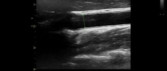
is an improved diagnostic method that combines standard ultrasound and Doppler ultrasound. This method allows you to diagnose vascular pathologies at the earliest stage of development. This is possible due to the fact that the doctor can evaluate not only the external characteristics of the vessels, but also the internal structure, which makes it possible to identify internal pathology.
Duplex scanning makes it possible to:
- Determination of blood flow speed in vessels
- Definitions of any vascular pathology
- Determining the causes of blood flow disorders
Benefits of Duplex Scanning
Sonography of this kind is carried out using modern equipment, which combines standard ultrasound and Doppler scanning in this method. Main advantages in:
- maximum information content and accuracy;
- speed of obtaining final results (in general, the duration of the entire process is approximately half an hour to an hour);
- reliability;
- complete safety;
- comfort during the study;
- no age restrictions, as well as special necessary preparation measures.
If a duplex scan of the brachiocephalic vessels was carried out, the norms of indicators or their deviations, allowing one to determine the earliest, preclinical symptoms of vascular diseases, will also allow assessing the degree of blood flow disturbance or vascular damage, identifying the presence of blood clots and atherosclerotic plaques, understanding the condition of the walls of blood vessels (their stretching; structure – abnormal or not; presence of valvular insufficiency).
Triplex scanning
The words “duplex” and “triplex,” as you might guess, come from the numbers 2 and 3, respectively. These numbers indicate in how many dimensions the arteries are examined. With triplex, the doctor assesses their condition using three ultrasound modes, while with duplex - only two. However, triplex does not have a big advantage, since even with a two-dimensional image you can get enough information about the patient’s health. Here, the quality of medical equipment and the skill of the doctor come to the fore, rather than the number of measurements. Therefore, everything described below can be equally attributed to both duplex and triplex.
Indications
Duplex testing is recommended for both healthy people for preventive purposes and those who are at risk, namely, with factors predisposing to the development of vascular diseases such as smoking, diabetes, age over 40 years, obesity, impaired lipoprotein and lipid metabolism, elevated cholesterol levels, sedentary lifestyle, as well as chronic stressful conditions.

This type of examination is also prescribed if there are indications such as impaired coordination of movements, tinnitus, constant dizziness, etc. Duplex scanning is carried out either before or after operations. Even if the norm of duplex scanning of brachiocephalic or other arteries corresponds to your indicators after the study, this check and exclusion of violations will never be superfluous. In general, such timely diagnosis makes it possible to identify many diseases in the early stages and, therefore, receive correct and effective treatment, preventing many complications in the future.
Direct scanning of the brachiocephalic arteries has a narrower range of indications, namely:
- pain (sometimes throbbing) in the neck and head when changing their positions (turns, etc.);
- sleep disorder (hypersomnia or vice versa - insomnia);
- cognitive impairment (deterioration of mental capacity, memory, etc.);
- visual impairment (loss of visual fields, flickering spots) and hearing (feeling of ear congestion, noise);
- fainting, migraines and severe, frequent dizziness;
- weakness in the limbs, temporary numbness of the body or face (sometimes paralysis);
- unsteadiness when walking, as well as vestibulopathy;
- lethargy, weakness, apathy, emotional disorders;
- seizures;
- changes in blood pressure (hyper- and hypotension).
In addition to the above signs of the need to perform duplex scanning of the vessels of the brachiocephalic arteries, there are also specific pathologies and abnormalities that imply such a diagnosis. These are tumor processes in the neck, dyscirculatory encephalopathy, post-stroke condition, dystonia, vasculitis, arrhythmia, atherosclerosis, hematological and systemic vascular pathologies, as well as osteochondrosis and injuries of the cervical spine.
Preparation for the procedure
For the procedure to produce results, you need to know which vessels need to be checked. You should not immediately sign up for this type of diagnosis: you must first make an appointment with a vascular surgeon. This is a highly specialized doctor who, based on the patient’s complaints and information from his medical record, will determine the scope and type of diagnosis. He can confirm or refute some diseases without scanning, so to save time and money, a preliminary consultation is necessary.
Based on the results of the examination, the doctor will prescribe a specific option. In most cases, the surgeon writes out a referral for diagnostics of the lower or upper extremities or analysis of the condition of the vessels of the head and neck. These procedures allow you to identify many diseases of the cardiovascular system and do not require additional preparation. This is due to the fact that these vessels are easily accessible to ultrasound, and the image on the screen is clear.

Another thing is the diagnosis of the abdominal cavity and pelvis. This type of study is used less frequently, but if it is prescribed, the patient requires a special diet for three days. During this time, it is prohibited to eat a whole list of foods: meat, milk, brown bread and, in general, any foods that contain a large amount of fiber. During the period of preparation for diagnosis, you should take medications that reduce the formation of gases in the intestines.
Procedure process
Duplex scanning of the brachiocephalic arteries, decoding, norm and deviations - all this is known by an experienced phlebologist, who, if necessary, refers for a study of a similar plan. By the way, no special preparation is required, the only thing is that the day before it is important to avoid alcohol and smoking, as well as taking vascular medications.

Duplex scanning is carried out using a special ultrasound machine, in comfortable conditions, in a specially equipped room and in a position where the patient lies on his back. The specialist moves the ultrasound sensor itself over the head and neck, and may also ask you to either take rapid breathing, or hold it, or turn your head in one direction or another. Thanks to the corresponding sensor, signals are sent to the computer, which, in fact, are converted into an image. During the scanning process, it is not recommended to turn your head or actively move without the request of a specialist. In general, this manipulation does not cause discomfort or pain in the patient. The only very short-term discomfort that may be present during duplex scanning is slight pressure on one or another area of the neck (for example, where the carotid artery is located). However, this is necessary for even greater information content and correct display of the final results.
Preparation for duplex scanning of cerebral vessels
Duplex scanning of blood vessels does not require special preliminary preparation. The only requirement for the patient on the day of the upcoming examination will be to abstain from taking substances that affect vascular activity (tea, medications, nicotine and coffee). Failure to comply with these requirements may result in receiving inaccurate information during the examination. Medicines that cannot be interrupted for a while must continue to be taken. The possibility of taking a break should be discussed with your doctor. Before the actual scan, the patient needs to remove jewelry and hairpins from his head and neck.
Duplex scanning of neck vessels: norm, deviations and interpretation of results
Pathology can be determined in almost any area of the vascular basin, and the assessment of certain changes within the lumen will even make it possible to identify diseases at an early stage. The decoding is based on the characteristics obtained from ultrasound examination in the form:
- the thickness of the artery along with its homogeneity;
- mobility and its diameter;
- its forms;
- patency, as well as the presence of occlusions or narrowings (stenosis or blockage of the artery, respectively);
- the presence of any changes in the type of bends; crimps.
Speaking about what the norm is for duplex scanning of the brachiocephalic arteries, we note that for different vessels in this zone they are different - they all have completely individual indicators. For example, the normal diameter of the carotid artery is considered to be 4-7 mm, and the blood flow to the brain is 55 ml per 100 g of tissue. Indicators are also determined in the form of an index of vascular resistance, diastolic and systolic speed of their blood flow, diameter of the external/internal branches, etc.
Duplex scanning of blood vessels
Duplex scanning of blood vessels is a highly accurate method for diagnosing diseases and pathologies of the vascular system at the earliest stage of development. This examination helps the doctor more accurately diagnose the disease and develop treatment tactics.
Duplex scanning of blood vessels is performed for the following diseases:
Conclusion
Duplex scanning of the arteries, as an accessible, fairly detailed and, most importantly, safe study necessary to determine the condition of the vessels and identify the cause of certain symptoms, can be supplemented by other tests similar to the same ultrasound examination of the lower extremities or abdominal organs. This is usually necessary to identify a blocked or narrowed vessel in other places and make an even more informative diagnosis.
According to statistics, an annual preventive examination of blood vessels makes it possible to predict the development of a stroke by almost 90%.
The appointment is conducted by doctors
Pryalukhin Alexander Nikolaevich
Honored Doctor of the Russian Federation, doctor of the highest category, ultrasound diagnostics doctor
Karimova Galiya Mikhailovna
Ultrasound diagnostic doctor
Belova Daria Nikolaevna
Ultrasound diagnostic doctor
Petrova Natalya Sergeevna
Ultrasound diagnostic doctor
- Prices for ultrasound of blood vessels of the head, neck and extremities
Note! Prices are for adult patients. Please see the “Pediatrics” section for the cost of children’s appointments.
| Ultrasound of cerebral vessels (duplex scanning) | 2420 |
| Ultrasound of neck vessels (extracranial) | 2420 |
| Ultrasound (duplex) of veins of the extremities (two limbs) | 2180 |
| Ultrasound (duplex) of the arteries of the limbs (two limbs) | 2670 |
Duplex scanning is a relatively new development that has replaced the outdated Doppler ultrasound. While Doppler allows you to study only the direction of blood flow and its speed, duplex gives the doctor more information, including data on the condition of the blood vessels. The procedure is based on the work of ultrasonic waves, which the human ear cannot perceive. Depending on the state of the vessels, they are reflected and refracted differently; fall on the sensor, which forms an image on the monitor screen.
Some patients are wary of this procedure because they are afraid of the harmful effects of vibrations. In reality, there is nothing wrong with the procedure. Various studies have not revealed any negative effects of ultrasound on the human body. And if we take into account the fact that the patient does not feel discomfort during the session, then the procedure becomes one of the most effective diagnostic methods. Unlike X-ray diagnostics, scanning can be used several times with short breaks.

Order of conduct
Duplex is a safe and painless procedure that does not require anesthesia. The only preliminary operation is to apply a special gel that improves the passage of ultrasound. The examination is carried out in a standing, sitting or lying position, depending on the purpose of the procedure. The doctor places a sensor on the patient's body and moves it over the skin, observing a detailed picture on the screen. He may also ask the patient to perform simple exercises: hold his breath, move his arm or leg, etc. The whole procedure takes a little over half an hour.
Once completed, the patient needs to wash off the gel and can go home and continue with their business. There are no restrictions on lifestyle, nutrition or exercise. They are determined only by the rules of treatment or recovery after an identified disease.
In many cases, the procedure is the only reliable method for diagnosing diseases of the cardiovascular system. In other cases, duplex is used in combination with other research options - X-ray examination, magnetic resonance angiography, etc. The big advantage is complete safety for the patient. Any ultrasound examination, including duplex, causes complications extremely rarely. Duplex can be used frequently, which is especially important for assessing the dynamics of recovery.


