Ultrasound in the CIR group of companies (Center for Immunology and Reproduction)
Average size of the uterus in the first trimester of pregnancy
| Date of last menstruation (weeks) | Time at conception (weeks) | Length (mm) | Width (mm) | Anterior-posterior size (mm) | Perimeter (mm) | Area (mm2) | Volume (mm3) |
| 5 | 3 | 71 | 50 | 40 | 150 | 2170 | 74000 |
| 6 | 4 | 80 | 57 | 45 | 158 | 2330 | 94000 |
| 7 | 5 | 91 | 68 | 49 | 167 | 2770 | 119000 |
| 8 | 6 | 99 | 74 | 52 | 179 | 3086 | 152000 |
| 9 | 7 | 106 | 78 | 55 | 187 | 3828 | 183000 |
| 10 | 8 | 112 | 83 | 58 | 199 | 4368 | 229000 |
| 11 | 9 | 118 | 89 | 62 | 210 | 5138 | 287000 |
| 12 | 10 | 122 | 95 | 66 | 222 | 5794 | 342000 |
| 13 | 11 | 135 | 102 | 70 | 234 | 6809 | 365000 |
Average size of the fertilized egg in the first trimester of pregnancy
| Date of last menstruation (weeks) | Time at conception (weeks) | Inner diameter (mm) | Area (mm2) | Volume (mm3) |
| 5 | 3 | 18 | 245 | 2187 |
| 6 | 4 | 22 | 363 | 3993 |
| 7 | 5 | 24 | 432 | 6912 |
| 8 | 6 | 30 | 675 | 13490 |
| 9 | 7 | 33 | 972 | 16380 |
| 10 | 8 | 39 | 1210 | 31870 |
| 11 | 9 | 47 | 1728 | 55290 |
| 12 | 10 | 56 | 2350 | 87808 |
| 13 | 11 | 65 | 3072 | 131070 |
Is it necessary to carry out
Throughout pregnancy, the expectant mother undergoes 3 scheduled functional and biochemical screenings. The first is carried out between the 11th and 13th weeks of gestation, the second occurs at 18–20 weeks, and the third at 3–34 weeks. During this time, the woman undergoes an ultrasound, gynecological smears, blood and urine tests. All studies are aimed at early detection of pathologies of fetal development and the course of pregnancy.
CTE is measured during the first screening. Later, this indicator loses its information content. Ultrasound examination does not harm the unborn child, but helps to detect deviations from the norm in its development in time. Therefore, it needs to be carried out. Based on the results of the diagnostician’s conclusion, additional laboratory and instrumental studies, observation and treatment may be prescribed.
Screening should be carried out throughout pregnancy.
Average size of the embryo in the first trimester of normal pregnancy
| Date of last menstruation (weeks) | Time at conception (weeks) | Coccygeal-parietal size (mm) | Biparietal size (mm) | Yolk sac diameter (mm3) |
| 5 | 3 | 3 | — | — |
| 6 | 4 | 6 | — | 3 |
| 7 | 5 | 10 | — | 4 |
| 8 | 6 | 16 | 6,0 | 4,5 |
| 9 | 7 | 23 | 8,5 | 5,0 |
| 10 | 8 | 31 | 11,0 | 5,1 |
| 11 | 9 | 41 | 15,0 | 5,5 |
| 12 | 10 | 53 | 20,0 | 6,0 |
| 13 | 11 | 66 | 24,0 | 5,8 |
Why is CTE different from the norm?
It happens that the data differs from the norm. Almost all women in this case begin to worry. In most cases, unnecessary worries are in vain. Minor deviations do not pose a danger to either the fetus or the mother.
If the indicators are above or below the norm, which do not normalize over a long period of time, it can be judged that the baby is small or, conversely, large in size. If there is a significant deviation, the doctor may suspect the course of pathological processes.
If abnormalities are suspected, a woman is recommended to undergo a comprehensive examination. It is important that the doctor determines the exact root cause of the CTE disorder.

If deviations are detected, a comprehensive study will be ordered
Why is CTE less than normal dangerous?
The first thing a doctor suspects when CTE is less than required is a non-developing pregnancy. In this case, the consequences are extremely unpleasant for the mother. The violation indicates that the child died. The condition requires urgent medical attention.
If you ignore the current condition, the process of decay may begin in the body. Any delay can lead to infertility. Sometimes there is a possibility of death in a woman. Curettage is extremely necessary.
During a frozen pregnancy, a woman’s health deteriorates significantly and internal bleeding begins. Possibly an infectious-toxic condition. The risk of shock is also high.
A reading less than required may also indicate hormonal deficiency. This can be dangerous due to spontaneous abortion.
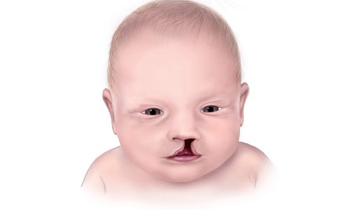
If a violation is suspected, a woman should urgently consult a doctor and undergo a comprehensive examination. A figure less than normal is a clear sign of genetic abnormalities. In this case, there is a risk of needing a medical termination of pregnancy due to diseases incompatible with the life of the fetus. Such a complication is difficult for a woman to endure on a moral level.

If the CTE is below normal, this may be a sign of fetal pathology
Why is CTE more dangerous than normal?
There is no need to worry if the child is several mm larger than normal and is steadily growing. This usually indicates that the baby is large. In this case, all you need is to reconsider your diet. There is no danger to the expectant mother or baby.
A higher than normal figure is also often found when a woman does not remember the exact date of her last menstruation. This suggests that the preliminary gestational age is set incorrectly. The situation does not pose any danger.
The condition can be dangerous when the fetus enlarges due to the presence of diabetes mellitus in the expectant mother. Such a woman needs careful monitoring of the progress of her pregnancy. Otherwise, the risk of premature birth is high.
From this video you will learn about the features and parameters that are noted on ultrasound in early pregnancy:
Root causes of slow growth of readings
Sometimes the increase in CTE occurs slowly, but at the same time the child has both a heartbeat and motor activity. The baby is falling behind the required norm. In most cases, the condition is caused by the presence of pathological processes.
Growth retardation is caused by genetic diseases. It is difficult to establish this at the initial time.
High risk of presence:
- Down's disease;
- Patau syndrome;
- Turner's disease.
To confirm or refute, you should donate blood for analysis or resort to a prenatal DNA test. If necessary, ultrasound is recommended again.
If the indicator deviates, Patau syndrome may appear
Another root cause of growth deficiency is endometrial insufficiency. This is likely if the pregnant woman has a history of abortion, which was performed relatively recently. Increased risk of miscarriage. The woman needs urgent hospitalization.
Relationship between the coccygeal-parietal size (CPR) and the duration of pregnancy (based on the last menstrual period)
| KTE, mm | Weeks + days | KTE, mm | Weeks + days | KTE, mm | Weeks + days |
| 7 | 6+3 | 32 | 10 | 57 | 12+2 |
| 8 | 6+4 | 33 | 10+1 | 58 | 12+3 |
| 9 | 6+6 | 34 | 10+2 | 59 | 12+3 |
| 10 | 7 | 35 | 10+2 | 60 | 12+4 |
| 11 | 7+2 | 36 | 10+3 | 61 | 12+4 |
| 12 | 7+3 | 37 | 10+4 | 62 | 12+5 |
| 13 | 7+4 | 38 | 10+5 | 63 | 12+5 |
| 14 | 7+5 | 39 | 10+6 | 64 | 12+6 |
| 15 | 7+6 | 40 | 11 | 65 | 12+6 |
| 16 | 8 | 41 | 11 | 66 | 13 |
| 17 | 8+1 | 42 | 11+1 | 67 | 13 |
| 18 | 8+2 | 43 | 11+2 | 68 | 13+1 |
| 19 | 8+3 | 44 | 11+2 | 69 | 13+1 |
| 20 | 8+4 | 45 | 11+3 | 70 | 13+2 |
| 21 | 8+5 | 46 | 11+3 | 71 | 13+2 |
| 22 | 8+6 | 47 | 11+4 | 72 | 13+3 |
| 23 | 9 | 48 | 11+5 | 73 | 13+3 |
| 24 | 9+1 | 49 | 11+5 | 74 | 13+4 |
| 25 | 9+2 | 50 | 11+6 | 75 | 13+4 |
| 26 | 9+3 | 51 | 11+6 | 76 | 13+5 |
| 27 | 9+3 | 52 | 12 | 77 | 13+5 |
| 28 | 9+4 | 53 | 12+1 | 78 | 13+5 |
| 29 | 9+5 | 54 | 12+1 | 79 | 13+6 |
| 30 | 9+6 | 55 | 12+2 | 80 | 13+6 |
| 31 | 10 | 56 | — | — |
Decoding the results
In the first trimester of gestation, embryos grow approximately the same, regardless of race, heredity and environmental conditions. But this happens only until the 14th week, when the genetic characteristics of the future person appear. Therefore, the growth rate is changing.
To correctly assess CTE, it is important to conduct screening before the 14th week of pregnancy, then the calculations will be quite accurate. The specialist compares the obtained dimensions with tabular indicators and makes a conclusion about the absence or presence of deviations.
The conclusion of a functional diagnostics doctor is not a diagnosis.
And the detected deviations should become an indication for additional research to confirm or refute the pathology.
Relationship between biparietal size (BDS) of the fetal head and gestational age (based on last menstruation)
| BPR (mm) | Duration on average (weeks) | Fluctuation range (week) | BPR (mm) | Duration on average (weeks) | Fluctuation range (week) |
| 17 | 10,6 | 9,6—11,5 | 58 | 23,5 | 22,7—24,4 |
| 18 | 10,9 | 9,9—11,8 | 59 | 23,8 | 23,0—24,7 |
| 19 | 11,2 | 10,2—12,1 | 60 | 24,1 | 23,3—25,0 |
| 20 | 11,5 | 10,5—12,4 | 61 | 24,4 | 23,7—25,4 |
| 21 | 11,8 | 10,8—12,7 | 62 | 24,7 | 24,0—25,7 |
| 22 | 12,1 | 11,1—13,0 | 63 | 25,1 | 24,3—26,0 |
| 23 | 12,4 | 11,4—13,3 | 64 | 25,4 | 24,6—26,4 |
| 24 | 12,7 | 11,7—13,6 | 65 | 25,7 | 25,0—26,7 |
| 25 | 13,0 | 12,0—13,9 | 66 | 26,0 | 25,3—27,0 |
| 26 | 13,3 | 12,3—14,2 | 67 | 26,3 | 25,6—27,4 |
| 27 | 13,6 | 12,6—14,5 | 68 | 26,7 | 25,9—27,7 |
| 28 | 13,9 | 12,9—14,9 | 69 | 27,0 | 26,3—28,0 |
| 29 | 14,2 | 13,2—15,2 | 70 | 27,2 | 26,6—28,4 |
| 30 | 14,4 | 13,5—15,4 | 71 | 27,6 | 26,9—28,7 |
| 31 | 14,7 | 13,8—15,7 | 72 | 27,9 | 27,2—29,0 |
| 32 | 15,0 | 14,1—16,0 | 73 | 28,2 | 27,5—29,4 |
| 33 | 15,3 | 14,4—16,4 | 74 | 28,5 | 27,9—29,8 |
| 34 | 15,6 | 14,6—16,7 | 75 | 28,9 | 28,2—30,3 |
| 35 | 15,9 | 14,9—17,0 | 76 | 29,2 | 28,5—30,8 |
| 36 | 16,2 | 15,2—17,3 | 77 | 29,5 | 28,8—31,3 |
| 37 | 16,5 | 15,5—17,6 | 78 | 29,8 | 29,2—31,8 |
| 38 | 16,8 | 15,8—17,9 | 79 | 30,6 | 29,5—32,3 |
| 39 | 17,1 | 16,1—18,3 | 80 | 31,1 | 29,8—32,8 |
| 40 | 17,4 | 16,4—18,6 | 81 | 31,6 | 30,0—33,3 |
| 41 | 17,7 | 16,7—18,9 | 82 | 32,1 | 30,5—33,8 |
| 42 | 18,0 | 17,0—19,2 | 83 | 32,6 | 31,1—34,2 |
| 43 | 18,5 | 17,3—19,5 | 84 | 33,6 | 31,6—34,8 |
| 44 | 19,0 | 18,2—19,7 | 85 | 33,7 | 32,1—35,3 |
| 45 | 19,4 | 18,5—20,0 | 86 | 34,2 | 32,7—35,8 |
| 46 | 19,7 | 18,8—20,4 | 87 | 34,7 | 33,2—35,9 |
| 47 | 20,0 | 19,2—20,7 | 88 | 35,2 | 33,7—36,1 |
| 48 | 20,3 | 19,5—21,0 | 89 | 35,7 | 34,2—37,1 |
| 49 | 20,6 | 19,8—21,4 | 90 | 36,4 | 34,7—38,1 |
| 50 | 20,9 | 20,1—21,7 | 91 | 37,3 | 35,3—39,1 |
| 51 | 21,3 | 20,5—22,0 | 92 | 38,1 | 35,8—40,1 |
| 52 | 21,6 | 20,8—22,4 | 93 | 38,9 | 36,9—41,1 |
| 53 | 21,9 | 21,1—22,7 | 94 | 39,7 | 37,6—42,1 |
| 54 | 22,2 | 21,4—23,2 | 95 | 40,5 | 38,3—43,1 |
| 55 | 22,5 | 21,7—23,6 | 96 | 41,3 | 39,6—44,1 |
| 56 | 22,8 | 22,1—23,7 | 97 | 42,1 | 39,6—45,1 |
| 57 | 23,2 | 22,4—24,0 | 98 | 42,9 | 40,3—46,1 |
Examination of the mother for abnormalities
If the ultrasound CTE indicators indicate obstetric period, and there are deviations in them, then it is necessary to find the reason. For this, the doctor prescribes additional studies. First, the woman’s health is checked, examined for the presence of infection, chronic and hereditary diseases. To do this, you will need to do blood and urine tests, and study the pregnant woman’s medical record.
Then they begin to identify fetal pathologies. Several examination methods can be offered to choose from:
- Mniocentesis is the collection of amniotic fluid to study its parameters.
- Cord blood analysis.
- Chorionic villus biopsy.
After identifying the reasons that served as the impetus for stopping the child’s development or slowing it down, appropriate treatment is prescribed, which involves maintaining or terminating the pregnancy, depending on the specific situation.
In the period from 10-14 weeks, a pregnant woman undergoes an ultrasound scan. There is no need to prepare for the procedure, and it is safe. Since during this period the fetus, apart from the heart and the body itself, has no internal organs, development can be predicted only by CTE indicators, which must meet the standards characteristic of a certain obstetric stage of pregnancy. If the size of the baby deviates, a woman should not panic; perhaps it is just an incorrectly determined period. In this case, after the research, the doctor will adjust the indicators.
Average sizes of the fetal chest at different stages of pregnancy (according to the last menstruation)
| Duration (weeks) | Dimensions (mm) | ||||
| transverse | anterior-posterior | average | perimeter | area (mm2) | |
| 15 | 28 | 30 | 29 | 90 | 588 |
| 16 | 34 | 32 | 33 | 99 | 768 |
| 17 | 38 | 36 | 37 | 111 | 972 |
| 18 | 41 | 38 | 39 | 117 | 1200 |
| 19 | 44 | 44 | 44 | 132 | 1452 |
| 20 | 48 | 44 | 46 | 138 | 1587 |
| 21 | 50 | 48 | 49 | 147 | 1875 |
| 22 | 53 | 50 | 52 | 156 | 2028 |
| 23 | 58 | 53 | 56 | 168 | 2352 |
| 24 | 59 | 56 | 57 | 181 | 2523 |
| 25 | 62 | 60 | 61 | 183 | 2724 |
| 26 | 65 | 63 | 64 | 192 | 3072 |
| 27 | 69 | 66 | 67 | 201 | 3468 |
| 28 | 73 | 70 | 72 | 219 | 3888 |
| 29 | 76 | 72 | 74 | 228 | 4107 |
| 30 | 78 | 74 | 76 | 234 | 4332 |
| 31 | 81 | 77 | 79 | 243 | 4563 |
| 32 | 83 | 79 | 81 | 249 | 4810 |
| 33 | 85 | 82 | 83 | 255 | 5292 |
| 34 | 88 | 85 | 86 | 264 | 5547 |
| 35 | 91 | 89 | 90 | 273 | 6075 |
| 36 | 94 | 90 | 92 | 282 | 6348 |
| 37 | 97 | 92 | 94 | 291 | 6627 |
| 38 | 98 | 94 | 96 | 294 | 6912 |
| 39 | 99 | 96 | 98 | 297 | 7203 |
| 40 | 101 | 97 | 99 | 303 | 7498 |
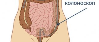
How to do it
The medical procedure is performed by a doctor using an ultrasound scanner using a special sensor - a transputer. CTE is measured in the only way - scanning the sagittal plane. With this method, the axial guide of the ultrasound sensor is directed along the body, that is, the orientation follows the type: from the head to the feet of the embryo. The manipulation is absolutely painless. Ultrasound examination of fetal CTE proceeds as follows:
- you need to lie on the couch and completely expose your stomach;
- the doctor will apply an acoustic contact paste-gel to the abdomen, which will provide vacuum contact with the body and facilitate the sliding of the sensor;
- Using an ultrasound scanner, the specialist will select the appropriate plane and take several frames on the monitor;
- After that, he will print them out and hand them over to the patient.
How is CTE of the fetus performed?
The main task of the doctor is to fix the frame in an unbent, extended position of the fetus. To more accurately capture a still image, you have to be patient and wait until the baby calms down in the womb. The sagittal plane runs in the direction of absolute symmetry on both sides, and after taking a freeze frame, it visually shows it on the monitor screen.
The calculation occurs automatically. This is done by a program that is built into the scanner. The CTE distance obtained in millimeters is compared with existing standards. For this, there is a special table that indicates the correspondence of measurements to a specific stage of pregnancy. The resulting numbers are immediately written on the screen, after which a protocol with the data is printed and handed to the patient.
Average dimensions of the abdominal cavity of the fetus (gestational age - at the last menstruation)
| Duration (weeks) | Dimensions (mm) | ||||
| transverse | anterior-posterior | average | perimeter | area (mm2) | |
| 14 | 26 | 20 | 23 | 72 | 396 |
| 15 | 30 | 24 | 27 | 78 | 548 |
| 16 | 34 | 25 | 32 | 96 | 760 |
| 17 | 40 | 32 | 36 | 108 | 972 |
| 18 | 44 | 36 | 40 | 120 | 1210 |
| 19 | 50 | 40 | 45 | 138 | 1452 |
| 20 | 52 | 41 | 47 | 140 | 1600 |
| 21 | 55 | 43 | 49 | 144 | 1728 |
| 22 | 59 | 49 | 54 | 162 | 2180 |
| 23 | 63 | 52 | 57 | 168 | 2350 |
| 24 | 68 | 55 | 62 | 186 | 2880 |
| 25 | 73 | 60 | 66 | 198 | 3260 |
| 26 | 77 | 62 | 69 | 204 | 3675 |
| 27 | 81 | 65 | 73 | 216 | 3888 |
| 28 | 84 | 68 | 75 | 228 | 4107 |
| 29 | 86 | 71 | 79 | 240 | 4330 |
| 30 | 89 | 75 | 82 | 248 | 5042 |
| 31 | 93 | 79 | 86 | 259 | 5547 |
| 32 | 97 | 82 | 89 | 270 | 6074 |
| 33 | 100 | 84 | 92 | 278 | 6348 |
| 34 | 102 | 86 | 94 | 288 | 6625 |
| 35 | 104 | 88 | 96 | 290 | 6912 |
| 36 | 107 | 92 | 99 | 300 | 7495 |
| 37 | 110 | 94 | 102 | 306 | 7803 |
| 38 | 113 | 97 | 105 | 310 | 8425 |
| 39 | 116 | 98 | 107 | 324 | 8748 |
| 40 | 118 | 100 | 109 | 325 | 9074 |
| 41 | 119 | 101 | 110 | 327 | 9192 |
| 42 | 119 | 102 | 111 | 330 | 9408 |
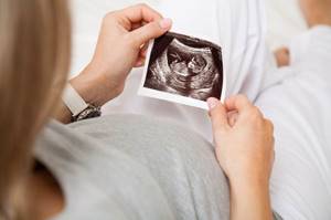
What does an ultrasound show?
For an expectant mother, an ultrasound at 12 weeks of pregnancy is also interesting because you can see your baby on the monitor. The child is very small, but can already suck the thumb of his small hand, and it is quite possible that you will be able to see it. For a gynecologist who is caring for a woman’s pregnancy, an ultrasound examination at the 12th week of pregnancy will show the condition of the pregnant woman’s uterus, the place of placenta attachment and the state of vaginal muscle tone will be determined. During pregnancy ultrasound, much attention is paid to examining the occipital region of the fetus, since thickening of the nuchal translucency space can be a bad sign and indicate a developmental defect.
An ultrasound at 12 weeks of pregnancy will also show the number of fetuses in the uterus. All established indicators will subsequently serve as the basis for monitoring the correct development of the fetus. At this stage of a woman’s pregnancy, the obstetrician-gynecologist will be able to draw a conclusion about how the pregnancy process will proceed, whether there are complications, and, if necessary, make appropriate appointments.
Average length of long bones at different stages of pregnancy (according to last menstruation)
| Duration (weeks) | Length (mm) | |||
| femoral | humeral | ulnar and radial | tibia and tibia | |
| 14 | 16 | 14 | 14 | 14 |
| 15 | 19 | 18 | 16 | 16 |
| 16 | 22 | 21 | 19 | 18 |
| 17 | 24 | 24 | 22 | 21 |
| 18 | 28 | 27 | 24 | 23 |
| 19 | 31 | 30 | 27 | 26 |
| 20 | 34 | 33 | 30 | 29 |
| 21 | 37 | 36 | 32 | 32 |
| 22 | 40 | 39 | 33 | 34 |
| 23 | 43 | 42 | 35 | 36 |
| 24 | 46 | 46 | 37 | 39 |
| 25 | 48 | 46 | 39 | 41 |
| 26 | 51 | 48 | 41 | 43 |
| 27 | 53 | 48 | 43 | 45 |
| 28 | 55 | 50 | 45 | 47 |
| 29 | 57 | 53 | 47 | 50 |
| 30 | 59 | 54 | 49 | 52 |
| 31 | 62 | 55 | 52 | 54 |
| 32 | 63 | 56 | 54 | 55 |
| 33 | 65 | 58 | 56 | 57 |
| 34 | 66 | 60 | 58 | 59 |
| 35 | 67 | 62 | 61 | 60 |
| 36 | 69 | 64 | 62 | 62 |
| 37 | 70 | 65 | 64 | 63 |
| 38 | 72 | 67 | 66 | 65 |
| 39 | 74 | 69 | 67 | 67 |
| 40 | 77 | 71 | 69 | 70 |
Tags: ultrasound
Checklist for the ninth week of pregnancy
- It's time to take care of a new bra: your breasts are enlarged and may become engorged, so choose natural fabrics and models designed for pregnant women.
- To cope with constipation, drink enough fluids and eat foods high in fiber: vegetables, fruits, whole grains.
- Do not get out of bed abruptly in the morning: this way you will avoid dizziness.
If you still have questions related to the well-being of the mother and baby in the ninth week of pregnancy, you can consult a gynecologist at the Women’s Medical Center at any time convenient for you. Appointments are available 24 hours a day.
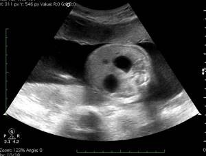
Determination of embryo viability
A fetal ultrasound in the first trimester of pregnancy shows the baby's heartbeat. It begins at week 5 and only at the first screening can you make sure that there is no possibility of freezing.
The full viability of the embryo is determined by the heart rate. Normally they should be from 90 to 110 beats/min. These are important indicators that include the size of the fetus, ultrasound at 11 weeks. Modern equipment allows you to view all the baby’s organs and exclude the presence of pathologies or congenital defects. For example, Down syndrome or Edwards syndrome. Therefore, women are sent for a full and in-depth diagnostic study.