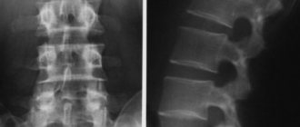
MRI of the spinal cord and spine is a method for diagnosing the condition of the spinal column and surrounding tissues using an electromagnetic field. It provides complete information about bone and soft tissues, the spinal cord, and allows you to obtain a detailed image of the vertebrae and intervertebral discs. The price of a diagnostic procedure varies not only depending on what type of equipment will be used in the study. The cost of MRI of the spine depends on a number of factors: the number of parts of the spinal column examined, the use of additional methods in the diagnostic process to obtain a clearer picture of the disease, the use of sedatives for patients with claustrophobia.
The Moscow MRI Center offers inexpensive MRIs of the spine and other organs. The 1.5 Tesla tomograph takes high-quality images, which allows you to obtain more accurate diagnostic results.
Types and costs
| Service | Cost, rub. | Action in the center on Musa Jalil |
| MRI of the craniovertebral junction and atlantoaxial joint | 4500 rub. | 3950 rub. |
| MRI of the cervical spine | 4500 rub. | 3950 rub. |
| MRI of the thoracic spine | 4500 rub. | 3950 rub. |
| MRI of the lumbosacral spine | 4500 rub. | 3950 rub. |
| MRI of the sacrococcygeal spine | 4500 rub. | 3950 rub. |
| MRI of the sacroiliac joints | 4500 rub. | 3950 rub. |
| COMPLEXES (profitable) | ||
| MRI of two parts of the spine | 7500 rub. | 6800 rub. |
| MRI of three parts of the spine | 9900 rub. | 8950 rub. |
| MRI of 3 parts of the spine and coccyx | 10900 rub. | 9950 rub. |
| MRI of the lumbosacral spine and iliosacral joints | 7500 rub. | 6800 rub. |
The prices indicated on the website are not a public offer (according to Article 435-437 of the Civil Code of the Russian Federation). You can find out the exact cost of studies and additional services from the administrators of our MRI centers by calling the numbers listed on the website or using the feedback form.
General information about MRI
Magnetic resonance imaging (MRI) is a modern, high-precision diagnostic method that allows one to visualize pathological changes in body structures using high-quality images.
Diagnostics of this kind involve obtaining exceptionally clear and detailed images of organs and tissues using the phenomenon of nuclear magnetic resonance. Under the influence of radio signals, atomic nuclei respond in the form of various signals. The combination of excited electromagnetic waves depends on the nature of the lesion in a particular area under study. There are open-type (no tube) and closed-type MRI scans. Each option has its own advantages and disadvantages. In an open device, the procedure is carried out both for the youngest children and for elderly weakened patients. But (unlike a device with a pipe) diagnostics are contraindicated for people whose weight exceeds 120 kg. Also, the open type of tomograph is not able to evaluate moving organs - the heart, lungs. The advantages of closed-type MRI include the high accuracy of the resulting images (regardless of the area being examined). The disadvantage of the procedure is the high noise level. It also requires complete immobility to obtain high-quality images, which is impossible to achieve, for example, with the smallest patients.
Medical in Moscow has modern equipment for MRI. The examination is a universal method: it is carried out both for young children and elderly people with severe concomitant pathology. Moreover, it will not take much of your time. Medscan implements a large number of multi-directional complex programs that solve the problems of diagnosing a wide range of diseases.
How is an MRI of the back muscles done?
The study is carried out both on an outpatient basis and when the patient is hospitalized in an inpatient department. The patient is changed into a hospital gown (in some cases it is allowed not to change clothes if they are loose enough and there are no metal elements on them), placed on a retractable table, which is later pushed into a magnetic capsule. Using special belts and bolsters, the arms, chest and head are secured, since movement is strictly prohibited during the examination.
The device itself contains a two-way intercom so that the radiologist can talk to the patient, give him the necessary instructions, and also so that the patient can report changes in his condition, ailments, nausea, dizziness, difficulty breathing or any strange sensations . In such cases, scanning is forced to stop. But do not be alarmed by the sensations of slight tingling or warmth in certain areas of the body - this is considered absolutely normal.
Since the magnet makes quite a lot of noise, for the comfort of patients, headphones are most often provided to drown out the noise. Some models of tomographs have the ability to turn on calm music, which will not only relieve the patient from noise, but also help to calm down and relax.
All medical personnel must leave the room before starting the scanner.
What does an MRI show?
Doctors recommend doing an MRI to confirm diseases of the brain and spinal cord, internal organs (with the exception of hollow organs), articular apparatus, spinal column, and organs of the reproductive system. Scanning is used to determine:
- spinal hernias;
- inflammatory processes;
- benign and malignant neoplasms;
- vascular pathologies (diseases of the cardiovascular system, changes in blood flow);
- presence of bone fragments;
- infectious processes of the osteoarticular apparatus;
- traumatic lesions;
- liquid in cavities.
The procedure is not relevant for identifying problems of the stomach, intestines, lungs (contain air), as well as bone structures.
The accuracy of MRI can be improved by using a contrast agent. A gadolinium-based agent is used as a contrast, administered intravenously. The drug accumulates in areas with increased blood flow (inflammation or tumor). MRI with contrast determines the pathological process in the initial stages of the disease. The substance introduced into the bloodstream is absolutely harmless to the body and is eliminated within 24 hours.
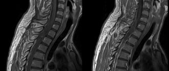
MRI of the cervical spine
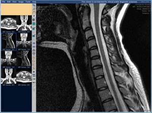
Cervical spine, sagittal projection
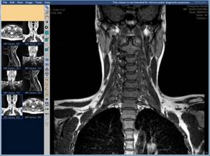
Cervical spine, frontal view
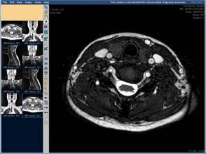
Cervical spine, axial projection
It should be noted that diseases of the cervical spine and lumbosacral spine are noted by experts as the most common.
Indications for MRI of the cervical spine:
- deforming spondylosis, osteochondrosis of the cervical spine;
- protrusion and herniation of intervertebral discs of the cervical spine;
- metastases of various tumors in the cervical spine;
- spinal canal stenosis;
- injuries of the cervical spine (fracture, dislocation, displacement of vertebral bodies);
- short neck syndrome (Kliepel-Fayle syndrome);
- the presence of various pathological changes in the cervical spinal cord.
Indications for the procedure
A specialist of almost any profile can prescribe a diagnostic procedure, since the procedure helps make a diagnosis for many pathologies. There are the following indications for diagnostics using MRI:
- Brain diseases: if a stroke is suspected; neoplasms and their relapses; assessment of the condition after surgery; demyelinating disorders (multiple sclerosis); inflammatory processes; congenital features of the development of the craniovertebral zone; convulsive readiness and epilepsy; DEP (dyscirculatory encephalopathy); hypertension of unknown origin; metastatic foci; traumatic brain injury (if the injury is not recent).
- Cervical vessels and cerebral vessels: anomalies in the development of the vascular bundle; aneurysms; signs of vertebrobasilar insufficiency due to cervical osteochondrosis; vascular stenosis (narrowing); migraine pain.
- Diseases of the temporal and jaw joints, their discs; preparation for dental prosthetics.
- Problems with the eye orbits and extraocular muscles.
- Diseases and inflammation of the lacrimal glands.
- Problems with the paranasal sinuses: cysts, sinusitis, preparation for surgery.
- Diseases of the thyroid gland (including oncology).
- Spinal column and spinal cord: osteochondrosis, osteoporosis, injuries, etc.
- Osteoarticular system: rheumatoid arthritis, inflammatory, traumatic lesions, conditions after surgery.
- Organs of the thoracic cavity: lung tumors, mediastinum, myocardial infarction, coronary angiography, pleurisy.
- Organs of the abdominal cavity and retroperitoneal space: problems with the liver, pancreas, gall bladder, intestines, spleen, kidneys.
- Pelvic organs: diseases of the bladder, prostate gland, ureters, uterus, etc.
MRI of the spinal cord
Indications for MRI of the spinal cord:
- suspected focal lesion, tumor of the spinal cord or its membranes;
- assessment of cerebrospinal fluid spaces of the spinal cord (spaces filled with cerebrospinal fluid), identification of syringomyelia (formation of cysts in the spinal cord);
- assessment of the results of surgical interventions on the spinal cord;
- suspicion of a spinal cord pathology of vertebrogenic origin, i.e. caused by trauma, tumor lesions of the spine, herniated intervertebral discs.
Depending on the expected diagnosis, the patient may be prescribed an MRI of the entire spine or its parts:
- MRI of the cervical spine (neck MRI);
- MRI of the thoracic spine;
- MRI of the lumbosacral spine (lumbar MRI);
- MRI of the coccygeal area of the spine (MRI of the coccyx).
Let's consider each type separately.
Contraindications
When choosing a clinic where it is better to have an MRI, you need to remember the main contraindications for the procedure:
- first trimester of pregnancy;
- the patient has a pacemaker;
- there are metal particles or structures in the body (including fragments after injuries).
Modern orthodontics and orthopedics (dentistry) use in their practice to install braces and dentures such materials (made of titanium and various alloys) that do not interact in any way with the magnetic field. Therefore, in this situation, performing a diagnostic procedure is not a contraindication.
Who is prohibited from MRI with contrast?
Intravenous administration of a contrast agent may be required before the study begins. Most often, it does not have a negative effect on the body, but certain allergies or bronchial asthma can be a good reason for refusing to administer contrast.
In case of kidney failure or other serious kidney diseases, the contrast agent may cause side effects or poisoning of the body. Therefore, such patients are prescribed a biochemical blood test, the results of which will determine the possibility of using contrast in the study.
Tomography with contrast is not recommended for pregnant and lactating women; it is prescribed only in cases where the benefits outweigh the possible risks. The final decision remains with the attending physician. If a nursing mother is still prescribed a test, then she must stop feeding for 24–48 hours after the procedure and express milk until it is completely renewed so that all harmful substances leave the body.
Principle and safety of the procedure
Tomography helps to determine the nuances of the location and structure of the internal organs and structures of the body. Scanning the body in thin layers helps with this. Safe contrast can be used, but there is no harmful x-ray exposure. The MRI machine recreates an extremely powerful magnetic field based on interaction with atomic nuclei. Most often we are talking about atoms of the chemical element hydrogen. Their excitation leads to a change in position, which allows the equipment to build many detailed layer-by-layer “slices” of various organs.
The magnetic field does not carry a negative radiation load on the body. There are also no other unfavorable factors that could harm human health. Therefore, the diagnostic procedure can be carried out the required number of times (according to indications).
Preparing for magnetic resonance imaging
Examination of the pelvic organs (organs of the female reproductive system, prostate gland, bladder) requires the following measures:
- the procedure is carried out with a full bladder: you should not urinate for several hours, and an hour before the scan you should drink half a liter of clean water;
- It is recommended to cleanse the intestines with an enema or laxatives;
- half an hour before the study, it is advisable for the patient to take an antispasmodic drug (“No-spa”);
- women of childbearing age indicate the day of the menstrual cycle.
Magnetic resonance imaging of the abdominal organs is performed on an empty stomach or after a light snack if the procedure is scheduled for daytime or evening hours. A few days before diagnosis, you should exclude from your usual diet foods and dishes that contribute to increased gas formation. These include legumes, whole milk, and sweets. Also avoid foods that cause constipation (flour, rice, chocolate). It is advisable to take enzymes and sorbents (activated carbon) during this period. On the eve of the study, the patient is recommended to take an antispasmodic.
Examination of other parts of the body does not require special preparation. Immediately before scanning, you must remove keys and other metal items from your pockets and remove jewelry.
How to prepare?
MRI for back pain does not require special preparation or adherence to a special diet. Immediately before the examination, the patient must remove and deposit all metal objects that may affect the correct operation of the magnet: piercings, rings, chains, bracelets, earrings, watches, bank cards, dentures, keys, pens, belts, etc. If necessary, you are also asked to remove your everyday clothes before the examination.
Be sure to tell your doctor if you have fillings, braces, or tattoos, as there is a risk that heat may occur. The doctor must be prepared for such a situation in order to quickly respond if this happens.
If necessary, a contrast agent, sedatives or anesthesia are administered at the preparation stage.
Advantages of MRI at Medscan
The diagnostic clinic in Moscow performs many MRI options at quite affordable prices: joints, internal organs, soft tissues, peripheral nerves, spine, etc.
Diagnostic testing is carried out as prescribed by a doctor. Screening programs are also being implemented that involve analyzing the state of certain body systems. Some of the most relevant are cancer screening and screening of the cardiovascular system.
The leading advantages of our clinic:
- High information content of diagnostic images. The contrast agent increases the information content of the images.
- The radiology department is headed by one of the best radiologists in the entire post-Soviet space, Kirill Sergeevich Petrov.
- There is a medical center. It helps to understand controversial or complex clinical cases and clarify the diagnosis.
- The diagnostic center operates under international standards. The scanning is consistent with the recommendations of European professional societies.
- Diagnosis is carried out by highly qualified doctors with sufficient practical experience.
- Our professionals work with expert-level equipment. The equipment meets modern international standards.
- The results of the scanning are recognized by leading specialists of the Russian Federation and foreign countries.
- An MRI in Moscow can be done in one of several branches of our clinic, the addresses of which are listed on the website.
Medscan doctors will help identify health problems at the initial stages and prevent their development.
Sign up
Sources and literature
- Akberov R. F., Yaminov I. Kh., Safiullin R. R., Puzakin E. V. The effectiveness of magnetic resonance imaging in the diagnosis of brain tumors. [Electronic resource] // Kazan Medical Journal. 2011
- Kremneva E. I., Konovalov R. N., Krotenkova M. V. Functional magnetic resonance imaging. [Electronic resource] // Annals of Clinical and Experimental Neurology. 2011
- Kotlyarov P. M., Lagkueva I. D., Sergeev N. I., Solodkiy V. A. Magnetic resonance imaging in the diagnosis of lungs. [Electronic resource] // Pulmonology. 2021