Breast examination using MRI is often carried out to identify all kinds of neoplasms in the mammary glands.
Compared to other diagnostic methods, MRI in this area is 2 times more effective: the percentage of detection of pathological foci in breast tissue with MRI is 90%, with mammography in combination with ultrasound (US) – up to 45%.
Doctors recommend that women at high risk of developing cancer undergo an annual MRI examination of the mammary glands:
- If genes responsible for the growth of cancer cells (BRCA 1 or BRCA 2) are detected in the woman herself or her close relatives (mother, daughter, sister);
- If a woman has undergone radiotherapy (irradiation) for malignant diseases at an early age;
- If there are other risk factors for developing breast cancer (hereditary Cowden or Li-Fraumeni syndromes).
Oncologists routinely prescribe breast MRI to patients with a tumor in one breast in order to find out whether the second gland is affected by cancer. Magnetic resonance imaging without surgery or X-rays provides a detailed image of the internal structures of the mammary glands and blood vessels, and allows you to detect even small (up to 3-5 mm) foci of pathological tissue at the earliest stages of development.
MRI with contrast
MRI of the mammary glands can be performed with or without contrast. Contrast (gadolinium is a hypoallergenic dye) is used to visualize the vasculature of the gland. Cancerous lesions are intensively supplied with blood, which is clearly visible on MRI images.
A non-contrast examination is performed to determine tissue density and study the structure of the mammary glands.
Indications for MRI of the breast
Breast MRI is prescribed not only for suspected cancer. A mammologist may refer a woman for this examination for:
- determining the condition of the mammary glands and the integrity of silicone implants;
- differential (comparative) diagnosis of benign formations when other studies are ineffective;
- monitoring the dynamics after anticancer treatment;
- diagnosis of any other breast diseases in pregnant women and in the presence of contraindications to radiography;
- identifying various pathologies with poor visualization by other methods (proliferation of scar tissue after surgery or injury).
Often, the results of an ultrasound or mammography do not provide enough information to satisfy the woman - to calm her down or encourage her to take certain actions. Therefore, many patients come for examination without a doctor’s referral, on their own initiative.
Important! In women who undergo breast MRI once a year, the possibility of death from cancer is minimized.
MRI of the abdomen with contrast
MRI of the abdominal cavity with contrast increases its information content. It is indicated if there were any suspicions on the ultrasound.
Specialists of the University Breast Center regularly improve their professional level, attend domestic and foreign conferences to keep abreast of all modern trends in oncology and plastic surgery.
Author: Chizh Igor Aleksandrovich head, kmn, oncologist of the highest qualification category, surgeon of the highest qualification category, plastic surgeon
Start your treatment right now: sign up for a consultation by phone or through the form on the website
Contraindications
Although MRI is a safe method, this procedure has contraindications. Absolute and relative – conditions in which MRI is strictly prohibited, and conditions that can be overcome, i.e., with proper preparation, the examination becomes possible.
Absolute contraindications
- The presence of metal objects in the patient’s body (implants, clips, braces, etc.): exposure to a magnetic field can provoke their movement and cause internal bleeding or other negative consequences.
- Hyperkinesis (involuntary movements), convulsions, muscle spasms: the patient must remain completely still during the entire procedure, otherwise the examination result may be inaccurate (the pictures will be blurry, smeared).
- Allergy to contrast agent: gadolinium (intravenous contrast) rarely causes allergic reactions, but in case of individual intolerance, its administration is strictly prohibited - the substance can cause the development of allergies of varying severity, up to anaphylactic shock.
- Chronic renal failure or other diseases of the genitourinary system: the contrast agent can provoke an exacerbation of the disease, since it is removed from the body through the kidneys.
- Having a pacemaker or insulin pump: exposure to magnetic waves can affect the stable operation of the device.
- The patient's obesity: a weight of more than 120-150 kg prevents the procedure from being performed, since the diameter of the MRI capsule does not correspond to the woman's waist circumference.
Relative contraindications
- Pregnancy and lactation: the magnetic field can have a negative impact on the intrauterine development of the fetus, and the contrast agent, if released into breast milk, can cause allergies and other reactions in the child.
- Claustrophobia: Fear of enclosed spaces can cause the patient to panic and provoke chaotic motor activity.
- Mental disorders of the patient: various unpredictable reactions to being in an MRI scanner and undergoing medical procedures are possible.
- Tattoos made using inks containing metal: a burning sensation occurs in the area of the skin design. Minor damage to the skin in the form of a burn may also occur.
Safety
MRI does not involve radiation and is safe for the patient. But there are important points to consider:
- Metal implants or foreign bodies are potentially dangerous and can cause injury or death during an MRI scan. Patients must be carefully questioned before the study to eliminate the risk of injury.
- Foreign metal objects that could injure the patient as a result of their strong (and sharp) attraction to the magnet should not be allowed into the room where the MRI is being performed.
- It is important to inform your doctor about any previously identified allergic reactions to medications.
- In case of emotional stress, anxiety and restlessness due to moderate claustrophobia, it is permissible to use sedatives or anesthesia during the examination.
Preparing for the study
Experts recommend performing an MRI of the mammary glands on days 8-12 (in the first phase) of the menstrual cycle.
At this time, the woman has no swelling and infiltration around the large ducts in the mammary glands (unlike the premenstrual period), so the examination result will be more informative. If menopause has occurred, then any other time will do.
If necessary, the doctor will prescribe a special diet or limit the intake of certain medications.
Tomosynthesis
Tomosynthesis uses X-rays like mammography and is performed layer by layer like MRI. The result is a set of X-ray images, with the help of which the program recreates a three-dimensional picture of the soft tissues of the breast:
✅ Advantages
takes a picture of the entire breast in a hundred planes at different angles,
suitable for women with dense breasts,
microcalcifications are visible on the photographs,
does not require special preparation and injections of contrast agents,
one study can be analyzed by several specialists.
❌ Flaws
not suitable for pregnant women,
Available in few places in Russia.
Technology
Before starting the diagnosis, the patient removes all objects that are made of metal (jewelry, piercings, hair clips, watches, etc.).
During the procedure, the patient lies on her stomach on a movable tomograph couch (special holes are provided for the mammary glands). The procedure lasts from 30 minutes to 1 hour, so the woman needs to make sure that the position is comfortable and that she does not feel the urge to move during the scan.
During a breast MRI with contrast, the nurse administers a contrast agent through an intravenous catheter. This is a mandatory procedure for diagnosing cancer. When contrast enters the blood, the patient may experience cold or slight heat spreading along the vein.
To ensure the greatest comfort and protection of hearing from the noise effects of the operating device (hum, clicks, crackles), the use of headphones is provided.
The table moves directly into the ring of the MRI scanner, where the scanning takes place. The woman may feel a burning or tingling sensation in the area being examined. These signs are considered normal. If unforeseen situations arise, severe pain or a sharp deterioration in condition, the patient can inform the doctor about this through the built-in feedback system.
How is the procedure done?
MRI of the mammary glands is performed both in outpatient and inpatient settings. The patient is placed on a special platform, lying on her stomach face down. Inside the platform there are holes for the mammary glands. Thus, MRI images are clear, since there is no displacement or compression of the mammary glands. Electronic sensors are built into the treatment table. It is important to relax and not move. Before you turn on the device, you should make sure that you are in a comfortable position, as tension in the chest or arm muscles can lead to interference in the image.
For screening MRI examination of the mammary glands, as well as checking the integrity of implants, no contrast is required. In other cases, especially for differential diagnosis of various forms of mastopathy and cancer, the introduction of contrast is mandatory.
Research results
The patient receives a report containing the results of an MRI of the mammary glands within half an hour after the procedure. In the description, the doctor indicates the identified changes in the patient’s chest, the nature and extent of their distribution, localization (location), the exact size of the lesion, other parameters and information, and the final diagnosis. Based on the examination data, the attending physician predicts the further development of the disease and plans the most effective treatment regimen.
Diagnostics with contrast agent:
- detects the presence of pathological processes in the breast;
- establishes the size and exact location of the neoplasm;
- determines the size of lymph nodes;
- determines the degree of malignancy of the tumor.
Diagnostics without contrast agent:
- determines the density of the mammary glands;
- studies the condition of the milk ducts;
- identifies formations in the form of cysts;
- detects hematomas;
- determines implant ruptures and the degree of damage to surrounding tissues.
Stages of the disease
Stage 0 This is the stage when the primary tumor is not identified or cannot be assessed, and also in the case of non-invasive breast cancer (which means the tumor does not extend beyond its appearance, so-called in situ cancer).
Stage 1 Cancer cells in this stage invade or grow into neighboring tissues. The tumor node is no more than 2 cm, the lymph nodes are not affected at this stage.
Stage 2 In this stage, the tumor node exceeds 2 cm and can reach up to 5 cm. At this stage, lymph nodes may be affected, but the damage to the lymph nodes is single, they are not fused to each other and are located on the same side as the tumor. In case of lymph node involvement, the tumor size may be less than 2 cm.
Stage 3 Invasive cancer, more than 5 cm or with obvious and significant damage to the lymph nodes. In this case, the lymph nodes can be fused together.
Stage 4 At this stage, the tumor grows into the skin of the chest, chest wall, or internal mammary lymph nodes. It can be of any size.
Stage 4 breast cancer is inflammatory cancer; it occurs in up to 10% of all cases. Symptoms of the inflammatory form of breast cancer are redness of the skin, the gland becomes warm, and there is an enlargement and/or hardening of part or all of the breast. The skin takes on the appearance of an orange peel. This form of cancer must be differentiated from inflammation of the mammary gland - mastitis.
Also, at stage 4, the tumor can spread beyond the chest, into the axillary region, and internal mammary lymph nodes. Metastases to the supraclavicular lymph nodes, as well as to the lungs, liver, bones or brain are possible.
What breast cancer looks like in different stages:
Alternative research methods
Carrying out MRI of the mammary glands in the complex diagnosis of various diseases is advisable and desirable, since:
- MRI perfectly visualizes the soft tissues of an organ, much better than CT, radiography and ultrasound;
- MRI does not contain ionizing radiation, which is used in radiography and computed tomography;
- MRI scanning allows you to get the most complete and clear picture, unlike mammography, which is less informative;
- MRI makes it possible to study an organ both from the outside and from the inside (in ultrasound diagnostics it is only possible to assess the condition of the outer shell);
- MRI allows you to determine the presence of even small formations (unlike mammography).
The possibility of repeated repetition and the absence of negative effects on the woman’s body makes this type of research the most popular in modern diagnostics.
Alternative
Other methods may be prescribed:
- Mammography. It is carried out using a special device - a mammograph. Refers to radiographic methods that produce small ionizing radiation. Pictures are taken in two projections. Detects cancer, dense nodes, metastases. In women under 40 years of age it is ineffective due to the high density of the gland. Often requires additional techniques. Does not visualize lymph nodes.
- Ultrasound of the mammary glands. Scans the gland in different projections, revealing the structure of the organ and its anatomical features. This method makes it easy to detect cysts and other benign formations. Allows you to monitor their dynamics. Shows the condition of blood vessels. Can be done many times.
- Tomosynthesis. With it, several images are taken in a certain sequence with a fixed interval between slices. This produces from 11 to 90 3D photographs of varying depths. This helps to achieve maximum information about the condition of the breast and nearby organs. Poorly recognizes low-contrast anomalies.
- Laser CT. Newly developed method. Laser radiation in the infrared spectrum helps to identify areas with abnormal accumulation of blood vessels. This may signal an oncological process. Requires expensive equipment and additional training of employees. This means that its cost is very high. Determines only the condition of blood vessels.
What does a chest MRI show?
Using this method, the following pathologies are identified:
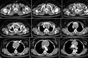
- blood supply problems, ischemic damage, condition before a heart attack;
- thromboembolism, dissection of the aortic wall;
- MRI-sensitive inflammation;
- cystic formations with different contents;
- benign and oncological tumors;
- bronchiectasis;
- change in tissue density.
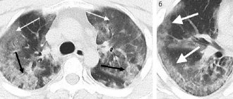
After an MRI of the chest, doctors and the patient are left with black and white images with a description of the diseases identified on them. Radiologists at the diagnostic center do not make a final diagnosis. MRI serves only as an imaging method, on the basis of which the location, type and stage of the disease are established.
Sign up for diagnostics
You can undergo a scan at any center you like, presented on the MRI registration portal.

The hotline number for free consultation is also posted here. Apply search filters, study offers based on your request, read reviews, learn about doctors and treatment conditions from customer reviews. For a convenient appointment for diagnostics, contact the service administrators using the number on the page, ask any remaining questions, receive recommendations for preparing for an MRI, and book a session for a convenient day with discounts.
Sources used:
- Vorotyntseva N. S. X-ray pulmonology [Text]: strategy and tactics for obtaining and analyzing X-ray images in pulmonology: textbook. allowance / N. S. Vorotyntseva, S. S. Golyev. - M.: MIA, 2009. - 280 p.
- Erokhina, A. V. Peculiarities of interpretation of chest radiographs in pathologies of the respiratory system in newborns [Text] / A. V. Erokhina, A. V. Gorbunov, M. V. Degtyareva // Questions of practical pediatrics. - 2012. - No. 4. - P. 50-56. — Bibliography: p. 55-56 (34 titles).
- Lange S. Radiation diagnosis of diseases of the chest organs [Text] = Radiology of chest diseases: manual / S. Lange, D. Walsh; lane from English edited by S.K. Ternovy, A.I. Shekhter. - M.: GEOTAR-Media, 2010. - 432 p.
- Radiation diagnostics [Text]: textbook. T. 1 / ed. G. E. Trufanova. - M.: GEOTAR Media, 2007. - 416 p.
- Radiation diagnostics of heart and vascular diseases [Text]: national. hands / ch. ed. S. K. Ternovoy, L. S. Kokov; ASMOK. - M.: GEOTAR-Media, 2011. - 688 p.
- Digital technologies in the department of radiation diagnostics [Text]: manual / ed. G. G. Karmazanovsky, A. I. Leichenko. - M.: Vidar-M, 2007. - 200 p.
When to check your chest organs with an MRI
Diseases of varying nature and severity can develop in this area, since many vital systems are located here.

In modern realities, when a patient complains of inflammation and pain in the sternum, one must first undergo a test for Covid, and then a full tomographic scan to rule out infectious pneumonia and its complications on neighboring organs. In addition, tissue degeneration and atrophy, tumors, blood supply problems, focal extrapulmonary and pulmonary infections, injuries, and congenital changes can occur in the chest. Typically, MRI is used as a “strike force” when the previous diagnostic method or their complex did not cope with the task - did not reveal the pathology.
The main reasons for seeking MRI are:
- acquired and genetic cardiopathologies;
- atherosclerotic disorders, aneurysms and stenoses;
- attacks of suffocation, constant cough, fever, discharge of purulent sputum with bloody spots;
- signs of tumor growth against a background of systematic chest pain, progressive weight loss, and general weakness of the body;
- decreased sensitivity in the arms/legs, irradiation around the sternum;
- enlargement of lymph nodes in the search area.