You may already know these 5 facts about breast cancer:
- 98% — survival rate of women with breast cancer (BC) if the tumor is detected and treatment is started at the initial stages T0─T1. Such neoplasms have a diameter of up to 20 mm, in most cases they can only be detected by hardware examination (mammography, ultrasound, MRI)
- 55% — survival of patients with breast cancer stages T2, T3, T4. At these stages, tumors begin to be palpated, create discomfort, cause changes in the skin, shape or color of the nipple, discharge, pain and other signals appear, with which the woman ultimately goes to the doctor ─ however, the moment when oncology could be cured with a 98% probability , irreversibly lost
- 40 years is the age at which for the average woman, according to WHO, the risk of breast cancer begins to increase annually
- 9 years - on average, during this time the tumor reaches a size of 10 mm and begins to be palpated (all patients have different growth rates)
- One in eight women will develop breast cancer in her lifetime
And 5 more facts you definitely need to know:
- Every second woman under 50 years of age (and every 3rd woman aged 50 years and older) is at particular risk for breast cancer due to the dense x-ray background ─ individual structural features of the breast tissue, which makes it difficult to detect a tumor on mammography
- Up to 30% of breast cancer cases are associated with a dense radiological background. For comparison: only 5-10% of cases are associated with mutations in the BRCA1 and BRCA2 genes (“Angelina Jolie gene”)
- On average, the detection of breast cancer in patients with a dense x-ray background increases by 2 times
- According to various international studies, the use of 3D ultrasound in patients with a dense x-ray background makes it possible to detect 30% more breast cancer. In Moscow there are only 3 machines for 3D breast ultrasound ─ and we have one of them, at Hadassah Medical
Breast mammography
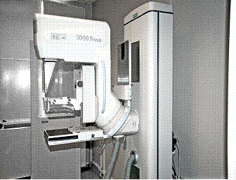
Mammography is the main screening method, and remains today the only way to early diagnose breast cancer. In European countries, annual examination of women after 40 years of age has significantly reduced mortality from this disease, as well as minimized surgical treatment. Modern digital equipment allows for precise examinations of the breast with minimal radiation exposure in 5-10 minutes. During mammography, the mammary glands are compressed in special plates, which can cause moderate discomfort.
To increase reliability and information content, mammography is recommended to be performed from the 5th to the 12th day of the menstrual cycle.
Our Center's mammologists perform mammography during consultation. Cost of mammography:
- mammography of one breast in 2 projections 3400 rubles.
- mammography of both mammary glands in 2 projections 4000 rubles.
Our doctors perform all medical and diagnostic procedures and work for you from Monday to Friday from 8.00 to 20.00. You can sign up for a consultation by calling: (495) 942-40-20.
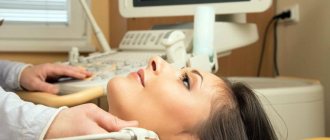
How safe is breast ultrasound?
Unlike X-ray mammography, breast ultrasound does not use X-rays, which means the patient's body is not exposed to any radiation. Therefore, ultrasound has no contraindications; it can be done an unlimited number of times and combined with any type of diagnosis, be it CT or MRI.
The patient does not experience discomfort either. During the scan, the doctor simply moves a special sensor over the patient’s skin, which is in contact with the body and performs two functions. It emits sound waves (echoes) at a specific frequency and picks up the reflected sounds. The frequency of the wave returning to the sensor is converted into a digital formula and appears on the screen in the form of echoes or dots, from which the ultrasound image is constructed.
Ultrasound of the mammary glands
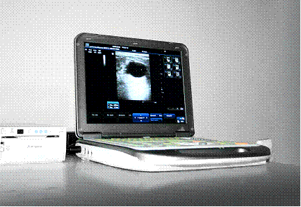
Ultrasound diagnostics of the mammary gland allows you to freely obtain images of the mammary gland in various projections, visualize the ductal and ligamentous systems of the mammary glands, and clearly distinguish tissue formations (fibroadenomas, fibrolipomas) from liquid formations (cysts, abscesses, etc.). Ultrasound is the leading method for examining the mammary glands during pregnancy and breastfeeding, with short-term planned monitoring of patients at intervals of 2-3 months, because does not have radiation exposure and is considered absolutely harmless.
It is most often performed on women under the age of 35 - at this age, a large amount of dense glandular tissue remains in the mammary glands, which is of little information for x-ray techniques - mammography.
In our Center, breast ultrasound is performed by a doctor during a consultation. To increase the reliability and information content of ultrasound (as well as other examinations of the mammary glands), doctors recommend conducting it from the 5th to the 12th day of the menstrual cycle.
Our doctors perform all medical and diagnostic procedures and work for you from Monday to Friday from 8.00 to 20.00. You can sign up for a consultation by calling: (495) 942-40-20.
When should the study be performed?
The doctor prescribes a breast ultrasound when the patient complains of the appearance of certain, previously uncharacteristic symptoms. As a rule, pathological manifestations in our body occur if there is a disruption in the functioning of internal organs. The mammary gland is no exception to the general trend. If the doctor identified a lump during the examination or felt a violation of the tissue structure, he will refer the woman for an ultrasound diagnosis of the mammary glands. The examination does not involve radiation exposure and does not require special preparation. The procedure can be prescribed by a gynecologist, mammologist, endocrinologist or oncologist.
Alarming symptoms that may indicate a violation of women's health regarding the breast are the following:
- pathological discharge from the nipples;
- painful sensations in the chest;
- change in the normal size, contour, shape of the breast;
- during lactation, pregnancy, or at the level of pregnancy planning, a preventive ultrasound examination of the mammary glands is prescribed, so that if pathology is present, it can be quickly identified and timely treatment initiated;
- to confirm suspicion of the formation of neoplasms;
- assessing the condition of the mammary glands after traumatic exposure or an inflammatory process;
- when inflammation occurs in the supraclavicular or axillary lymph nodes;
- to monitor breast condition after breast augmentation surgery;
- as an evaluation method for the dynamics of a patient’s therapeutic treatment, adjusting the treatment regimen if the positive effect is insufficient.
Indications for performing ultrasound of the mammary glands also exist for men. Warning signs include breast enlargement in a man or the appearance of hard lumps. Ultrasound of the mammary glands at the Alfa Health Center clinic is a good diagnosis at an affordable cost!
While using the method, the doctor also performs an ultrasound of the mammary gland ducts, if the doctor who gave the referral suspects their pathology. Ultrasound of the mammary glands in women is an important study that they should undergo once every 6 months from the age of 30 years. In addition, it is necessary to perform mammography with the same regularity.
For girls under 30 years of age, it is recommended only to undergo an ultrasound scan of the mammary glands once every six months, as a preventive measure to identify asymptomatic pathology. The price of breast ultrasound at the Alfa Health Center clinic is an acceptable figure for every person!
On what day of the cycle should a woman have a breast ultrasound? Doctors say that in patients under 35 years of age, ultrasound of the mammary glands in women should be performed in the first 14 days of the cycle. If the patient is over 35 years old and also takes oral contraceptives, the day of the cycle does not matter.
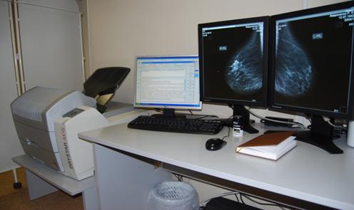
What does a breast ultrasound show? Based on the results of the examination, the picture of a paid ultrasound of the mammary glands allows the doctor to see the complete structure of the mammary gland and ducts, changes in the size, function, and shape of the gland, determine the patient’s diagnosis and prescribe the correct treatment.
MRI OF THE BREASTS using special setups and dynamic contrast enhancement
MRI is the most modern method of examining the mammary glands, allowing to detect microtumors of the mammary glands and conduct spectroscopy of suspicious areas of tissue. MRI of the mammary glands is performed using contrast agents, and the mammologist assesses the extent of the disease based on the degree of their accumulation in the tissue. This technique is indispensable for detecting cancers in situ - accumulations of cancer cells at the earliest stage - before the formation of a node. MRI of the mammary glands is the most effective method of examination after aesthetic surgery for breast correction using endoprosthetics. This method allows you to see the implant from all sides, assess the integrity of the capsule, exclude the presence of micro-tears, and also identify difficult-to-diagnose tumors against the background of endoprostheses.
Doctors recommend MRI of the mammary glands to be performed from days 5 to 12 of the menstrual cycle.
In our Center, MRI of the mammary glands is performed on the recommendation of a mammologist by appointment.
The cost of MRI of the mammary glands is 13,000 rubles.
Our doctors perform all medical and diagnostic procedures and work for you from Monday to Friday from 8.00 to 20.00. You can sign up for a consultation by calling: (495) 942-40-20.
How often should a breast ultrasound be done?
To prevent breast cancer, doctors recommend that a woman undergo a preventive breast examination using ultrasound with the following frequency:
- woman from 30 to 39 years old - once every 3 years;
- woman 40-50 years old - every year;
- woman 50 years and older - 2 times a year.
However, a preventive analysis of the condition of the mammary gland in women, even young women, should be carried out annually if the patient is at risk. Doctors include factors that increase the chances of developing breast cancer:
- hereditary predisposition;
- excess weight;
- nicotine use and alcohol abuse;
- frequent abortions.
Author: Telegina Natalya Dmitrievna
Therapist with 25 years of experience
Breast punctures. Types, indications, cost
A puncture is a puncture of the breast tissue. A small number of tissue cells are collected from the puncture to determine the cellular composition of the tissue (cytological examination). It is carried out when nodular formations in the mammary glands are detected by ultrasound, mammography or palpation.
Palpable (determined by hand) formations are punctured without instrumental support.
Non-palpable (not detectable by hand) formations are punctured under ultrasound or x-ray control in order to most reliably take material directly from the node.
When puncturing cysts or liquid formations, liquid contents are evacuated from the cavity of the formation. This is not only a diagnostic, but also a therapeutic manipulation. Against the background of subsequent therapy, the empty cyst cavity grows together
Preoperative interstitial marking is the staining of an area of breast tissue before surgical treatment. It is carried out under the control of ultrasound or mammography for the most accurate and less traumatic surgical treatment. To do this, a special coloring marking substance is injected into the location of the tumor.
Our Center's mammologists perform all types of diagnostic and therapeutic puncture techniques. You can make an appointment with mammologists by calling: (495) 942-40-20 from 8.00 to 20.00
Ultrasound of the mammary glands in honey
Ultrasound examination in medical care is a non-invasive, no contraindications, no preliminary preparation procedure, performed using the latest equipment that allows you to examine both superficial and deep layers of tissue that are inaccessible to x-rays. The data displayed on the monitor is deciphered by the best mammologists in Moscow working at Elegy. Their vast practical experience and professionalism, combined with the accuracy of diagnostic ultrasound equipment data, make it possible to detect breast cancer, as well as other formations, in the early stages, and, based on the diagnosis, select the most effective treatment method.
As for the cost, ultrasound of the mammary glands in honey is one of the cheapest in Moscow and is available to every woman who cares about her health.
Contrast spectral mammography
An upgraded version of regular mammography. Before the examination, the patient is injected into a vein with a contrast agent, which is opaque to x-rays.
In areas where the contrast agent accumulates, bright white spots are visible on the images. The accumulation of contrast agent is a sign of a tumor in the breast.
✅ Advantages
suitable for women with dense breasts,
takes a picture of the entire breast in two planes,
microcalcifications are visible on the photographs,
one study can be analyzed by several specialists,
lends itself well to analysis by artificial intelligence.
❌ Flaws
not suitable for pregnant women,
requires injection of contrast agent,
Available in few places in Russia.
The advantages of mammography with contrast compared to conventional mammography are evident in the example of a patient with very dense breasts:
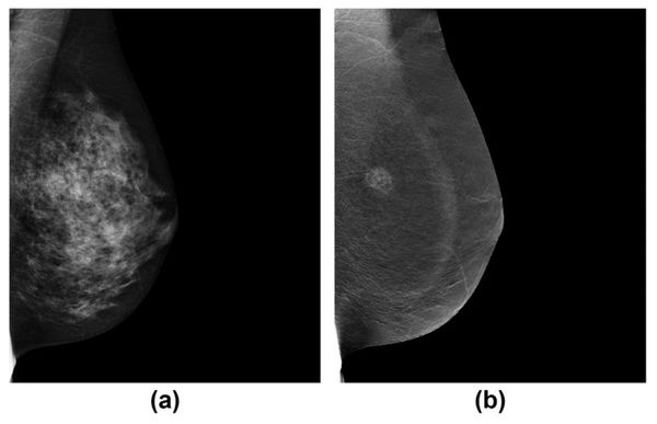
On the left is a conventional mammogram, on the right is a contrast-enhanced spectral mammogram ©JJ James, SL Tennant / Clinical Radiology
In the image on the left, dense breast tissue makes it difficult to distinguish the tumor. The image on the right clearly shows an accumulation of contrast agent. Histological analysis of the tumor showed grade 3 ductal adenocarcinoma.
Breast MRI with contrast
MRI uses radio and magnetic waves to scan the soft tissue of the breast layer by layer.
A breast MRI is done with a contrast agent that is injected into the patient intravenously.
✅ Advantages
suitable for women of any age,
suitable for women with dense breasts,
takes a picture of the entire breast in hundreds of planes,
one study can be analyzed by several specialists.
❌ Flaws
not suitable for pregnant women,
not suitable for patients with metal magnetic elements in the body (plates, prostheses),
microcalcifications are not visible on the photographs,
requires injection of contrast agent,
high percentage of false-positive tumors compared to other methods.
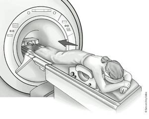
Breast MRI ©Sam and Amy Collins
Breast MRI is prescribed as an additional test to mammography and ultrasound for patients at high risk of developing breast cancer:
The risk of developing breast cancer can be calculated online using a calculator based on the Gale model.
- The risk of developing cancer according to the Gale model exceeds 1.7% over a five-year period or 20% for the rest of life.
- Patients with mutations in the BRCA1, BRCA2, CHECK, NBS1 or tP53 genes.
- Patients who underwent radiation therapy between the ages of 10 and 30 years.
- Patients diagnosed with lobular carcinoma in situ or atypical hyperplasia.
- Patients with high radiological density of the mammary glands.