Author: Sozinova A.V., obstetrician-gynecologist, has been in continuous practice since 2001.
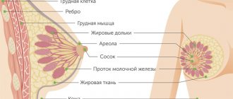
Ductography (galactography) is an X-ray examination of the milk ducts of the mammary gland with the introduction of a contrast agent into them. This method is a type of mammography.
Ductography is carried out to identify changes in the milk ducts (their expansion or narrowing), to identify tumors (intraductal papilloma, cancer).
As a rule, women with bloody spontaneous discharge or that occurs when the nipple is compressed (less often with serous discharge) are sent for ductography.
Ductography allows you to determine the pathological area of the mammary gland, the number and location of cystic formations. The patient is sent for such a study after mammography and/or MRI by an oncologist, radiologist or mammologist.
What is breast ductography?

Breast ductography is a method of x-ray examination of the milk ducts by artificially contrasting them (that is, introducing a special contrast agent into them). This method can also be called “galactography”.
Ductography is carried out to identify a number of pathologies of the mammary glands in the presence of specific signs of disease. This procedure is preceded by a cytological examination of the fluid released from the mammary gland. The complex of these procedures helps with a 96% probability to identify diseases such as intraductal papilloma, Mintz disease, intraductal cancer and others.
Advantages and disadvantages of ductography
| Advantages | Flaws |
| Early detection of cancer | Possibility of false positive results |
| Detection of small tumors that cannot be diagnosed with conventional mammography | Risk of duct injury |
| Low radiation dose | Risk of developing sensitization of the body to iodine |
| High price |
To fully diagnose the condition of the breast, other tests, such as MRI, may be needed.
With this we say goodbye to you. We hope we helped you understand when and how ductography is done, whether the procedure is painful and when it should not be done. We are waiting for you on our website again. You will be glad if you share this article with your friends via social networks.
Indications
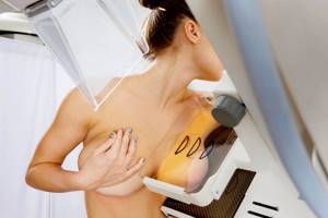
Almost every woman knows the procedure of mammography, which is an X-ray of the breast. It is often prescribed for preventive purposes and helps to detect dangerous pathologies in the early stages. Ductography may be prescribed for additional and more detailed diagnostics in cases where mammography has given controversial results or if the patient complains of frequent discharge from the nipples (there are other indications).
Discharge from the nipple of bloody or brown fluid (sometimes serous discharge)
Nipple discharge is normal when it occurs during pregnancy or lactation. If these factors are not present, but discharge is present, this should be a reason to go to a mammologist for examination and to identify the cause of the alarming symptom.
The discharge may be colorless, or it may have a specific shade - yellowish, greenish, brown or brown, red. The last two are the most serious symptoms, since most often these colors are caused by breast cancer, mastopathy or intraductal papilloma. The appearance of such discharge is explained by the fact that neoplasms appear in the milk ducts, which provoke the rupture of small blood vessels. Bloody discharge can appear directly when you press on the nipple or spontaneously.
Suspicion of intraductal papilloma
Intraductal papilloma, in addition to pathological discharge from the nipples, may have another symptom. On palpation, a large lump is felt in the area around the nipple. At the same time, on the periphery of the gland, compactions and outgrowths of the milk ducts are almost impossible to recognize. In this case, ductography of the mammary gland (breast) must be prescribed, since it is important to identify how far the disease has progressed, where the tumors are located and what their size is.
Suspicion of intraductal cancer
Intraductal cancer is one of the most complex and dangerous breast diseases. Dark-colored discharge from the nipples is a symptom of this pathology, but besides them, there are others: pain, redness, changes in breast shape, color and structure of the skin.
In this case, ductography is an event that will help determine the stage of oncology development, the localization of tumors, their size, structure, which in the future will help to correctly prescribe therapy or plan an operation.
Breast adenoma
Breast ductography is perhaps the most informative method for identifying mammary adenoma. The symptoms of this pathology are often not clearly expressed. When examined by a doctor
or during self-examination, an elastic formation with clear contours can be detected in the chest (sometimes there may be several such formations). The woman does not feel pain when pressing on the sites where adenomas form, and the skin of the breast does not change color or structure. In other words, ductography helps to identify a disease that is difficult to determine externally, by palpation, and even by mammography.
Breast cyst
Against the background of a hormonal imbalance in a woman’s body, as well as due to the lack of pregnancy, frequent abortions, cysts (one or more) may appear in the mammary gland (in the milk ducts). These are neoplasms filled with liquid contents. In the early stages of development, cysts are difficult to identify (they do not have clear symptoms), but with the help of ductography, mammologists receive fairly reliable information about the presence/absence of the disease.
Nodular or diffuse mastopathy
Nodular or diffuse mastopathy can develop together with existing fibrous mastopathy. Its symptoms are similar to those of many other pathologies of the mammary gland: neoplasms, thickening, increase in size, change in shape, pain on palpation, discharge from the nipple. In this case, ductography will help to accurately make a diagnosis.
Cost of the examination and reviews about it

As a result of diffuse mastopathy, ductography can give false positive results.
Reviews of ductography are mostly positive. This is due to the fact that the technique has an important advantage: the possibility of early diagnosis of tumor disease. Some of the tumors are detected at small sizes, which significantly increases the effectiveness of subsequent therapy. If the malignant tumor does not extend beyond the mucous membrane, its timely removal makes it possible to avoid re-development in the future or the formation of metastases.
The disadvantages of the procedure include obtaining false positive results. Often, defects in duct filling are the result of compression by surrounding tissue, while there are no volumetric formations inside them. Compression occurs due to the development of periductal fibrosis, diffuse mastopathy, and sclerosing adenosis. Since the diagnosis of oncological pathology requires an integrated approach, a positive result is confirmed or excluded using other studies: computed tomography, magnetic resonance imaging, biopsy.
The price of ductography differs in different private medical institutions. This is due to the fact that the cost is influenced by several factors, which include:
- equipment with which the research is carried out;
- radiopaque compound;
- qualifications of the radiologist who carries out the study;
- pricing policy of a medical clinic.
The study is included in the list of services that are provided free of charge under the insurance policy in the relevant public medical institution after a doctor’s prescription. The necessary equipment is available in central clinical hospitals.
Preparation for the procedure
Immediately before undergoing ductography, it is prohibited to massage the breasts or apply mechanical pressure to the nipples in order to avoid spasms of the milk ducts. Three days before the x-ray diagnosis, the patient should take an antispasmodic drug (the name and dosage are named by the attending physician).
It is necessary to carry out ductography between the 5th and 12th days of the menstrual cycle if the woman’s reproductive system has not entered the menopause stage. During menopause, ductography can be performed any day.
Carrying out ductography
Before ductography, a woman is strictly prohibited from pressing on the nipple to obtain discharge in order to prevent spasm of the milk ducts.
It is recommended to take antispasmodics (papaverine, baralgin) 2-3 days before.
Ductography is carried out on days 5-12 of the menstrual cycle, when the gland is maximally relaxed and there are no painful phenomena or lumps in the mammary gland. During menopause, ductography is performed any day.
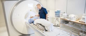
The duration of the procedure is on average 30 minutes. The woman undresses to the waist and removes all body jewelry.
After treating the nipple, areola and surrounding skin with a 70% alcohol solution, the doctor injects 1-2 ml of 1% novocaine into the base of the nipple (pain relief and relaxation of the milk ducts). The radiologist then applies gentle pressure to the nipple to identify the milk duct from which the secretion is obtained. A thin flexible catheter is inserted into this duct perpendicular to the nipple. The catheter should be inserted freely and without force.
An aqueous solution of a contrast agent (cardiotrast, verografin, urotrast) in an amount of 0.25-0.5 ml is injected through the catheter (up to 8 ml can be administered). Then the mammary gland is placed on a special mammograph stand and pressed on top with a plate. This allows the contrast to be evenly distributed throughout the duct. X-ray images after contrast administration are performed in two projections (direct and oblique). After the ductography procedure is completed, the contrast is removed from the duct.
Usually 1-2 ducts are examined.

Contraindications
Like any medical procedure, this diagnostic technique has a number of limitations. Let's consider the main contraindications.
Absolute
Absolute contraindications serve as a reason to refuse diagnosis and not carry it out under any circumstances, except for a direct threat to life.
Pregnancy
Breast ductography is contraindicated in pregnant women. In addition to the fact that the image will be uninformative, since the milk ducts will be filled with liquid in any case, the woman will receive a dose of ionizing radiation (albeit small, but capable of harming the unborn baby).
Lactation
The explanations for this contraindication are the same as in the previous case - lack of information and the negative impact of radiation (in this case, on the composition of the secreted milk).
Acute inflammation of the breast (abscess, mastitis, etc.)
The introduction of a contrast agent into the milk ducts can worsen a woman’s condition if her mammary gland is inflamed (for example, mastitis, mastopathy, etc. are diagnosed). In addition, without anesthesia, contrast injection may be accompanied by severe pain.
Relative
Relative contraindications leave the possibility of diagnostics, but impose serious restrictions.
Allergy to iodine
Since the contrast agent contains an iodine component, ductography is not recommended for patients who are allergic to iodine.
Palpable formation in the excretory duct area
Breast ductography may be contraindicated if a neoplasm is detected during palpation of the breast. This may interfere with the injection of contrast into the milk ducts.
Does it hurt to do
The ductography procedure is considered relatively painless and is usually easily tolerated. In some cases, patients report some discomfort, which does not last long and is associated with dilation of the duct.
Why can our articles be trusted?
We make health information clear, accessible and relevant.
- All articles are checked by practicing doctors.
- We take scientific literature and the latest research as a basis.
- We publish detailed articles that answer all questions.
If discomfort increases, local anesthesia may be used. Unpleasant and painful sensations are often associated with a negative psychological attitude, fear of the procedure and the individual level of a woman’s pain threshold.
Ductography sometimes has a therapeutic effect - in 50% of women, after this diagnostic method, discharge from the nipples completely stops due to washing of the ducts. The amount of iodine contained in the contrast solution is insignificant and does not have a negative effect (in the absence of an allergy to iodine).
Interpretation of results
Ductography (what it is and how it is done, you can watch in the video) helps to visualize the ducts and their branches. In the pictures you can see tumors and determine their type - malignant or benign. Most often, ductography helps to recognize defects:
- filling of the milk ducts, indicating the presence of intraductal papilloma;
- milk ducts – short, rigid, dilated, with uneven contours or with neoplasms, these are signs of intraductal cancer;
- filling of the duct in the area of the contrasted cystic cavity is a sign of epithelioma of the cyst wall.
If it is not possible to administer a contrast agent because the excretory ducts are narrowed or swollen, we can talk about the patient having papillomatosis or mastopathy.
Purpose of galactography

The purpose of the study is to identify changes in the condition of the milk ducts:
- narrowing;
- extension;
- twist;
- growth of neoplasms.
Interestingly, sometimes flushing the ducts with contrast has a therapeutic effect:
- drainage;
- bactericidal.
Together with the iodine-containing substance, the secretion located in the duct is removed and its secretion stops. Iodine gives contrast antiseptic properties. After the procedure, about 60% of patients note a cessation of secretion.
Service cost
Ductography, as well as ductography with double contrast of the mammary ducts, are included in the compulsory health insurance program.
In other words, citizens of the Russian Federation have the opportunity to have contrast mammography done for free: by referral from a doctor, by appointment, on a first-come, first-served basis, if they have a compulsory medical insurance policy and SNILS.
The self-supporting cost of breast ductography varies in different regions and clinics: from 3,500 to 16,000 or more rubles. The price of the service depends on the quality and class of equipment, the qualifications of the doctor, the cost of the X-ray contrast agent, and the individual price list of the clinic.
- Tags:
- tests
- ductography
,
.
Save the article for yourself!
Olga23.04.2017 at 21:20
They did this to me. Unpleasant, but not painful.
Rita05/28/2017 at 21:52
Thanks for the clear article. I was prescribed ductography. I was very afraid, but I read it and already calmed down.
Love05/04/2018 at 12:49
Girls, it doesn’t hurt at all, I don’t tolerate pain very well, I didn’t feel it at all, don’t be afraid!!!!!
Ekaterina07/10/2018 at 07:39
Is the procedure done on both breasts or just one?
Aptekins.ru07/10/2018 at 18:24
Ekaterina, hello, ductography is performed on the breast that is suspected of pathology. If there is reason to believe that BOTH mammary glands are affected, both breasts will be examined. Sincerely.
Ekaterina20.10.2018 at 17:56
Thank you very much for the article! Before the appointment, I will definitely smear my nipples with Emla and take some antispasmodics)))
Anonymous06.12.2018 at 15:51
Does ductoscopy hurt or not?
Irina Vasilievna05/20/2019 at 15:22
What is Emla?
Snezhana09.29.2020 at 17:26
Based on the results of mammography and ultrasound, we are referred to ductography. However, I had an ultrasound 1 day before my period. Diagnosis: Ductectasia. Does it make sense to do another ultrasound on another day of the cycle before doing ductography? Thank you in advance
Aptekins.ru09.10.2020 at 20:01
Snezhana, hello Before menstruation, the milk ducts can be dilated naturally, which is within the physiological norm. The optimal time for performing an ultrasound of the mammary glands is from the 5th to the 8th day of the menstrual cycle. If possible, then of course the study should be redone. Sincerely.
Add a comment Cancel Reply
Read more…

Norm and interpretation of general urine analysis in adults