Computer scanning helps to establish a diagnosis in the most difficult cases
Chest CT is an advanced X-ray imaging technique that uses radiation to create detailed images of the sternum, sternoclavicular joints, ribs, adjacent soft tissues, and organs (primarily the lungs). The study is carried out if, after a routine x-ray, the cause of the complaint remains unclear or there is a suspicion of a pathological process in this area. A CT scan of the chest may be performed as an alternative to an MRI if the patient has a contraindication to an MRI scan. For better visualization, a contrast agent may be injected. The main advantage of CT is its ability to simultaneously demonstrate bone, soft tissue (with contrast), and blood vessels.
What is CT or computed tomography
Computed tomography (CT) or multislice computed tomography (MSCT) is a modern diagnostic method that consists of a layer-by-layer examination of the human body using X-rays to obtain a detailed image of internal organs and structures.
CT imaging is based on the varying densities of organs and tissues through which the X-rays pass. During the examination, an X-ray tube with a narrow beam of X-rays rotates around the patient. At the same time, many sensors record the slightest changes in density. Information from the sensors is processed by a powerful computer, after which it is sent to the monitor in the form of successive longitudinal and transverse sections (“tomography” literally means “image of a slice”). The thinner the section, the more accurate the diagnosis.
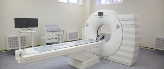
The University Clinical Hospital No. 2 has modern equipment - a high-class computed tomograph, which allows obtaining high-quality images with minimal radiation exposure.
Today, CT is one of the main diagnostic methods and is widely used both for making a diagnosis and for clarifying it. One of the main advantages of computed tomography is the speed of the study and the ability to scan several organs simultaneously. Modern tomographs can significantly reduce the duration of the examination and reduce patient exposure to a minimum, while obtaining hundreds of sections up to 0.5 mm thick. Thanks to high-precision three-dimensional projections, which make it possible to examine the position, shape, size and structure of various organs and structures of the body, the results obtained from CT examination are the key to success in making an accurate diagnosis, selecting the most effective treatment and planning surgery if necessary.
A significant advantage of this method is the ability to examine patients with pacemakers, artificial heart valves, metal clips in the body, etc., that is, those for whom MRI is contraindicated.
CT scanning is used to examine almost all parts of the body and organs: chest, abdomen, pelvis, limbs, liver, pancreas, intestines, kidneys and adrenal glands, bladder, lungs, heart, as well as blood vessels, bones and spine.
The range of diseases for which this research is necessary is extremely large. In each individual case, the need and feasibility of this study is assessed by your attending physician.
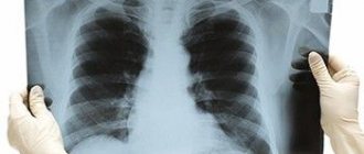
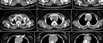
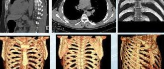
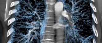
Chest tomography photo
The images are decrypted on the same day. The resulting images are analyzed by a radiologist, who issues a conclusion. Determining the correct diagnosis is not easy, because among several dozen shades of black, gray and white, it is necessary to isolate the one that does not correspond to the norm and interpret the result. Let us present to your attention several photos of chest tomography:
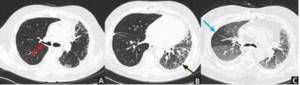
A non-contrast CT scan in a patient with a history of interstitial lung disease and a right lung transplant shows a narrowed area of the right bronchial anastomosis (red arrow). The left lung is reduced in size, with signs of bronchiectasis and bronchiolectasis (black arrow). Narrowing of the central airway during expiration in a transplanted lung (blue arrow).
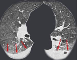
Effusion (red arrows) in both pleural cavities.
At the Magnit diagnostic center in St. Petersburg, you can do a CT scan of the chest organs, pre-registration through the website form or by phone.
What is the difference between CT and MRI
This is the most common question asked to a radiologist. CT and MRI are diagnostic methods. They have similarities and differences. The similarity lies in the external similarity in the design of the devices themselves - tomographs. In their center there is a tunnel into which the table slides.
The main difference is a completely different principle for obtaining images: CT uses x-rays, MRI uses a magnetic field. Which method is right for you is determined by your doctor, taking into account the specifics of the disease and the purpose of the study.
Important!
Please prepare in advance and bring with you all extracts, protocols or recordings (disks) of previous studies. The more information the doctor has before the study, the clearer the task assigned to him. In addition, previous results will allow us to assess the dynamics of the disease.
Before the procedure, be sure to notify your doctor if you have:
- Pregnancy
- Are you allergic to medications, including iodine in the contrast agent?
- Cardiovascular disease (eg, heart failure)
- Diabetes mellitus or you are taking metformin (Glucophage). You may need to avoid taking this medication the day before and for the day after your procedure.
- Kidney diseases
- Bronchial asthma
- A pacemaker or insulin pump is installed
- Multiple myeloma.
- You have had an X-ray examination within the previous 4 days using a barium contrast agent (irrigoscopy) or you have used medications containing bismuth. Barium and bismuth, appearing on X-ray film, reduce image quality.
- There is a fear of closed spaces (claustrophobia). Because you will have to lie still inside the scanner during the procedure, you may need sedatives. In this case, you should ask someone to take you home after the procedure.
The Department of Radiation Diagnostics of the University Clinical Hospital No. 2 operates within a multifunctional clinic, therefore, if any possible complications are suspected, the decision on conducting the study, its type and methods of preparation is made together with medical specialists (nephrologist, endocrinologist, etc.).
Low-dose screening MSCT of the lungs
If there are no symptoms, and the purpose of the study is a preventive examination of the condition of the lungs, it is advisable to perform a low-dose screening MSCT study of the lungs.
The dose for this type of study is less than 1 mZr. This threshold (1 mZr) is defined by the regulatory documents of the Ministry of Health of the Russian Federation as an acceptable annual dose for preventive examination of healthy people.
Low-dose screening MSCT of the lungs can detect gross pulmonary pathology (i.e., detect the very fact of the existence of lung pathology). The diagnostic value of this study is much higher than chest radiography. Therefore, it is prescribed for diseases such as pneumonia, pleurisy, tuberculosis. With its help, a lung abscess, the presence of effusion in the pleural cavities (pus, blood, ichor), pneumotrax (the presence of air in the pleural cavity), and malignant formations are detected.
The study is carried out without the introduction of a contrast agent. The procedure takes up to 15 minutes.
Preparing for an abdominal CT scan
If you are scheduled for a CT scan of the abdominal organs, refrain from eating solid food the evening before the examination. Before the procedure, you may be asked to drink a contrast agent, and in some cases, take a mild laxative or have a barium enema.
CT and MSCT of the abdominal organs are performed on an empty stomach or no earlier than 3-4 hours after the last meal. It is allowed to take medications (with a small amount of water). Immediately before the examination, the department may ask you to drink about 500-600 ml of water.
MSCT examination is performed after preliminary contrasting of the intestine with a diluted contrast agent. The gastrointestinal tract is filled either on the day of the study or the day before: On the eve of the study, drink 0.5 liters of solution in the evening, in the morning (06:00-07:00) drink the remaining 0.5 liters. The study is carried out with a full bladder. Women must have a sanitary tampon with them.
Attention! After an X-ray examination of the stomach, colon with barium (gastric fluoroscopy, irrigoscopy), CT examination of the abdominal organs is recommended no earlier than 5-7 days (at least 3 days). In cases of emergency - after cleansing enemas.
Is it possible to do a CT scan on your own initiative without a doctor’s referral?
Children will not be given a CT scan without a doctor's order. There are no rules for adults that would prohibit them from signing up for examinations and taking tests on their own. However, there is a risk of examining the wrong thing or not. For example, a woman, unknowingly, can undergo a pelvic ultrasound herself on any day and only then go to the gynecologist. The doctor may be alarmed by some points in the ultrasound results, and he will ask you to repeat the examination during the 5-10th day of the cycle, when, according to the standards, this diagnosis is carried out. As a result, the patient will simply pay for the same procedure twice, but without harm to health.
With a self-ordered CT scan, the risk isn't just double payment. As we wrote earlier, due to X-ray radiation, this examination must be done correctly the first time and with good equipment. Only an experienced doctor will determine which organ needs to be examined and to what extent. Plus, like any medical procedure, there are contraindications that the patient may not be aware of, even if he has read several articles on the Internet. Therefore, in this case, self-medication is doubly dangerous.

Functional diagnostics doctor Inna Petrovna Aleshova:
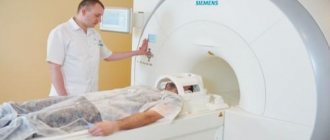
We invite you to the Filatov Clinic to undergo a safe examination using our new SOMATOM go.Up computed tomograph.
The device is an advanced model with low radiation exposure. IVR technology allows you to obtain 64 high-quality image slices per revolution of the ring, which guarantees more accurate diagnosis of the lungs and other organs. You can sign up for the study by phone. or online registration.
How the research works
We ask you to come to the radiology department (9th floor) 10-15 minutes before the scheduled start time of the study and, having contacted the office, inform the staff of your arrival. Given the possibility of emergency research, there is a possibility of waiting for the procedure for some time. Experienced doctors and x-ray technicians will do everything to make you feel comfortable and safe throughout the entire examination.
Computed tomography is an absolutely painless procedure that takes no more than 30 minutes (including changing clothes and, if necessary, intravenous contrast). The scanning itself lasts several minutes. If you need a CT scan with contrast, you will first be invited to the procedure room, and a nurse will install an intravenous catheter for you to administer a contrast agent during the examination.
Before the examination, you will be asked to remove all jewelry and metal objects that fall into the scanning area; the x-ray technician who will carry out the procedure will explain to you the rules of conduct during the examination and will help you sit comfortably on the tomograph table.
If the study is carried out using a contrast agent, it can be introduced into the patient’s body in various ways depending on the purpose of the study: intravenously - in CT scans of the chest, abdomen and pelvis.
- orally - for some abdominal examinations, the contrast agent must be drunk
- through a special catheter into the bladder or intestines
During the procedure, the patient lies on a special table connected to a CT scanner, which is a large ring-shaped machine. The scanner rotates around the area of the patient's body being examined, taking layer-by-layer images of the corresponding organ. A faint hum or clicking sound may be heard.
Try not to move during the procedure, as moving your body may reduce the quality of the image.
Given that a CT scanner emits X-rays, the machine is usually located in a special shielded (protected) room. The device is controlled automatically from the adjacent office, which houses the tomograph’s computer unit, monitors and equipment for monitoring the patient’s condition. The doctor performing the examination sees and hears you. You will be able to talk to him through a special intercom. The specialist may ask you to hold your breath for a few seconds to get a clearer picture.
Based on the images obtained, the doctor at the radiology department gives a medical opinion. In addition, your doctor or surgeon can comment on the results of the study.
When should a CT scan be done instead of an X-ray?
X-ray is a well-known and more affordable diagnostic procedure. The price of CT scans is usually higher. A logical question arises: why pay more if you can get an x-ray?
A regular X-ray produces a two-dimensional picture, so images of organs overlap each other, which reduces the accuracy of diagnosis. The result will depend on the doctor's experience and ability to read radiographs. X-ray is more suitable for primary diagnosis, since it does not allow a full assessment of the situation, the condition of tissues, inflammation, formations, etc. Therefore, often with “suspicious” results and other related indications, the doctor refers the patient to the most accurate and unambiguous diagnosis - computed tomography.
The CT machine provides a three-dimensional volumetric image of the examined organ. It can be studied in small details from different angles. A computed tomogram clearly shows the consequences of injuries, benign and malignant neoplasms, stones, cysts, developmental defects, etc. A high-quality three-dimensional image helps to identify pathologies even at the earliest stages of development.
What is safer - a CT scan of the lungs or an X-ray?
During a routine X-ray of the lungs, a person receives a radiation dose of 1.2 mSv. While a modern computed tomograph allows you to do a low-dose CT scan of the lungs, in which the radiation dose will be from 0.3 to 0.7 mSv, depending on the patient’s build. Plus, the radiologist will be able to evaluate the lung tissue in 3 projections at once, which is impossible with X-rays. Therefore, examining the lungs with a modern tomograph is several times safer and more informative than taking an X-ray.
What sensations does the patient experience during the CT scan procedure?
The procedure itself is absolutely painless. Some discomfort may be caused by the hard surface of the table, the inability to move, and the temperature in the CT room (the room may be cool).
Some patients feel nervous while inside the scanner. If contrast material enters a vein, you may experience a feeling of warmth, heat, or a metallic taste in your mouth. Sometimes patients experience nausea or headache. Be sure to tell your doctor or x-ray technician how you feel.