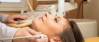
What is a vaginal ultrasound?
Vaginal (transvaginal) ultrasound examination of the pelvis is carried out by inserting a special device equipped with a sensor into the vagina.
The device is a rod with a handle, which is made of plastic, about 10-12 centimeters long and up to three centimeters in diameter. A special groove may be built into it to insert a needle to take biopsy material.
The examination allows you to determine the presence of pathologies, neoplasms or diseases in the following female genital organs:
- Uterus
- Fallopian tubes
- Ovaries
- Cervix
It is considered the most effective for studying these parts of the reproductive system, as it allows one to identify various health problems of the patient in the early stages. A pelvic ultrasound with a sensor can show the presence of abnormalities even at a time when other studies do not show any problem areas.
Detailed analysis of M-echo indicators by thickness and cycle days and possible pathologies
| 8 mm | In women of childbearing age, this indicator is normal on days 10–15 of the cycle. If the patient has no history of hormonal therapy and the 8 mm mark remains after the 15th day of the cycle, this indicates hypoplasia. In this case, there is a high probability of rejection of the fertilized egg. For postmenopausal women, this indicator is evidence of pathology. |
| 9 mm | Typical for days 15–21 of the standard cycle. With a shorter cycle, 9 mm is the norm in the early period, with a longer cycle - in the late period. |
| 10 mm | The norm for the second phase of the cycle. The detection of such a value in the first phase indicates the development of a hyperplastic or inflammatory process or other pathology. For postmenopausal women, this is the norm only when undergoing hormone replacement therapy. |
| 11 – 12 mm | Also normal for the second phase of the cycle. This is the minimum value of endometrial thickness for successful implantation of the fertilized egg. A detected thickness of 11–12 mm in the first phase of the cycle or in postmenopausal women indicates pathology. |
| 13 – 14 mm | Normal thickness after day 15 of the cycle. Reaching this value earlier indicates pathology. Additional examination is required. |
| 15 mm | Limit value of the thickness of the lining layer for childbearing age. His early achievement is a suspicion of pathology. |
How is the procedure done?
The study is organized as follows:
- The patient should remove clothing from the lower part of the body (from the waist down)
- She sits on a special couch in the same way as during a regular gynecological examination.
- The doctor prepares the sensor: puts an individual condom on it, lubricates it with a special gel for the procedure
- The physician then inserts the device shallowly into the patient’s vagina.
- To get a complete picture of the state of the organs, he can move the sensor from side to side
- All data is recorded and processed by a doctor
The gel is necessary to facilitate the penetration of the sensor (and thereby reduce the likelihood of negative sensations) and enhance the ultrasonic effect by increasing conductivity.
This type of examination lasts no more than 10 minutes. It is painless and gives the most complete picture even in cases where an abdominal ultrasound shows nothing or cannot be performed.
Reasons for development
The root causes of the development of the pathology have not been fully established. But doctors put forward various theories with the help of which they try to explain the etiology and pathogenesis of adenomyosis. A popular theory is the implantation of separated endometrial cells during retrograde reflux of menstrual blood through the fallopian tubes.
There are theories that speak of the spread of pathological cells by hematogenous and lymphatic routes. The genetic factor may also play a significant role, given the concordance of the manifestation of adenomyosis in twins. Many medical studies have been conducted in search of the exact root cause of the pathology, but doctors have not received a clear answer to this question.
When is a pelvic ultrasound with a sensor necessary?
There are symptoms in which the doctor must refer the patient for a transvaginal examination:
- Pain in the lower abdomen (not related to the menstrual cycle)
- Suspicion of the presence of neoplasms
- Periods of menstrual bleeding that are too short, too long or absent
- Impossibility of pregnancy
- Bloody discharge that is not menstruation
- Presence of fallopian tube obstruction
- Nausea, vomiting and weakness with bloody discharge from the vagina
Doctors recommend using this type of examination for preventive purposes, since not every ailment may have symptoms at an early stage, just as pregnancy in the first trimester may not manifest itself with classic symptoms (nausea, etc.).
In this case, vaginal ultrasound is used for:
- Diagnosis of infertility
- The need to determine the presence of changes in the size of the ovaries and uterus
- Diagnosis of pregnancy
- Pregnancy monitoring (first trimester only)
- General monitoring of the condition of the uterus, fallopian tubes and ovaries
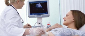
A pelvic ultrasound can be performed simultaneously with two sensors. In this case, an abdominal ultrasound examination is performed first, and then a transvaginal one. The use of two types of analysis at once is necessary to identify disorders in the high-lying pelvic organs.
What pathologies does the examination reveal?
Echography is a valuable procedure that can detect serious pathologies of the endometrium at an early stage of development. The main ones:
- Hyperplasia is a benign disease in which the endometrium grows and, as a result, thickens and increases its volume.
- Hypoplasia is underdevelopment of the endometrium, its insufficient growth.
- Endometrial cancer is an extremely dangerous pathology, in most cases affecting postmenopausal women. With timely diagnosis, there is a good prognosis for recovery - 95%.
What does a vaginal ultrasound show?
This examination allows you to evaluate the following parameters of the reproductive system organs:
- Dimensions of the uterus. In normal condition, it should be about seven centimeters in length, six in width and 4.2 in diameter. If it is significantly less or more, then this indicates the presence of pathology
- Echogenicity. The structure of the organs must be homogeneous, uniform, have clearly defined, clearly visible edges
- General picture of internal organs. The uterus should be slightly tilted forward. And the fallopian tubes may be slightly visible, but should not be clearly visible without the use of a contrast agent
Norm
The concept of normal ovaries depends on the stage of the cycle and the age of the patient.
At the beginning of reproductive age, the ovaries are the same size in all women. By the beginning of natural decline, by the age of 40, the size of the glands decreases. Normally, the ovaries have lumpy outlines due to the follicles. During normal reproductive activity of the body, there should be 9-10 follicles. If there are significantly fewer of them, this makes us think about pathological changes in the reproductive sphere. The diameter of the follicles is about 3-5 mm. During maturation, the dominant follicle increases to 24 mm. It consists of a fully mature egg, which is released from the follicle at the end of ovulation. Author: Telegina Natalya Dmitrievna
Therapist with 25 years of experience
Diagnosed diseases
Transvaginal ultrasound allows you to identify a number of diseases and problems in the reproductive system at an early stage. It allows you to detect:
- Fluid and pus in the uterus and fallopian tubes. The cause of their appearance may be infections, viruses, mechanical damage
- Endomentriosis is an excessive proliferation of cells in the inner layer of uterine tissue into other layers and organs. It can occur due to inflammatory processes, damage (surgery, abortion), the appearance of neoplasms, disruption of the endocrine system, too frequent use of certain medications and substances
- Myoma is a benign neoplasm in the tissues of the uterus or its cervix. May occur due to chronic diseases, frequent abortions, hormonal imbalances, constant stress, pathologies, excess weight, or with hereditary predisposition
- Cysts and polycystic ovaries are tumors filled with fluid. Occurs with endocrine disorders, chronic diseases of the genitourinary system
- Various polyps on the walls of the uterus are benign formations in the endometrium of the organ. They can reach several centimeters in diameter. Their appearance may be associated with polycystic disease, chronic diseases, mastopathy, fibroma
- Inflammation and enlargement of organs can occur due to both infection and injury.
- Hydatidiform mole - appears instead of a full-fledged embryo during the process of conception, filled with fluid. Occurs due to duplication of male chromosomes with the loss of female chromosomes, sometimes due to fertilization of an egg that does not contain a nucleus. This disease is rare
- Fetal development disorders during pregnancy
- Defects and pathologies in the development of the fallopian tubes: obstruction, spiral-shaped or too long tubes, blind passages, duplication of organs
- Ectopic pregnancy occurs when an egg implants outside the uterine tissue after fertilization. Occurs due to blockage of the fallopian tubes, congenital anomalies in them, as well as after an inflammatory process, abortion
- Cancer is a malignant tumor in various organs:
- Uterus
- Ovarian
- Cervix
New generation diagnostics at the VIP Academy
- High accuracy and information content. The Voluson E10 General Electric device, fully equipped, takes ultrasound of the uterus and appendages to a new level. The doctor sees a high-quality image of the organs, can study the tissue structure in detail, and build a three-dimensional model. The equipment is designed for women, and various types of sensors are provided to obtain maximum data.
- Specialized diagnosticians. We see doctors who specialize in radiation diagnostics specifically in gynecology. Many years of experience allow them to identify pathologies in the most difficult situations.
- Multidisciplinary approach. "VIP Academies" is a multidisciplinary medical center. If necessary, specialists from related fields are involved in the diagnosis and additional examinations are carried out.
After the examination, the patient receives a detailed transcript of the results (it is possible to record video and photos from the procedure on a digital medium) and recommendations for further actions.
Stages of preparation for the study
To conduct a pelvic ultrasound with a sensor, no special preparation is required, but there are several mandatory requirements:
- Unlike an abdominal examination, with a transvaginal analysis the patient should not drink liquid one to two hours before the examination.
- If she emptied her bladder more than an hour before the test, she needs to do it once immediately before the procedure
- With increased flatulence, the patient needs a drug that will help normalize the processes of gas formation in the gastrointestinal tract. She can consult with her doctor about the choice of medication
Doctors also recommend using such a study on certain days of the cycle, depending on which organ needs to be diagnosed and for what purpose:
- In the case of a preventive examination, it should be done in the first days after the end of menstruation
- If there is a suspicion of an increase in the endometrial layer in the uterus, then in the second half of the cycle
- When it is necessary to monitor the development of a disease or the progress of treatment, the study can be carried out several times in one cycle, at different stages.
- An ultrasound is performed urgently if there is bleeding that is not menstrual, regardless of the day of the cycle
It is important to remember to maintain personal hygiene before the examination, use wet and other wipes.
If you plan to conduct a pelvic ultrasound with two sensors, then you should pay attention to preparing for the abdominal examination.
This includes:
- Follow a diet for at least three days before the test to reduce the likelihood of symptoms of flatulence and bloating
- The last meal should be completed by six o'clock in the evening on the eve of the test.
- It is recommended to do an enema after eating
- If there is still a risk of flatulence, you need to use special medications that reduce gas formation
- An hour before the test, drink at least 400 ml of water
The diet involves excluding a number of foods from the diet:
- Sweets
- Flour (bread, cookies, etc.)
- Legumes
- Cabbage
- Milk and dairy products
- Vegetables and fruits that have not undergone heat treatment
- Coffee and strong tea
- Carbonated drinks
- Fast food
- Fatty foods (meat, fish, oils)
You can eat porridge cooked in water, lean boiled beef, poultry and fish, and hard cheeses. It is recommended to drink lightly brewed, slightly sweetened tea.
It must be remembered that since you need to drink liquid before the abdominal examination, you must empty your bladder before the transvaginal analysis.
Indications: symptoms and prevention
A woman should be prompted to undergo an ultrasound of the uterus with an echo not only by relevant indications. Regular examination for the purpose of prevention is an irreplaceable and useful thing that will help protect yourself from problems of the reproductive system. Median echo is necessary for women of any age category. The procedure is prescribed for the following indications:
- pregnancy planning;
- irregular menstrual cycle with long delays;
- acute and chronic diseases of the reproductive system;
- pain in the lower abdomen;
- bleeding outside the cycle;
- infertility;
- premenopause;
- postmenopausal period.
Contraindications
Vaginal ultrasound examination has a small number of contraindications:
- It is never performed if the patient is a virgin, so as not to violate the integrity of the hymen. In this case, such a patient can undergo a transrectal examination, in which a sensor is inserted into the rectum
- The study is prohibited from being carried out in the second or third trimester of pregnancy, because it may provoke premature contractions or uterine contractions before the expected due date.
- This test is not used if the patient is allergic to latex
- If the patient has epilepsy, since the examination requires that she lie still
M-echo when planning pregnancy predicts the possibility of miscarriage
When planning a pregnancy, ultrasound with echo is a mandatory procedure, since it will allow you to assess readiness for implantation of the fertilized egg, i.e. to pregnancy.
The optimal endometrial thickness for this event is 11 – 14 mm. Insufficient thickness will prevent the penetration and fixation of the egg to the uterine wall. Excessive thickness increases the risk of rejection of the fertilized egg, which as a result is observed as a miscarriage.
At the same time, the structure of the layer lining the uterus should be more loose and spongy, which will allow the fertilized egg to attach to it without any problems.
Risk factors
- Risk factors for the occurrence of pathology include:
- higher than normal estrogen levels;
- early onset of menstruation, their abundance, duration more than 8 days;
- surgical intervention on the uterus, appendages;
- abuse of bad habits;
- obesity;
- immune dysfunction;
- chronic stress, nervous overload;
- unfavorable environmental conditions;
- genetic predisposition;
- history of abortions.