When is a jaw x-ray taken?
Tell the patient exactly whether he needs and can have x-rays of his jaws
, should his attending physician. General indications for such a diagnostic procedure are:
- Suspicion of jaw development abnormalities (in childhood or adulthood). These may be congenital clefts of the hard/soft palate, impaired growth and proper development, or malocclusion.
- Suspicion of infectious-inflammatory and specific pathologies. These include osteomyelitis, periostitis, syphilis, actinomycosis, tuberculosis or necrosis of the jaw bone.
- To identify various acquired defects resulting from injury or removal of oncological formations.
- Suspicion of the presence of a false joint.
- Suspicion of a benign or malignant neoplasm.
What can you see in the photographs?
The following can be seen on x-rays of the upper and lower jaws:
- defects - cracks, fractures, presence or absence of bone fragments;
- pathological changes in bone tissue - areas of tissue thinning, compaction, thickening or thinning of the cortical plate;
- areas of necrosis - sequestra;
- sclerotic process;
- various periosteal layers, osteophytes (bone growths);
- tumor-like neoplasms.
When is an X-ray prescribed for a child?
If X-rays in adolescents and adults do not cause concern, then X-ray diagnostics of young children worries many parents.
The devices used in modern medicine do not harm the body, but the study is still carried out only if indicated. Dental X-rays are recommended for children for the following indications.
- Deep caries. The procedure is carried out to determine the lesions and affected area. The image allows the specialist to assess whether the roots and rudiments of permanent teeth are damaged.
- Developmental anomalies. X-rays make it possible to determine the causes and characteristics of irregular teething, as well as delays in their growth.
- Control of permanent teeth. Diagnostics allows you to see all the features of the rudiments of permanent teeth, as well as predict the period of their eruption and identify possible pathologies.
- Malocclusion. X-ray is the most accurate diagnostic method for determining abnormalities in tooth growth and jaw position.
- Abscess. The study is aimed at confirming or refuting the development of an abscess if signs are present.
- Jaw injuries. In case of mechanical damage, an x-ray is taken to assess the damage to bone tissue that has occurred.
X-rays are often used to monitor the treatment being carried out. Diagnostics is also used when restoring teeth using temporary prosthetics or installing orthodontic structures.
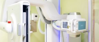
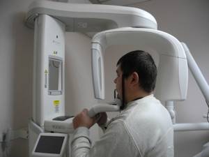
How is the procedure performed?
X-ray of the jaw of an adult or child is a simple procedure. It can be done in different projections in order to better examine the structure of the bone tissue and make an accurate diagnosis.
X-ray of the lower jaw
To obtain general information about the condition of the lower jaw, an X-ray is taken in a direct projection. It allows for primary diagnosis of inflammatory, oncological and traumatic diseases. The patient is positioned as follows: lying on his stomach, face down, resting the tip of his nose and forehead on the cassette. The X-ray machine sensor is installed at the occipital protuberance.
A lateral view of the lower jaw is taken to assess the condition of the body, ramus and teeth of the desired side. The patient lies on his side, with his cheek on the cassette located at a slight angle.
To diagnose diseases of the lower jaw, images are also taken in the axial projection. The patient takes a position lying on his stomach, stretching his head forward with his chin as much as possible. At the same time, it is pressed against the cassette by the front surface of the neck and lower jaw.
X-ray of the upper jaw
To assess the condition of the bone tissue of the upper jaw and the chin area of the lower jaw, a nasomental placement is performed. To do this, the patient lies on his stomach, face down, resting the tip of his nose and chin on the cassette. The sensor is installed perpendicular to the cassette, 2 pictures are taken - with the mouth open and closed.
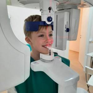
How is a dental x-ray performed?
An X-ray of a child’s jaw with baby teeth is a painless procedure that takes 3 to 5 minutes.
The course of dental radiography in children consists of:
- the pediatric dentist gives a referral for examination;
- the child, with or without a parent, is sent to a special room;
- a special apron with lead plates is put on him;
- clamps a plastic plate with his teeth;
- the specialist fixes the position of the head and leaves the room;
- The device turns on and takes a photo.
If there is a need for a targeted X-ray, the child is seated on a chair, a film is placed in the mouth, and an X-ray tube is placed near the mouth and held for several seconds. During the procedure, the child must sit still, otherwise the images may be distorted.
The results are delivered in 20–30 minutes. With the received images, the patient is sent to the attending physician. You can get an X-ray of a child in Moscow at the Novodent dental clinic. There is advanced and modern equipment with minimal radiation doses, but high information content. There are doctors who will find an approach to every little patient, inspire trust, and listen to their recommendations. More detailed information about the clinic’s capabilities can be found on the website or by phone.
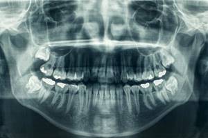
Panoramic shot of the jaw
A panoramic X-ray, or orthopantomogram, displays the entire upper and lower jaw in a direct projection. In such a picture you can see almost all the anatomical features of the patient’s dental system, all kinds of neoplasms, defects, fractures of tooth roots, and so on. An orthopantomogram allows you to identify the following pathologies:
- damage to tooth roots by caries;
- hidden carious cavities;
- cysts, granulomas, tumors;
- pathological changes in the roots and peri-root space.
What can a dental x-ray show?
Carrying out X-rays of a child’s teeth or jaw is aimed at identifying pathologies and determining the anatomical features of the development of the dental system. Dental x-rays are performed before treatment, during treatment or upon completion for control.
X-rays may show:
- decrease in tooth weight due to inflammation of soft tissues;
- foci of carious lesions in any area;
- changes occurring in dental roots;
- purulent processes flowing into an abscess;
- rudiments of permanent teeth;
- anomalies in the structure and development of teeth and jaw;
- impacted teeth;
- supernumerary teeth;
- damage to the jaw bones;
- and other.
The result of the image is assessed exclusively by a specialist. Based on the information received, the doctor can determine the diagnosis, prescribe or adjust treatment, or evaluate the effectiveness of the measures taken.
Decoding the results
An x-ray of an adult's jaw is usually interpreted by a radiologist.
Other specialists may also be involved in this work: dentist, facial surgeon, otolaryngologist. Specific pathologies of the upper and lower jaws have certain radiological features:
- Chronic osteomyelitis. Depending on the type of disease, the images may show foci of resorption of various shapes with a shadow of necrotic masses inside. In more severe cases, communication of the sequestra with the oral cavity is visualized.
- Acute osteomyelitis. When examining the image, you can see areas of bone tissue resorption that do not have clear boundaries.
- Fractures or cracks in the jaw bones. This pathology reveals itself by the fact that thin, elongated shadows are quite clearly visible in the photographs. In such cases, it is important not only to identify the fracture, but also to see the bone fragments, if any, and to understand how much they are displaced.
- Chronic periostitis. In the image with such a pathology, periosteal thickenings will be visible. If the disease has become severe, areas of ossification of the periosteum and new bone tissue along the edge of the jaw are visualized.
X-ray TMJ
X-ray TMJ
X-ray of the TMJ is performed to identify pathologies of hard structures in this area. This scanning method is non-invasive and does not cause pain or discomfort. Radiation exposure is minimal and safe for humans. After the diagnosis, a conclusion is drawn up with a description of the area and conclusions about the condition of the joint. The essence of radiography of the temporomandibular zone With the help of modern X-ray machines, you can obtain images on electronic media, which are highly informative and clear. This method is most relevant when diagnosing joint diseases. This study is often prescribed if jaw mobility has decreased or pain has appeared in this area. A specialist can give a referral for examination if there is facial asymmetry, extraneous sounds appear when moving the jaw, or headache. X-rays show the effects of the area after surgery, conservative treatment or injury. A scan is required before surgery. X-ray placement of the TMJ does not take much time. To obtain clear and reliable images, the patient only needs to fix the oral cavity in a closed and open position in turn. If there is a suspicion of serious diseases such as tumors, an additional illuminating substance is injected. Checking is often required when a tooth is damaged, when baby teeth are replaced with permanent ones, if there are symptoms of oncology, or when blood flow is impaired. Scanning is required when planning the installation of implants, in case of pathological abrasion of teeth. To avoid serious complications and identify abnormalities in the early stages, it is recommended to carry out diagnosis at the first symptoms of the disease. Algorithm of the procedure No complex preparation is required before the examination. Diagnosis can be difficult if the patient cannot take a stationary position and suffers from involuntary movements of the head and limbs. If necessary, a contrast agent is administered in advance. It is completely eliminated from the body after 1-2 days and, in the absence of contraindications, does not harm health. The procedure is prescribed for pain, swelling, and clicking in the joint. It is possible to identify layers, stones in the submandibular gland, evaluate the relationship of the jaws, the correctness of the closure of the teeth, their relationship, and the development of atrophic processes. The ratio of the articular head to the glenoid cavity is assessed. X-rays in dentistry An instrumental examination is required, since not in all cases the doctor can fully assess the picture of the disease or check the condition of the teeth and joints. Scanning allows you to identify hidden pathologies and make a correct diagnosis. X-rays may be prescribed for preventive purposes in order to identify dangerous diseases in the early stages. An X-ray examination allows you to evaluate the root canal, the condition of the filling and crown, the quality of its installation, detect carious lesions, and monitor the filling of the canals. The condition of the tooth tissue is determined. Research is required for joint diseases and planning prosthetics and implantation. To find out the cost of performing an x-ray of the jaw joint and jaw, contact our specialists at Ortolaym. We will help you calculate the cost of diagnosis and treatment.
Features of the shadow picture of temporary teeth
On x-rays, the roots of primary teeth are shorter and the bifurcation angle of molars is greater than that of permanent teeth. The root canals are wider, and the dental cavity is larger in volume. The size of the periodontal gap in a child may vary, and this is considered a normal variant.
The dental follicle appears on an x-ray as a rounded clearing and has a clear rim of compaction. In its cavity there is a rudiment, which changes at different stages. First, points are visible along the cutting edge, which gradually form a single contour of the crown.
Examination at SM-Doctor
If you need to take an X-ray of the jaw or temporomandibular joint, contact a medical specialist. Our advantages:
- High quality photos. The details of baby teeth, follicles and periodontal tissue are so small that they can only be examined on technically ideal radiographs with high-resolution apparatus.
- Minimum radiation dose. We use digital x-ray. It produces less radiation and also allows you to save images in digital format, process them, and record them on digital media.
- Comfort. We use X-ray methods that do not cause anxiety or discomfort to the child.
In our clinic, after an x-ray, you can get a consultation with any pediatric specialist.
If necessary, you can undergo additional studies aimed at clarifying the nature of the pathology identified on the x-ray. You can make an appointment or ask questions around the clock by calling +7 (495) 292-59-86