Reasons for conducting the study
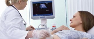
Ultrasound diagnostics is a highly accurate and safe examination for humans. The signals sent by the sensor are reflected from the internal organs of a person and returned. Thus, a picture is created on the monitor, which the doctor explains.
This type of study is prescribed for examining the pelvic organs. Using ultrasound, the condition of internal organs is assessed, the presence of diseases and pregnancy is determined. You can have a pelvic ultrasound in Kaluga at the Elite Medical Center. A referral for examination can be obtained from a general practitioner; also, if menstruation is irregular, a gynecologist can refer you for the procedure. A pelvic ultrasound should be performed for the following indications:
- Frequent pain in the lower abdomen;
- With heavy bleeding;
- For unusual discharge;
- If a formation is suspected.
Ultrasound diagnostics can detect cysts, kidney stones, tumors, fibroids and other diseases. Preparation for pelvic examination is carried out in the following ways:
- Transabdominal;
- Transvaginal;
- For pregnant women - obstetrics.
Before the first type of examination, you need to fill your bladder and drink about a liter of liquid. It is necessary to empty the bladder during transvaginal diagnosis. There are no recommendations for screening and obstetric ultrasound.
You need to prepare for each type of examination differently, but there is a general rule: do not eat food that can cause gas for several days before the examination. The procedure will have to be postponed if you had an x-ray two or three days ago. This may affect the results. On the day you are examined, you need to cleanse your intestines.
How is an ultrasound performed?
First it is carried out through the abdomen. The procedure is performed while lying down; the part of the body being examined should be exposed. A neutral gel is applied to improve the conductivity of the device’s waves. The doctor moves the sensor over the body, periodically changing its inclination. The process is painless, the device does not make noise, and only a slight pressure from the sensor itself can be felt.
It then continues through the vagina. The patient lies on the couch. The specialist uses an oblong sensor, puts a condom on it, treats it with gel and inserts it into the vagina. The doctor moves the sensor to get clear projections of the organs from different sides. The method can cause pain only if there is internal inflammation.
Through the rectum. The patient lies on his side, knees pulled up to his stomach. A sensor with a special condom, lubricated with gel, is inserted into the anus 4-6 cm. The doctor examines the rectum from all sides. The method does not cause pain, discomfort occurs in the presence of illness.
Regardless of the type of ultrasound, the machine displays an image on the screen, which is later printed.
When is the best time to perform the procedure?
When examined by a gynecologist, it is important to know on what day of the cycle to do a pelvic ultrasound. It should be carried out in the first week after menstruation ends. If you have an allergy, you need to tell your doctor about it. There are no contraindications to the procedure; pelvic ultrasound cannot be performed only if you are menstruating.
A study in the first seven to ten days of the menstrual cycle will be indicative. If the doctor suspects you have fibroids, then a pelvic ultrasound should be performed immediately after menstruation has ended. When planning a pregnancy, a study is carried out to identify folliculogenesis on days 5, 9 and after two weeks of the menstrual cycle. The timing may vary depending on the specific case, depending on the duration of menstruation.
You should be examined by a female doctor at least once a year, including an ultrasound examination. If you experience discomfort or changes in your menstrual cycle, consult your doctor immediately.
Ultrasound during menstruation
Ultrasound examination of some organs requires special preparation. It is not recommended to perform diagnostics on women during menstruation. However, there are exceptions when an ultrasound is performed urgently.
Since women are recommended to regularly undergo pelvic ultrasound, and often make an appointment with a doctor several weeks in advance, it is not always possible to predict whether menstruation will then occur. Menstruation will not interfere with the research. The doctor is aware of the additional measures that are required in this case.
Is it possible to undergo an ultrasound during menstruation?
If you have a scheduled ultrasound on the day of your period, it is better to reschedule it for another time.
There are at least 2 reasons for this:
- during menstruation, the uterus fills with clots and swells, so the doctor will not be able to correctly assess the condition of the endometrium; if there is a possible deviation, he may simply not notice it;
- A diagnostic procedure during menstruation causes psychological discomfort, because a vaginal sensor is often used during the examination.
It is better to postpone the ultrasound examination until after your period. At the same time, when there is a suspicion of diseases that require urgent diagnosis, an ultrasound scan is performed.
When is an ultrasound performed mandatory?
There are situations in which menstruation is not an obstacle to conducting research:
- Suspicion of diseases such as hyperplasia, polyps, fibroids. The doctor can pre-determine these pathologies during a gynecological examination. After this, you need to perform an ultrasound on days 1-3. Every day the endometrium thickens, which makes it difficult to recognize the disease.
- Significant menstrual bleeding that is not normal. In such a situation, you need to find out what condition the woman’s genitals are in. Heavy bleeding can be a sign of certain diseases, such as endometriosis. During an ultrasound, the doctor assesses the condition of the endometrium.
- Checking for ovarian cysts. Cystic formations are best visible before the 5th day of the cycle. When performing an ultrasound examination at a later stage, it is difficult to determine whether the cyst is pathological or physiological.
Preparing for an ultrasound during menstruation
The main thing that is required of a woman before performing an ultrasound during menstruation is to get rid of psychological discomfort. It should be understood that this is a standard procedure that many patients have to undergo. Under no circumstances should you douche, as this is a dangerous undertaking if there is bleeding. Nothing more is required from a woman.
Survey results
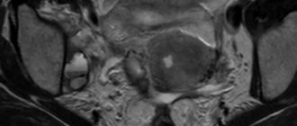
Based on the image displayed on the screen, the doctor explains the results, makes a diagnosis and assesses the size of the organs and their echogenicity. A full conclusion is made only by a specialist who examines the size, location of the female organs, and determines whether there are follicles, formations, or stones. After the procedure, the doctor explains in writing or orally all the information about the compliance of the size of the internal organs with the standards, their condition and the presence of formations. If there are deviations from normal values, this indicates the presence of diseases. Lumps that are detected during examination can diagnose cancer. If round structures appear on the image, this may indicate the presence of cysts. Changes in the size of internal organs occur with polycystic disease, and deviations in echogenicity indicate the presence of fibroids.
At the same time, as mentioned above, only a specialist can accurately diagnose the disease. The result of the ultrasound is provided on a photo card or disk, at the request of the patient. A pelvic examination protocol is also provided. The procedure is painless, with its help you can determine the presence of diseases and find out about pregnancy. A pelvic ultrasound is a mandatory procedure for every woman, but for correct diagnosis you need to choose a qualified specialist!
What does an ultrasound of the pelvic organs show?
Ultrasound of the pelvis in women allows:
- determine multiple pregnancy;
- determine the location of the fertilized egg (in the uterus, in the tube, in the cervix, in the ovary);
- diagnose and assess the condition of uterine fibroids;
- determine the degree and location of a disease such as endometriosis;
- diagnose polyps of the cervix and uterine cavity, cysts, tumors, space-occupying formations of the uterus and its appendages;
- malformations of the internal genital organs (in adolescents);
- chronic inflammatory processes;
- complications after abortion and childbirth.
Cost of ultrasound in gynecology
| Name of service | Price, rub.) |
| Primary appointment with an obstetrician-gynecologist (candidate of medical sciences; doctor of medical sciences) | 1900 rub. |
| Repeated appointment with an obstetrician-gynecologist (candidate of medical sciences; doctor of medical sciences) | 1400 rub. |
| Primary appointment with an obstetrician-gynecologist | 1800 rub. |
| Repeated appointment with the obstetrician-gynecologist | 1300 rub. |
| Consultation with an obstetrician-gynecologist on pregnancy | 2000 rub. |
| Ultrasound of the pelvic organs in gynecology (transabdominal) | 2000 rub. |
| Ultrasound of the pelvic organs in gynecology (transvaginal + transabdominal) | 2500 rub. |
| Ultrasound examination of the ovaries (control of the dominant follicle) | 1400 rub. |
| Ultrasound of the thyroid gland | 2000 rub. |
| Ultrasound of the mammary glands with regional lymph nodes | 2000 rub. |
| Ultrasound - pregnancy up to 12 weeks | 2500 rub. |
| Ultrasound - pregnancy (long term) | 3000 rub. |
| Ultrasound 3D (without recording to disk) | 3500 rub. |
| Doppler examination (in combination with obstetric ultrasound) | 5000 rub. |
| Women's health (ultrasound of the abdominal organs, ultrasound of the bladder, ultrasound of the pelvic organs, ultrasound of the mammary glands, ultrasound of the heart, ultrasound of the vessels of the neck, ultrasound of the thyroid gland, ultrasound of the kidneys and adrenal glands) | 15500 rub. |
All our services and prices
Features during pregnancy
When and why is a 3D ultrasound prescribed during pregnancy?
Nowadays, not a single pregnancy passes without ultrasound diagnostics of the condition of the mother’s reproductive organs and the development of the fetus. So, in addition to determining the presence of conception, ultrasound is scheduled at least three times during the gestation period - at weeks 11–13, 22–23 and 31–32. This allows you to keep under control the intrauterine development and growth of the embryo, as well as the possible formation of pathologies in the mother’s body.
- At 11–13 weeks, gross developmental disorders of the fetus are determined, the thickness of the nuchal zone is an important indicator of the presence or absence of Down syndrome, and anatomical features are assessed.
- At 22–23 weeks, it becomes possible to study the structure of the main organs and systems of the fetus - the cardiovascular, nervous, digestive tract and urinary tract. At this time, you can determine the gender of the unborn baby.
- At 31–32 weeks, ultrasound shows late developmental abnormalities of the heart, gastrointestinal tract, urinary and respiratory systems, as well as other important organs. In addition, the growth rate and its compliance with normal indicators are studied.
Diagnosis of fallopian tube patency using ultrasound
The fallopian tubes connect the ovaries to the uterus - mature eggs move through them into the uterine cavity after leaving the follicles. Sometimes, for certain reasons (chronic inflammation, STDs, adhesions after surgery, endometriosis, compression from other organs), the patency of the fallopian tubes worsens, caused by their blockage with inflammatory fluid. As a result of obstruction, the risks of infertility or ectopic pregnancy increase.
To check the patency of the fallopian tubes, echosalpingography is used - an ultrasound method in which the uterine cavity is filled with a contrast agent (saline) through a thin catheter. Echosalpingography makes it possible to observe the speed of fluid movement in the fallopian tubes during examination with an intravaginal sensor and identify blocked areas, if any.