The prostate ultrasound procedure is aimed at identifying pathological processes that can harm sexual life and disrupt everyday comfort. If there are complaints from the patient, the urologist, after collecting a preliminary history, may refer you for an ultrasound of the prostate.
The study is carried out using a special ultrasound sensor and allows you to accurately determine the size of the prostate gland with a detailed study of its echostructure.
Ultrasound of the prostate allows you to simultaneously examine the condition of the following organs:
- seminal vesicles;
- bladder neck;
- fiber located in close proximity to the genital organ;
- veins of the paraprostatic plexus.
The examination does not affect reproductive health at all, but rather helps maintain a normal sex life, increasing potency. Also, during ultrasound, inflammatory processes in the prostate and the presence of neoplasms are diagnosed, among which adenoma and malignant tumors can be identified.
Indications for prostate ultrasound
Pathological processes affecting the prostate are often found in men, both at a young age and at an older age. Ignoring characteristic symptoms can seriously threaten sexual life.
Diagnostic procedures using ultrasound are carried out in order to identify pathology in the early stages of its development. This allows you to complete the required course of treatment in a timely manner and avoid subsequent complications.
Direct indications for undergoing this procedure are:
- difficulty urinating in small proportions;
- urine takes on an uncharacteristic dark shade;
- problems with erection and ejaculation;
- acute pain in the groin area when trying to empty the bladder naturally;
- the analysis of seminal fluid revealed impurities of pus or blood;
- interest in the opposite sex disappears;
- causeless increase in body temperature, without characteristic symptoms of a cold;
- periodic vomiting.
For men over 45 years of age, it is recommended to undergo regular ultrasound examination of the prostate every year. Such manipulation will allow timely detection of the appearance of deviations and the development of pathologies in the early stages, which helps prescribe an effective course of therapy and prevent the occurrence of possible complications.
Make an appointment with a urologist by phone or by filling out the online form
| Select a clinic | Urological ultrasound | Urologist in the clinic | Calling a urologist to your home |
Contraindications to prostate ultrasound
The presented research method actually has no significant contraindications.
Diagnostics through ultrasound is a completely safe and painless method of obtaining information.
However, for some categories of patients, there are certain contraindications to undergoing the examination:
- the presence of third-party inflammatory processes;
- exacerbation of hemorrhoids;
- tumor of the ampulla in the rectum;
- complications after surgery associated with narrowing of the anal canal;
- anal fissure;
- increased sensitivity of the anorectal area.
Most of the above contraindications will not harm your health when undergoing an ultrasound scan, but will only reduce the accuracy and information content of the results.
When is TRUS prescribed without fail?
TRUS is prescribed by a urologist, andrologist or oncologist in the following cases:
- if cancer is suspected (after a digital rectal examination or transabdominal examination);
- in case of deviations in the results of blood or urine tests;
- for the purpose of clarification and monitoring during cryotherapy, brachytherapy, ultrasound ablation, drainage of abscesses, prostate cysts or cellular spaces of the pelvis;
- to monitor the extent of cancer during treatment or before other interventions. If a relapse is suspected after treatment;
- for male infertility , erectile dysfunction;
- for pain in the pelvis;
- any type of prostatitis.
The study is not prescribed in cases of exacerbation of hemorrhoids and in inflammatory processes of the rectum until the inflammation is relieved.
How to prepare for a prostate ultrasound
It is recommended 2 days before the procedure to exclude from the daily diet foods that affect increased gas formation in the intestines, which contributes to digestive disorders.
List of undesirable products:
- fresh fruits or berries;
- drinks containing a high concentration of gases;
- alcohol;
- brown bread and pastries;
- cakes, pastries and similar foods high in sugar.
If a prostate ultrasound is scheduled for the morning, then you should completely skip breakfast; if the examination is carried out in the afternoon, then food intake should be minimal.
A few hours before the procedure, you need to drink a large amount of liquid without gases in a volume of 1 to 1.5 liters. Plain drinking water or unsweetened tea will do.
After this, you must refrain from urinating. This is due to the peculiarities of the examination, which requires a completely filled bladder. If the desire to urinate is too strong, it is allowed to pass a small part of the urine, but without completely emptying the bladder. After this, you should drink another 1-2 glasses of water.
Preparing for the examination
After being referred for an examination, the doctor tells the patient about the procedure, benefits and possible complications.
Preparing for TRUS involves having a bowel movement. On the day of the examination or the day before, you must use a cleansing enema. It is also recommended to exclude foods that cause increased gas formation. The day before the procedure, avoid carbonated drinks, dairy products, cabbage, apples, pears and grapes. You can create a menu of cereals, lean meat or fish, and vegetables. Don't forget about personal hygiene.
As for the enema, it is not at all necessary to use “grandmother’s methods” and introduce a large amount of water into the intestines. Pharmacies sell special rectal suppositories called microenemas. Such microenemas, provided you follow a diet, are enough to completely prepare the intestines.
Another important point is psychological preparation. All exciting moments should be discussed with a doctor, because fear can cause additional spasm of the anal sphincter. This will interfere with the examination and time will be wasted.
Before the study, you need to relax and take a deep breath while inserting the sensor. If there are indications (increased suspiciousness, pain in the prostate area), the doctor can use a gel with lidocaine - this facilitates the insertion of the sensor and relieves pain.
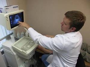
How is a prostate ultrasound done?
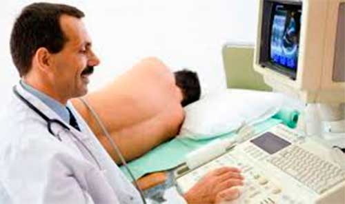
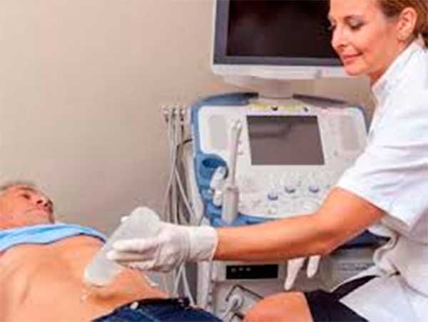
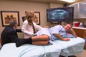
Research can be conducted in several ways. The abdominal method allows you to identify bulk pathology of the prostate gland. During the procedure, all nearby organs are diagnosed. This type of ultrasound is completely painless, and to perform the procedure, the patient lies on a couch with his stomach up. A special gel is applied to the skin of the abdominal cavity to better glide the sensor over the surface. The whole process takes no more than 20 minutes.
For a more accurate diagnosis, it is customary to use the rectal ultrasound technique of the prostate. This event is quite unpleasant and is based on the direct insertion of a sensor into the rectum.
The procedure is carried out as follows:
- the patient undresses to the waist and lies down on the couch sideways, turning his back to the doctor;
- the knees should be slightly pulled towards the chest;
- a sterile condom with hypoallergenic properties is placed on a thin sensor with a diameter of no more than 15 mm;
- gel is applied for lubrication and better penetration;
- the prepared device is inserted into the rectum and appropriate diagnostics are carried out.
When prescribing a rectal examination, it is imperative to notify the doctor about the presence of hemorrhoids or anal fissures. Completing this type of prostate ultrasound allows you to obtain more information about the structure of each lobe of the organ, blood vessels and general structure.
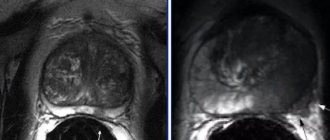
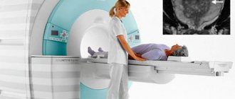
Transrectal diagnostics (TRUS)
TRUS of the prostate gland is considered a more informative diagnostic measure than the previous version of the study. This is due to fundamental differences in the procedure. The sound wave is delivered directly through the surface of the rectum, so the results will be more accurate, they will allow you to determine the location of the organ, diagnose the inflammatory process, the presence of cancer lesions and other problems. However, you should first determine the features of TRUS of the prostate gland, as we will discuss how the procedure is done below.
Accuracy directly depends on proper preparation. Strict adherence to preparatory measures will allow the specialist to obtain the most appropriate data, which will subsequently affect the overall course of treatment. That is why you should find out how to prepare for a prostate ultrasound and what rules need to be followed.
ORDER
Key recommendations include:
- a couple of days before the examination date, you must stop eating cabbage, beans and other foods that can lead to increased flatulence;
- it is important not to drink alcohol;
- before going to bed in the evening, you should cleanse the intestines with an enema, and then repeat the procedure immediately before the test;
- Preparation for TRUS of the prostate involves drinking 1 liter of clean water 1-2 hours before the diagnosis.
Depending on the patient’s condition and the results of previous studies, the doctor can make a number of additional recommendations and advise on how to prepare for a prostate ultrasound correctly. It is strictly forbidden to ignore advice. If you have problems with incontinence, it is better to drink water in the clinic than at home.
Now we should consider how a transrectal ultrasound of the prostate is performed. The indicator is inserted into the anus, which causes catastrophic fear and reluctance in many men to carry out the examination. It is recommended to fully follow the recommendations of the medical professional, relax, remain calm and not worry.
The event is divided into several stages and is performed in the following sequence:
- The patient undresses and puts on a robe.
- Take a position lying on your side on the couch, with your legs bent at the knees and moved closer to your stomach.
- The doctor puts a condom on the sensor to prevent the occurrence of an infectious disease, then lubricates the indicator and the anus with Vaseline.
- The sensor is inserted only 7 cm, so the patient does not experience any particular discomfort.
- Depending on the design of the device and the scanning method, the uzist makes inclined, translational, and rotational movements with the device.
The picture is broadcast on the screen; if necessary, the image is printed. The diameter of the indicator is about 2 cm, so the patient does not feel pain. Sometimes there may be minor discomfort or a urge to defecate, which you do not need to pay attention to. After 10-15 minutes, these sensations disappear without a trace. So we managed to determine what TRUS of the prostate gland is and how it is performed.
Diagnostics allows a specialist to accurately determine even a slight enlargement of organs, which is very important for making a diagnosis in the early stages of the development of adenoma or prostatitis. Thanks to the penetration of the sensor into the rectum, the uzologist can examine the neck of the bladder for the presence of an inflammatory process or dangerous formations.
What diseases are diagnosed with prostate ultrasound?
With the help of the study, certain types of pathologies can be identified, including:
- adenoma, manifested in the form of increased size of the prostate gland and the presence of formations in it with a certain weakly echogenic environment;
- asymmetry, which is considered a sign of cancer and manifests itself in the form of increased organ volume;
- chronic inflammatory processes;
- a cyst, manifested by bubbles with liquid and indicating the development of chronic prostatitis;
- the presence of stones in the ducts.
The most common cancer diagnosed by ultrasound is prostate cancer. It is observed mainly in men over 50 years of age. It is characterized by slow development without pronounced symptoms.
Useful information on the topic of urology:
- Appointment with a urologist at the clinic
- Calling a urologist to your home
- PSA tests
- Discharge in men
- Diagnosis of prostate diseases
- Urethral swab
- Prostate massage
- Circumcision
- Tests for STIs
- Treatment of sexually transmitted diseases
- Ultrasound at home
- Kidney ultrasound
- Pyelonephritis
- Ultrasound of the bladder
- Diagnosis of sexually transmitted infections
Prostate size by age
Most men of reproductive age have a prostate length and width of 30 mm and a thickness of 20 mm. The size changes as the concentration of sex hormones increases. For men over 20 years old, 25 cubic centimeters is the norm. By the age of 40, a volume of up to 26 cm³ is considered normal; by the age of 60, the figure reaches 28.1 cm³. By the age of 70, the norm for the prostate on ultrasound is 30 cm³. If the prostate enlarges above normal, there is a risk of adenoma, prostatitis or cancer. Making an accurate diagnosis is possible only after tests and ultrasound.
Norms of prostate size on ultrasound
Standards for survey results may vary slightly. This is due to different ages and individual characteristics of patients.
The main attention during diagnostic activities is paid to certain indicators:
- transverse size, the norm of which is from 2.8 to 4.9 cm;
- the length from the front to the back wall, 1.7 cm is considered a good indicator;
- length from top to base, within the normal range is considered to be from 2.4 to 4.6 cm;
- prostate volume should not exceed 25 cm3.
The parameters of the structure and echogenicity of the prostate gland should appear homogeneous. There are no clear indicators for prostate volume. This characteristic is calculated for each patient individually within acceptable values.
Normal indicators
The normal size of the prostate gland in adults depends on the age and weight of the subject. In a normal state, the prostate corresponds to the established size and shape, with no neoplasms, cysts or tumors.
The normal size of the prostate gland according to ultrasound corresponds to the following indicators:
- 2.4 – 4.1 cm – upper anterior;
- 2.7 – 4.3 cm – transverse;
- 1.6 – 2.3 cm – anteroposterior.
Normal prostate volume according to ultrasound varies from 24 to 30 cm3. In cross section, the seminal vesicles should be within 8-10 mm. Be sure to examine the appearance and indicators of the bladder, also taking into account the shape and dimensions. The wall thickness should be close to 3 - 5 mm. Normal prostate sizes according to ultrasound are usually accompanied by the absence of stones and other pathological formations. After urination, there should be no urine in the bladder; fluid should be efficiently transported into the bladder from the ureters.
It should be noted that the accuracy of the survey is determined by various factors. The normal size of the prostate according to ultrasound is distorted if there is an excessive amount of gas or feces in the intestines. The patient should take the correct position and relax. The fat layer or damage in the abdominal area leads to a significant error, which must be taken into account by a specialist. An enema must be given before the prostate ultrasound.
Who needs a prostate ultrasound?
The development of inflammatory processes in the prostate gland and closely related organs is more common in men over the age of 35 years. The close connection of the prostate with the genitourinary system contributes to disruption of the usual lifestyle when inflammation or neoplasms occur on the tissues of the gland.
It is possible to identify such abnormalities and concomitant pathologies using ultrasound. This diagnostic method has long proven itself due to the high accuracy of the results obtained and ease of implementation.
A referral for diagnostics is issued in certain cases:
- manifestation of characteristic symptoms that indicate the presence of pathology;
- unsatisfactory results of laboratory examinations;
- cancer screening for male patients over 45 years of age.
Prostate diseases can develop and manifest themselves at any age. Adenoma and malignant tumors are more common in men over 50 years of age, but prostatitis is not uncommon in young patients.
Patients with the following symptoms are required to undergo a prostate ultrasound:
- difficulty urinating;
- irradiation of pelvic pain to the rectum;
- regular premature ejaculation;
- discomfort during ejaculation;
- foreign impurities in sperm.
It is recommended to do a prostate ultrasound at absolutely any age once a year. Regular diagnosis of the prostate gland will identify all minor abnormalities and prevent the development of chronic pathologies in the future, avoiding serious complications.
Methods for diagnosing urological diseases:
- Calling a urologist to your home
- Diagnosis of kidney diseases
- Diagnosis of bladder diseases
- Diagnosis of prostate diseases
- Diagnosis of scrotal diseases
- Diagnosis of sexually transmitted infections
- Urine tests
- Tests for STIs
- PSA tests
- Ultrasound at home
- Kidney ultrasound
- Ultrasound of the prostate
- Ultrasound of the bladder
- Urological ultrasound at home
- Urethral swab
- Ultrasound of the penis
- Transrectal ultrasound
- Prostate juice tests
- Ultrasound of the genitourinary system
Advantages of the method
This type of diagnosis is based on the use of ultrasonic waves. The method allows you to visualize the prostate gland, identify its shape, size, and the presence of deviations from the norm. You can examine the condition of the bladder and other nearby organs.
The advantages of ultrasound are:
- safety - ultrasound examination does not cause any harm to health;
- painlessness - the use of the method is not accompanied by punctures or incisions;
- accuracy - allows you to study the organ in different planes, determine parameters and volume;
- no contraindications;
- accessibility – now it is possible to conduct a survey in every city for a relatively low fee;
- simplicity – the duration of the procedure is about 30 minutes, preparation is simple;
- the ability to collect tissue for histological analysis.
How often should you do a prostate ultrasound?
Studies have proven that prostate ultrasound is an absolutely safe examination method for men. During the procedure, the patient is exposed to the mechanical and thermal effects of ultrasonic radiation of high-frequency waves. If these effects are within normal limits and the doctor follows certain safety rules, they do not harm the human body.
Therefore, there are no strict restrictions on the number of ultrasound examinations of the prostate gland. It is worth noting that doctors do not recommend conducting such examinations frequently, but rather be guided by necessity. The number of diagnostic tests completed will not affect the accuracy of the diagnosis.
Based on this, it follows that one prostate ultrasound per year is sufficient for preventive purposes. All other studies are prescribed in the course of communication with the attending physician, who, based on individual characteristics and progression of the pathology, may prescribe an additional ultrasound of the prostate.
Ultrasound of the prostate at home
It is not always possible to visit a clinic in person. In this case, the modern method of prostate ultrasound at home will allow you to conduct the study in the comfort of your home and significantly save time.
This procedure is recommended for the following categories of patients:
- older people who have difficulty getting around using public transport;
- patients with serious limitations in mobility, which could be caused by injuries of various types or surgical intervention during an operation;
- people with limited time or simply those who do not like to be in medical institutions.
Most often, men experiencing intimate ailments hide it and do not want to visit the clinic. There may be personal reasons or lack of time, due to which a visit to the doctor is constantly postponed. This behavior has a detrimental effect on the general condition of the body subsequently, when the pathology becomes chronic.
When performing an ultrasound at home, equipment is used that is similar to stationary units in the clinic, but smaller in size. The process itself is no different from outpatient diagnostics and is carried out according to the same principles in several stages:
- the patient lies on a flat surface in a horizontal position with his stomach up;
- A special gel is applied to the skin of the study area for better and smoother glide of the scanner over the surface;
- The diagnostic process itself takes about 15-20 minutes;
- after completion, the remaining gel is removed with a damp sterile cloth;
- a patient card is filled out with a detailed description of all the data obtained.
Based on the results of a prostate ultrasound, the doctor makes a diagnosis, after which an effective course of treatment is developed, with reference to the individual characteristics of the patient.
Consultation with a urologist after ultrasound of the prostate
All patients should consult a urologist after undergoing a prostate ultrasound. During the appointment, the doctor may prescribe additional laboratory tests, if they occur in each specific situation.
Depending on the results of ultrasound examination of the prostate gland, the stage of development of the pathology and other associated disorders in the functioning of the body are determined.
This allows you to prescribe an effective course of therapy, which may include:
- prostate massage and ozone therapy;
- magnetic laser treatment method;
- laser coagulation;
- instillation into the bladder;
- medication course.
In case of serious diseases and advanced stages of development of inflammatory processes, surgical intervention is prescribed within the framework of urology. This is an extreme method of exposure and the doctor will take all measures to avoid it.
If the ultrasound did not reveal any abnormalities, the urologist can provide valuable advice on preventive measures for the prostate gland. Following the advice received will reduce the risk of problems and the formation of inflammatory processes in the prostate ahead of time.
Preparatory activities
The prostate gland is examined on an empty stomach. With both methods, to improve visualization, the bladder is filled with liquid before the procedure; for this, up to one and a half liters of water are drunk and the bladder is not emptied. Three days before the ultrasound, you should avoid eating foods that cause increased gas formation: fresh cabbage, carbonated drinks, milk, beans, peas. The dinner diet should not consist of difficult-to-digest foods. In addition, you should not use carminative medications or drink alcoholic beverages. Before performing a transrectal ultrasound of the prostate gland, 2-4 hours before, it is necessary to cleanse the intestines. To do this, you can use the Microlax microenema.

What factors can distort prostate ultrasound?
To obtain the most accurate and informative results from a prostate ultrasound, you must follow the recommendations for preparing for the examination. The following factors can distort the readings:
- ejaculation within 48 hours before diagnosis may cause a false increase in tumor marker;
- prostate massage;
- increased accumulation of gases in the intestinal tract;
- empty bladder;
- taking medications without consulting a doctor.
Accurate results obtained during prostate ultrasound contribute to the correct conclusion and diagnosis. This in turn allows you to prescribe the most effective course of treatment.
Answers to frequently asked questions about ultrasound diagnostics:
- Is ultrasound harmful?
- Where can I get an ultrasound done?
- How to properly prepare for an ultrasound?
- What organs are examined by ultrasound?
- What diseases are ultrasound used to diagnose?
- Is it possible to do an ultrasound at home?
- What symptoms require an ultrasound?
- How often should an ultrasound be done?
- Do they do ultrasounds for children in your clinic?
- Can I get the transcript and ultrasound images in hand?
- What organs are examined during an abdominal ultrasound?
- What organs are examined during a pelvic ultrasound?
- Ultrasound in Moscow
- Paid ultrasound
- What other diagnostic methods are used in your clinic?
- How to sign up for an ultrasound?
What does the diagnosis show?
First of all, ultrasound can be used to determine the size of the prostate; the norm during ultrasound is compared with the obtained values. If serious deviations are observed, then the problem may be hidden in a dangerous pathological process. At the next stages, the physician must check other pelvic organs, which will allow them to assess the condition and diagnose the causes of negative consequences. It is advisable to contact a qualified specialist so that he knows exactly how an ultrasound of the prostate gland is performed and can correctly interpret the results of the study.
The bladder is located directly behind the bony pubic joint; during the filling process, the organ rises above this element of the genitourinary system, its upper part is adjacent to the abdominal cavity. A correct ultrasound of the prostate and bladder shows the bladder as an oval structural formation with a demarcated edge, and the distant area is visible much better than the near one.
The seminal vesicles are located on the back side of the bladder on both sides, slightly higher than the prostate gland. Ultrasonic waves make it possible to identify them as interconnected formations in the form of bubbles. The prostate gland during ultrasound is visible as a glandular-muscular organ with a dense structure.
It should be noted that this is an important element, especially for a young man, since the quality and functionality of seminal fluid depends on it. The condition of the gland directly determines the reproductive capabilities of the stronger sex. On the screen you can see that the gland is located on top of the bladder. The normal volume of the prostate gland according to ultrasound does not exceed 30 cm3; the younger the man, the smaller this figure. The contours of the prostate are smooth and precise.
Connective tissue is responsible for the localization of organs; it acts as a binder. Each organ is located close to each other, that is, the elements are interconnected. That is why the failure of one organ entails the failure of another. To obtain an accurate assessment result, you should consider how you prepare for a prostate ultrasound.
Pathologies determined by ultrasound of the prostate
When examining an ultrasound scan of the prostate gland, certain chronic diseases can be identified that can significantly impair the functionality of the body.
Adenoma
Increased size of the gland and tissue density are the primary signs of the development of pathology. In some cases, nodes may appear on the surface of the organ. The diffuse form of the disease is characterized by pronounced heterogeneity, without any inclusions.
Fibrosis
The most common prostate disease. It is believed that the main reasons for the progression of the pathology are an advanced form of prostatitis or consequences after suffering an inflammatory process of the gland. A characteristic sign of the presence of fibrosis on ultrasound is a reduced size of the gland.
Prostatitis
The pathology is characterized by changes occurring in the echogenic background. Depending on the form of the inflammatory process, the prostate tissue becomes denser and the echogenicity decreases. The main features are a blurred contour and similarity of fibrous and glandular tissues. In some cases, abscesses may occur due to uncontrolled changes in echogenicity. An acute form of pathology is recorded when there is a simultaneous enlargement of the gland and a decrease in echogenicity, and vesiculitis is noticeable around the seminal vesicles.
Cysts and stones
Based on tissue structures, all inclusions in an organ can be determined. Often cysts appear as anechoic areas or areas of low echogenicity. Small cysts sometimes appear on a completely healthy organ. The size of such manifestations is no more than 5 mm. The presence of stones of different shapes and sizes is characterized by an increased echo signal.
Neoplasms
The loss of clarity of outlines and contours is determined by the presence of tumors of various types. Benign neoplasms are often located in the central regions of the organ. The presence of transformation and shifts along the edges may signal the formation of cancer cells.
How to prepare a prostate ultrasound protocol
During the procedure, the doctor draws up a special protocol in which all diagnostic information is recorded. The document consists of several points that must be completed:
- Name and address of the medical center where the prostate ultrasound was performed.
- List of equipment on which diagnostic procedures were performed.
- First and last name of the doctor and patient.
- Serial number of the study indicating the date of conduct.
- Diagnosed organ.
- A complete description of the echostructure of the prostate gland listing all indicators.
- Conclusion. All characteristics of the prostate are indicated with a description of the identified pathologies, if any.
- The protocol is handed over to the patient. With its help, you can get a consultation with a urologist with further recommendations and a course of therapy.
How long does a prostate ultrasound take?
The duration of the examination depends on the method of its conduct.
During the rectal method, the patient lies on his side, slightly pulling his knees towards his stomach. The doctor prepares the sensor to reduce the level of discomfort and ensure the best glide. Then the device is inserted into the rectum to a depth of 7 centimeters. This type of prostate ultrasound can last up to 25 minutes, taking into account 10 minutes of preparatory activities and 15 minutes of the diagnosis itself.
An abdominal ultrasound of the prostate is performed using a convex probe to scan tissue up to 25 centimeters deep.
The process looks like this:
- the patient lies with his back on the couch, having previously exposed his stomach and undressed to the waist;
- the doctor lubricates the surface of the abdomen in the required area with gel to improve the sliding effect and increase the area of contact of the sensor with the skin, which allows for more informative and accurate results;
- then the doctor slowly moves the device over the abdomen;
- the information is transmitted to the screen and subsequently recorded in the study protocol.
With this type of prostate ultrasound, the examination time does not exceed 20 minutes.
Where to do a prostate ultrasound
The “Your Doctor” clinic employs doctors who have many years of experience in various fields of urology. The choice of our medical center if there is a need for an ultrasound of the prostate is due to modern equipment, which carries out all specialized studies and subsequent diagnostic procedures:
- for ultrasound examinations of the prostate gland, separate rooms are allocated, equipped with all the necessary instruments for a comfortable position of the patient and obtaining the most accurate and informative results;
- doctors have all the modern technical components for effective work and safe interaction with the patient;
- It is possible to perform an ultrasound of the prostate at home, with a doctor visiting on the day of treatment.
In case of a personal visit, the urologist’s office is equipped with specialized furniture for comfortable placement of the patient. If additional research is necessary, the doctor has a wide range of tools that allows him to achieve the desired results.
Features of ultrasound examination
Indications for prescribing an ultrasound include disturbances in urination, compaction in the gland, an increase in its size, sexual dysfunction and changes in urine tests, blood tests and spectrograms. An ultrasound examination of the prostate gland has a number of important features:
- The accuracy of the result - the prostate tissue is carefully examined, its exact size and volume are determined.
- Safety – the wave effect does not have a negative impact on a man’s health.
- No pain - no incisions or punctures are made.
- Availability – the service can be used by any patient at a local clinic or for a small fee at a urology center.
- Versatility.
- Simplicity - the procedure is performed quickly and does not require complex preparation.
Where to go with prostate problems
Experienced doctors working at the “Your Doctor” clinic are ready at any time to conduct a full diagnosis of the prostate gland and make an accurate diagnosis, with subsequent treatment prescribed.
To understand when medical attention is needed, you need to know what symptoms to contact a medical center for:
- an inflamed prostate can compress the urinary canal, which leads to difficulty urinating;
- erection is impaired;
- the body weakens, resulting in constant lethargy and fatigue;
- accelerated ejaculation;
- pain and burning sensation in the groin area.
For patients suffering from prostate problems, psychological state is important. Any stress can significantly weaken the body and complicate the course of therapy. Therefore, in our clinic you can undergo a full range of diagnostic measures with subsequent treatment anonymously.
Which doctor does prostate ultrasound?
The condition of the prostate gland can be affected by any negative impact, from infectious inflammatory processes to hypothermia. In cases where the patient experiences characteristic symptoms of the development of prostate pathology, it is necessary to consult a urologist.
At your appointment, you will be given a referral for a prostate ultrasound to more accurately diagnose the problem with subsequent treatment.
An ultrasound of the prostate is performed by a doctor specializing in ultrasound diagnostics and having experience in various branches of urology with relevant diplomas. The procedure will be as comfortable as possible for the patient, thanks to modern equipment on which all diagnostic procedures are performed and the high level of training of doctors.
Publication date 2019-10-24
Surgical treatment of the prostate
Standard surgical procedures
- TURP: transurethral (through the urethra) resection (truncation) of the prostate gland through the urethra. This procedure is the "gold standard for effective treatment of BPH."
- Open prostatectomy (complete removal of the gland for cancer).
Transurethral resection of the prostate (TURP)
This surgery is used to treat problems with urination due to an enlarged prostate gland. This is the most common surgery to treat BPH. The doctor removes only the parts of the prostate gland that are obstructing the flow of urine. A combined visual and surgical instrument (resectoscope) is inserted through the tip of the penis and into the urethra.
Every male urologist must master diagnostic techniques and know how to calculate the volume of the prostate by size. The urologist’s tasks also include determining the most optimal treatment method, incl. determining the need for surgical intervention. It is important for men to consult a doctor on time and carefully monitor their condition.