Also, at approximately 1 to 2 months of age an ultrasound of the newborn's kidneys
to study the structure of the organ and identify abnormalities or changes
in the kidneys and urinary tract
.
Ultrasound for children over one year old
Doctors recommend that one-year-old children undergo an ultrasound of the kidneys and abdominal cavity after switching to a general diet for preventive purposes and to be able to detect any diseases at an early stage. In the future, for the same purpose, it is advisable to undergo examination once every three years.
How to prepare a child for a kidney ultrasound?
During an ultrasound of the kidneys, the child does not need to prepare for the procedure. The most important thing is to drink still water or tea about twenty minutes before the test (the volume depends on age), since the procedure is performed on a full bladder.
The day before the examination, do not give your child foods that promote gas formation: white bread, raw vegetables and fruits, carbonated drinks, legumes. If a child is overweight or has flatulence, then this diet should be started three days before the examination.
What criteria does the doctor pay attention to when making a diagnosis?
When performing a kidney ultrasound, the specialist pays attention to the following parameters:
- localization of the organ (in order to exclude nephroptosis);
- longitudinal and transverse size of the kidneys;
- state of the organ parenchyma. At the same time, its homogeneity and the occurrence of degenerative changes are assessed;
- structure of the renal pelvis (its size, absence of stones);
- blood supply to the organ (patency of arteries, absence of anomalies in the development of renal vessels).
How is an ultrasound of the kidneys performed on children?
In general, the kidney ultrasound procedure in children is approximately the same as in adults. The child undresses to the waist or simply exposes the abdomen and lower back. Then he lies down on the couch (the newborn baby remains in the mother’s arms), and a gel is applied to the area of the body being examined, which improves the glide of the sensor and helps better passage of ultrasonic waves. During the procedure, the doctor may ask older children to take a deep breath, exhale, or hold their breath. The examination can also be carried out from several angles: from the abdomen, side and back.
Main indications
One of the main indications for the procedure is cystitis. This disease occurs under the influence of various factors:
- hypothermia;
- failure to comply with personal hygiene rules;
- chronic kidney disease;
- wearing tight clothes.
An ultrasound examination of the bladder in a child should be performed if there are the following indications:
- the appearance of pain when urinating;
- the presence of oxalates in the urine;
- increased frequency of the urge to urinate;
- cloudy urine;
- discomfort in the lumbar region;
- the appearance of sediment in the urine.
Ultrasound examination not only helps to determine the contours and size of the bladder. When carrying out a diagnostic procedure, it is possible to establish a narrowing of the urinary canal, the presence of stones, and deterioration of blood circulation. Ultrasound is also informative in case of an inflammatory process occurring in the bladder.
Interpretation of ultrasound of the kidneys in newborns

The results are released after the procedure. As a rule, the specialist who performed the diagnostics only describes in the research protocol what he sees. The diagnosis is made by a doctor who refers you to an ultrasound scan of the kidneys. In children, the study is usually based on indicators such as length, width and thickness. Depending on age, these parameters may vary, as well as the size of the left and right kidneys. Decoding the results consists of comparing the obtained sizes and norms.
Normal kidney sizes on ultrasound in children of different age categories
The table shows the average normal sizes of kidneys on ultrasound in children, depending on age.
| Left kidney | Right kidney | |
| Newborns | ||
| Length | 36,2 — 60,7 | 36,9 — 59 |
| Width | 14,1 — 26,9 | 13,7 — 29,4 |
| Thickness | 13,6 — 27,4 | 16 — 27,3 |
| From 3 to 6 months | ||
| Length | 47 — 72 | 45,6 — 70 |
| Width | 17,2 — 31 | 18,2 — 31,8 |
| Thickness | 19 — 30,6 | 19,1 — 30,3 |
| 1-3 years | ||
| Length | 54,7 — 82,3 | 55,6 — 84,8 |
| Width | 20,9 — 35,3 | 19,2 — 36,4 |
| Thickness | 20,4 — 31,6 | 12,2 — 34 |
| 5-7 years | ||
| Length | 66,3 — 95,5 | 67 — 99,4 |
| Width | 26,2 — 41 | 23,5 — 40,7 |
| Thickness | 23,7 — 38,5 | 21,4 — 42,6 |
| 7-10 years | ||
| Length | 66,7 — 103,3 | 71,2 — 103,6 |
| Width | 24,5 — 44,9 | 26 — 44 |
| Thickness | 23,9 — 39,5 | 27 — 41 |
| 10-14 years | ||
| Length | 74,4 — 113,6 | 74,4 — 116 |
| Width | 28 — 48,7 | 27,2 — 47,7 |
| Thickness | 25,5 — 43,1 | 27 — 46,3 |
ULTRASOUND SCREENING OF NEWBORNS AND CHILDREN UP TO 1 YEAR OF AGE
at the EVO Specialized Medical Center.
WHAT IS A BABY ULTRASOUND SCREENING?
Ultrasound screening of newborns and children in the first year of life includes:
- examination of the abdominal organs and kidneys (deviations from the norm in shape, size, structure and location of organs) ;
- examination of the hip joints for orthopedic deformities (dislocation, dysplasia) ;
- examination of the brain for developmental defects.
WHY DO AN ULTRASOUND OF YOUR BABY?
For every mother and every father of a newborn son or daughter, the main issue is the health of the child. And the first year of a child’s life is the most critical stage for future health, during which a number of diseases and problems can be excluded. Modern parents have probably heard a lot about screening studies. For example, all expectant mothers in our country must undergo prenatal (antenatal) screening diagnostics during pregnancy. These studies are aimed at timely detection of abnormalities in the fetus. And the earlier the disease is detected, the greater the chance of a positive pregnancy outcome and the health of the newborn. Screening to identify hidden defects (congenital and acquired), hereditary pathologies, deviations from the norm in the structure and location of the internal organs of a newborn child allows you to calm the worried hearts of young parents or, if necessary, not to miss precious time to begin treatment for the baby.
WHAT DIAGNOSTICS DOES A BABY NEED?
Various disorders in the child’s body can occur both during intrauterine development and during childbirth. Diseases of newborns can be caused by hereditary chromosomal abnormalities, the influence of a number of negative factors during pregnancy, and birth injuries.
Diagnostics includes collecting patient complaints, assessing the presence of disease symptoms identified during an external examination + results of laboratory and instrumental studies. However, newborn children do not complain. Symptoms of the disease are often mild and sometimes completely absent. Laboratory tests and instrumental studies remain in our arsenal.
Currently, there is practically no problem with laboratory tests; in well-equipped child care institutions, any indicator in the blood or urine can be determined. Instrumental studies have some limitations, because screening of the baby must be carried out in the first 3 months of life, and it is during this period that the child’s body is extremely sensitive to any external influences.
The slightest carelessness during diagnosis can lead to serious and even irreparable consequences. The screening method for newborns should not only be highly informative, but also convenient and safe. Today, ultrasound examination (ultrasound) is the best diagnostic method for newborns and children under 1 year of age.
BENEFITS OF ULTRASOUND FOR CHILDREN UNDER 1 YEAR OF AGE:
- There are no harmful external influences on the child.
- Ultrasound is absolutely harmless even for a child’s body.
- Possibility of repeated research.
- Harmlessness will remain with repeated diagnostic procedures.
- Non-invasiveness of the method (ultrasound is not associated with damage to the skin, mucous membranes, or penetration into the internal environment of the body).
- The short duration of the study.
- There is no preliminary preparation for the study, in particular, ultrasound can be performed immediately after feeding, and not just on an empty stomach; there is no need for a cleansing enema, intravenous administration, ingestion of a contrast agent, etc.
- The method is completely painless. The child experiences absolutely no pain or discomfort, so there is no need for anesthesia.
IS ULTRASOUND HARMFUL FOR A NEWBORN?
Ultrasound waves, unlike X-rays, are completely safe - for both adults and children, even for the smallest inhabitants of the planet and the most precious people for you personally. Over several decades of ultrasound use, no side effects or negative effects on the body have been recorded. During an ultrasound examination, a special sensor comes into contact with parts of the child’s body. The ultrasonic wave emanating from it passes through the tissues without causing any harm or pain to the child’s body. The reflected signal is transmitted to the computer monitor in the form of an image.
WHY SHOULD CHILDREN BE PERFORMED IN EVO?
- Specialists. Diagnosis is carried out only by experienced and qualified doctors who can easily detect even the slightest deviation in the initial stage.
- Equipment. Our ultrasound machines meet all modern requirements and maximum resolution.
- Medical consultations. Upon completion of the diagnosis, if certain deviations from the norm are detected, you can immediately obtain advice from an appropriate specialist and recommendations for treatment and/or prevention. Find out more here.
- Time. Ultrasound is performed only at a time convenient for you, which can be agreed upon in advance with the administrators; queues and unwanted contacts are excluded.
NEURSONOGRAPHY in EVO.
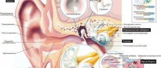
Ultrasound of the child's brain through the large fontanel. It is performed on children in the 2nd month of life, whose head bones have not yet fused, and between them there are “fontanelles” - canals with softer tissue. The presence of this natural bone defect will allow ultrasonic waves to easily penetrate deep into the brain structures, which means to qualitatively examine the internal structure of the brain, identify congenital anomalies, as well as pathology acquired during the prenatal period or during labor. As the “fontanelles” harden, this becomes difficult. Brain ultrasound can detect:
- brain tumors, cystic formations of the meninges, areas of hemorrhage in the medulla (intracranial hemorrhage);
- increased intracranial pressure;
- hydrocephalus due to the accumulation of cerebrospinal fluid (CSF) in the ventricles of the brain.
Untimely detection of circulatory pathology and brain disorders will not allow timely initiation of treatment and can lead to irreversible consequences such as brain dysfunction and neurological disorders.
Ultrasound of internal organs in EVO.
Allows you to identify congenital diseases, anomalies in the structure and location of organs, congenital neoplasms (cysts, tumors) in the abdominal cavity.
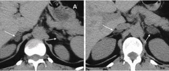
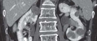
- Ultrasound of the kidneys and urinary tract is used to diagnose changes in the shape and size of the kidneys, underdevelopment of the kidneys, as well as various anomalies in the structure of the bladder and muscle valves (sphincters).
- Ultrasound of the liver, pancreas and gall bladder is performed to diagnose various structural and degenerative changes in the liver, pancreas and biliary tract.
Ultrasound of the hip joints at EVO.
This study will allow timely recognition of congenital hip dysplasia and its complication – congenital hip dislocation. Screening is carried out on infants aged 1 to 2 months and makes it possible to detect abnormalities that were not previously noticeable, which gives a high chance of complete normalization of the child’s musculoskeletal system. Early detection of orthopedic pathology allows immediate correction to begin, thanks to which the baby can be completely healthy within six months.
How much does ultrasound screening of a newborn and a child up to one year cost at EVO?
Currently, the EVO Specialized Medical Center is running a promotion: the total cost of systemic screening ultrasound examinations is 3,000 rubles instead of 5,050 rubles. Your savings are 2,050 rubles!!! And this is much lower than in other clinics!
FAQ:
My baby was born premature. Will an ultrasound harm him?
Ultrasound is harmless even for premature babies. Moreover, prematurity often entails an increased risk of a number of congenital diseases. Therefore, screening ultrasound is not only desirable for premature babies, but also indicated.
I did all the tests for the child. Everything is fine there. Why do I need an ultrasound?
Normal clinical indicators of a newborn’s tests do not at all exclude abnormalities in the internal organs, musculoskeletal system, and brain. Therefore, during screening studies, laboratory diagnostics are carried out only in combination with ultrasound.
Is it possible to limit yourself to one study (to look at the head or kidneys) and at the same time save additional money, and postpone the rest for later?
Yes, you can. But it's not necessary. The essence of screening diagnostics lies, firstly, in its timeliness, and secondly, in its complexity. “The miser pays twice” and, unfortunately, your savings today may cost you dearly in the future.
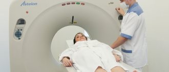
CT is a more informative method than ultrasound. Why don't you do it?
In terms of its diagnostic value, ultrasound is not inferior to computed tomography, and in some cases even surpasses it. It is worth noting that CT scanning is not safe for a child, since the baby is exposed to x-ray radiation and must lie still for a certain time, which entails anesthesia. In our opinion, without any specific evidence for this, such risks for a newborn and a child in the first year of life are unjustifiably unnecessary.
You can get answers to other questions, as well as sign up for an ultrasound scan, by calling the EVO Medical Center (8) 812 701 03 30.
Remember that “nature knows no stop in its movement and punishes all inactivity” (Johann Wolfgang von Goethe).
Information content of ultrasound

There is a need for ultrasound, at a minimum, because this type of research can determine, with an accuracy of a few millimeters, the structure of an organ, the features of its functioning and how such a structure affects the functioning of the whole organism. There may be a risk of the pathological organ influencing the functioning of the entire body system. For example, when the bile ducts narrow, the entire digestive system suffers. Therefore, the child is prescribed maintenance choleretic therapy from infancy. Ultrasound also very often reveals congenital cysts arising from organs. It is important to diagnose them as early as possible and monitor their dynamics. Such pathological formations are often combated with medications. If they are ineffective, then surgery is performed.
Ultrasound is a popular imaging test that is prescribed by pediatricians at the initial stage. At the Profimedica clinic, specialists carry out the procedure quickly, clearly identify the presence of the disease and select their own individual approach to each case.
Prices:
Ultrasound for children