Why is it worth choosing a diagnostic and treatment unit for EEG studies?
- In our work, we take into account all the latest recommendations and achievements of epileptology and neurology,
- You can receive a comprehensive examination and consultation in one center, which is currently difficult to achieve in regular clinics.
- Here you can get not only routine analysis of curves, but also the application of modern knowledge at the international level of clinical electroencephalography (“epileptological”).
- We take responsibility for the quality of the research, monitor the serviceability of all electrodes in the systems,
- Unlike most private and public clinics, our laboratory nurse monitors the progress of the study at a distance through another monitor - this allows us to improve the quality of the study and not create discomfort for the patient by continuously being in the same room.
- We may provide research data on a portable USB drive, particularly with a video stream. The need for video must be warned immediately at the time of the study.
- We store archival data from EEG studies for at least the last two years.
- We provide the result in a way convenient for you: it can be printed on paper in any branch of our clinic convenient for you, or sent to the patient’s email in the form of files with curves and a conclusion in PDF format.
- The Samara Medical and Diagnostic Center has modern high-tech hardware and diagnostic equipment, which allows, in addition to the common routine electroencephalography, to carry out video-EEG monitoring for a long time (from 1 – 4 hours to the whole night).
- Separate specially equipped comfortable rooms for EEG with a bed and a couch bed for sleeping;
- We conduct research on even the smallest patients (and in some branches with a head circumference of 36 cm or more).
EEG with sleep deprivation
EEG with sleep deprivation is a type of electroencephalography in which sleep time is limited or interrupted multiple times at night followed by falling asleep. This is used in cases where the patient’s EEG in a normal state does not reveal pathology in brain activity. This type of study is 30% more accurate than traditional EEG and is more effective in relation to the following types of pathologies:
- epilepsy;
- episyndromes;
- syncope;
- sleep disorders;
- insomnia;
- sleepwalking and dream-talking.
EEG with sleep deprivation can last from 1.5 to 3 hours.
How to choose duration
When a patient wants to conduct an EEG study on himself or a child, this question often arises.
First of all, patients should understand that the optimal type and duration of an EEG study should be recommended by a knowledgeable neurologist or neurologist-epileptologist. It depends on age characteristics, on the disease, on the issue being solved, etc.
Short types of research (up to 30 minutes) are used, as a rule, when the task is to assess the general state of rhythm: its maturity, its regularity, the correct localization of rhythms, etc., as well as for screening during professional medical commissions in some professions. The state of rest is assessed with eyes closed and special functional tests. In other cases, short studies without sleep recording are extremely rarely informative. When diagnosing epileptic disorders and observing their dynamics, studies require at least 1 hour: optimally from 3 hours. The older the person, the more difficult it is to detect epileptic activity, so in adults and adolescents there is often a need for a ten-hour overnight study.
Rhythms on the EEG of the brain
The main rhythm in the EEG of the brain after 7-8 years is the alpha rhythm. In younger children, the main rhythm is the rhythms of a lower frequency - theta and delta - range. Moreover, each of the rhythms has its own characteristic spatial organization and response to various external stimuli.
In various diseases, the ratio of these rhythms and their localization change; pathological forms of activity may occur, the main of which are epileptiform activity and slowing.
Methodology and conditions
All studies take place in a room where an operator is present with the patient for some time, recording information, and then he monitors the progress of the study from a remote monitor.
Video cameras provide constant surveillance.
The technical part of the work is carried out by a specially trained functional diagnostics nurse. Analysis of a completed study is carried out at a separate time by a neurophysiologist.
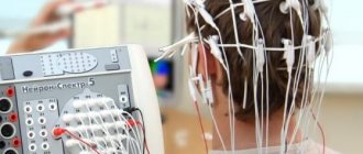
A certain number of sensor-electrodes mounted on special elastic helmets are applied to the scalp of the patient’s clean scalp. To create conditions for the normal passage of impulses from the scalp, a small amount of electrically conductive water-soluble gel is injected under the electrode.
At the time of registration, electrical impulses are transmitted to an amplifier box, then sent to a computer system, where they are converted into a graphical representation in the form of curved lines. After digital processing, the EEG is combined with the video recording and stored on the hard drive.
During the EEG, the patient lies on a couch or sits in a comfortable chair in a darkened room with his eyes closed. For long-term studies, the room is equipped with a bed.
The study includes standard functional tests: rest with eyes closed, opening and closing eyes, exposure to rhythmic light stimuli at different frequencies (usually from 1 to 30 - 60), deep breathing test for 3 minutes.
There are also additional tests for clenching the hand into a fist and for sound stimuli.
In case of deterioration of health during deep breathing or light stimulation, the patient informs the laboratory assistant, and this test can be stopped.
For long-term studies, after the standard part the patient is placed on a bed to sleep. After the end of sleep, all standard tests are repeated.
In the presence of significant events (conditions suspicious for an epileptic attack), the patient himself, his accompanying person or a laboratory nurse keep a diary of events. In our clinic in the branch on the street. Entuziastov-26 uses a special event marker button for the convenience of such monitoring. When you click on it at the appropriate moment in the recording, a note is set for the doctor.
During the entire study, a laboratory nurse monitors the recording progress on a remote monitor, monitors the quality of the electrodes and the visible events.
Prices
| Name | Price |
| EEG for medical examination and traffic police certificate 10 min | 600 |
| EEG monitoring 30 min | 1900 |
| EEG monitoring 1 hour | 3200 |
| EEG monitoring 2 hours | 4100 |
| EEG monitoring 3 hours | 5600 |
| EEG monitoring 4 hours | 6600 |
| EEG monitoring 3 hours (nightly from 20.00) | 6800 |
| EEG monitoring 10 hours (night from 20.00) | 11000 |
Why is an EEG needed and what does it show?
- The study shows the rhythmicity of the cerebral cortex in its different functional states: level of wakefulness (active wakefulness, rest, approaching sleep), sleep and depth of sleep, rhythmic response to various provoking activators.
- EEG is used mainly for diagnosing and monitoring treatment for epilepsy, for differential diagnosis between epileptic and non-epileptic seizures.
- To exclude epileptic mechanisms of impaired development and behavior of children.
- To assess the state of maturing rhythms in children, in particular with prematurity. But premature newborns usually undergo this study in specialized neonatology departments of hospitals.
- To diagnose the first signs of degenerative diseases of the nervous system and further assess the dynamics of the general rhythm.
- To assess the state of rhythm in severe brain diseases: traumatic brain injuries, encephalitis, strokes and much more.
- In intensive care and operating rooms, EEG should be used to monitor the depth of coma and depth of anesthesia.
Indications for EEG and EEG monitoring:
- Paroxysmal sleep disturbances (fears, nightmares, sleepwalking, enuresis, etc.) to exclude their epileptic nature;
- Various disorders of the child’s speech and mental development, some behavioral disorders;
- Suspicion of a first-time epileptic attack;
- Epilepsy – at different stages of diagnosis and monitoring the effectiveness of treatment;
- Migraine with aura for differential diagnosis with epileptic seizures;
- Fainting-like conditions for differential diagnosis with epileptic seizures.
- Suspicions of degenerative brain diseases to assess the dynamics of the general rhythm;
- Suffered severe brain damage (traumatic brain injury, stroke, encephalitis, etc.) to assess the state of the rhythm as a whole.
Advantage of EEG
EEG has an important advantage: it shows brain activity in real time, because the electrical signal travels almost at the speed of light. The time resolution of other methods is much worse - on the order of seconds, at best, fractions of tenths of seconds, and with their help only relatively slow processes can be observed. Most often, 19 electrodes are used for recording, which make up the standard installation in Russia, as well as in many other countries. Sometimes it is permissible to reduce the number of electrodes when recording in children under one year old or with a small head size, but it is advisable to use at least 16 channels.
What can be analyzed using EEG?
- presence or absence of a basic rhythm;
- background rhythm surrounding the main rhythm;
- changes in rhythms during special tests;
- changes in brain activity during various functional states, for example, when a person’s level of wakefulness changes, he begins to doze off or falls asleep.