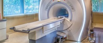
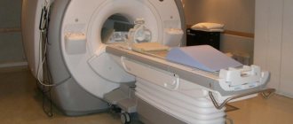
The examinations are carried out on a modern open-type magnetic resonance imaging scanner - Siemens Magnetom C!
Over the years, we have accumulated vast experience in conducting examinations of complex patients suffering from claustrophobia. The center’s specialists are always polite, patient and understanding about this problem, while always focusing on results. Our center has a high percentage of successfully performed examinations for claustrophobia, and the resulting tomograms are always of high quality.
MRI is a high-tech, highly informative method for diagnosing diseases. An examination with an open-type magnetic resonance imaging scanner allows one to identify the smallest changes in the tissues of the patient’s body, helping the attending physician make the correct diagnosis and timely prescribe effective treatment.
Open type tomographs are absolutely safe for humans. A constant magnetic field and a huge selection of scanning programs allow you to obtain images of the highest quality.
With the advent of an open-type tomograph, the procedure has become more comfortable in comparison with a closed (tunnel) type device. For people suffering from claustrophobia, the elderly, children, and patients who are unable to take a completely horizontal position due to injuries or physiological characteristics, open MRI will allow them to undergo examination.
Obese and overweight people can also be examined using an open-type tomograph. The tables of open devices with side loading are quite wide and comfortable; you can sit down and lie down on them comfortably. The open-circuit table of the device can support a weight of up to 170 kg, however, there are restrictions on the volume of the patient’s torso. These restrictions are always individual and depend on the person’s build (height, weight, height of the abdomen and chest when lying down) and the type of examination.
Brain tomogram with contrast
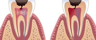
A contrast enhancer improves the quality of the image, the visibility of the contours and structural elements of organs. The advantage of MRI is the possibility of early detection of the development of oncological processes and the detection of microscopic anomalies within the body. To add precision, a gadolinium saline solution is injected. The drug is safe, but has certain limitations. Contrast is used:
- Intravenously immediately before scanning;
- Partially when performing a dynamic MRI examination (the necessary intervals between the substances entering the blood are observed).
The tool helps create images with the most accurate dimensions of tumor formations. Diagnostics is informative after chemotherapy, the results of treatment are clearly visible. The main advantage of the method is the absence of the harmful effects of X-rays, so the examination can be repeated again without the risk of harm to health.
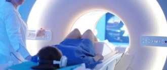
Video of MR scanning with a closed-type device
A closed-type tomograph is a classic equipment for magnetic resonance diagnostics. The procedure is done as follows: the object is placed on a special retractable table. The patient is moved into the device capsule, and the examination process begins.
The duration of an MRI of a joint is about half an hour. When scanning, you must remain completely still, or the image will be distorted, which will lead to loss of examination effectiveness.
Upon completion, the specialist gives tomograms or an electronic storage medium with images. A conclusion is drawn up describing the data obtained after the MRI.
Minuses:
- During operation of the installation, noise effects are constantly emitted, causing some inconvenience. It is usually suggested to use headphones (earplugs), but their use does not guarantee complete absence of sound;
- Feedback from the doctor is available using a microphone; direct contact is not possible.
In the video, the sound of the tomograph is very loud, don’t be afraid - this is how the installation works.
Main contraindications to undergoing the procedure
There are relative and absolute contraindications to MRI. This method is one of the most universal and suitable for most patients.
Absolute contraindications
These include those cases when a person cannot undergo such a procedure at all. Among them:
- Foreign devices in the human body. These include pacemakers, cardioverter-defibrillators, and insulin pumps. The magnetic field may prevent them from working correctly.
- Metal objects implanted on purpose or accidentally entered into the body. These are implants, special designs, such as Ilizarov apparatuses, prostheses, artificial joints.
- Personal intolerance to drugs administered during the process. These include components of contrast enhancement agents.
Also, the use of a contrast agent will be contraindicated if the patient suffers from renal and liver failure or allergies. Contrast-enhanced MRI is not recommended during pregnancy and lactation.
Relative contraindications
They say that for some time a person will not be able to undergo an MRI procedure or that special consultation is required from the doctor who makes the final decision.
These include:
- Fear of confined spaces. It is no coincidence that the question of how not to be afraid of closed-type MRI is popular online. If it is performed, the patient is placed in a tunnel with a device - many may experience psychological discomfort.
- Pregnancy in the first trimester. Although the magnetic field is safe, it is best not to risk exposing the fetus to it.
- Poor condition of the patient. This includes various types of heart and kidney failure, as well as other diseases in which it is very important for the doctor to monitor the patient’s condition.
- Inability to remain still. It can develop in different cases, including when a person has severe psychological illnesses, nervous tics and other problems. As a last resort, for such patients it is possible to use anesthesia.
- Discrepancy between the size of a person’s body and the parameters of tomographs. In most models, the tunnel width is 60 cm.
- Severe spinal deformities. During the examination, the person will have to lie on his back and remain completely still.
- Metal parts in the patient's body. In some cases, this includes filling large spaces on the skin with a tattoo containing a high percentage of metallic ink.
In some cases, problems may arise after various types of surgical intervention, for example, after removal of the gallbladder, puncture biopsy, sectoral resection and many other problems.
If you have any relative contraindications, you will need to further consult with your doctor. In some cases, the procedure can be postponed, in others it can be replaced with another diagnosis.
Sometimes the solution is to use alternative methods, for example, a vertical tomograph instead of a horizontal one.
If you still have foreign metallic inclusions in your body, you will need to provide documents confirming its complete safety or admissibility of use in magnetic resonance therapy.
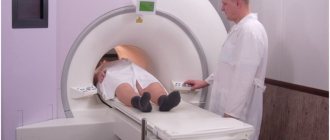
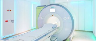
Filming the operation of an open (half-open) type apparatus
An open tomograph is a device that differs in shape from a standard tube-shaped device. The sides are not limited, nor are the head and lower extremity areas. The session takes place inside a room with the required force field strength. It is not necessary to stay in a confined space, which allows you to get rid of uncomfortable conditions.
The principle of the study is identical to scanning in a closed structure. The results are presented in the form of tomograms or on external media.
Advantages:
- In the diagnostic room, together with the patient, there are doctors (if necessary), relatives (if a feeling of fear overcomes or there are other objective reasons that require the presence of accompanying persons);
- The presence of damage to the spinal column, arms, legs does not create insurmountable obstacles for tomography;
- Administration of medications is possible throughout the entire procedure;
- Obese patients will be able to fit into the device, weight bearing is not a difficulty;
- The threshold of sound vibrations has been reduced, which eliminates unpleasant sensations when undergoing an MRI;
- People affected by claustrophobia will undergo the examination without hindrance;
- Having full contact with the doctor;
- The latest MR equipment makes it possible to form an optimally clear image; slight movement of the object under study will not become a hindrance;
- Ideal for performing MRI scans on children.
The installation has advantages in terms of ease of diagnostics, but in terms of technological parameters it is no better than tunnel-type equipment.
Who should be diagnosed with MRI using an open-circuit machine?
The open circuit of the tomograph allows you to examine patients of any age, including children over 4 years old, people suffering from claustrophobia and the elderly. The device is suitable for research in such areas as: neurology, traumatology, orthopedics, oncology, gynecology, urology and others.
Indications for MRI:
- frequent headaches;
- severe impairment of visual acuity;
- dizziness of unknown etiology;
- epileptic seizures;
- tumors;
- pain in the spine;
- numbness of the limbs;
- stiffness of movements;
- joint diseases;
- sports injuries;
- formation in the soft tissues of the body;
- gynecological diseases;
- infertility.
Which tomograph results are more accurate - open or closed type?
The results of magnetic resonance imaging are deciphered by the radiologist performing the examination. He gradually analyzes the resulting images and draws up a conclusion. The patient is given a diagnostic protocol, which describes the appearance, size and condition of the structural elements being scanned. At the end of the document, the doctor records the difference from the standard and the existing violations.
At the end of the session, the patient receives tomograms in film format and data on electronic media. A CD or flash drive is suitable for recording. A number of medical centers offer the second option exclusively upon request.
When preparing an epicrisis for scans, a clinic employee is not given the rights to assess the severity of the detected anomaly. He will be able to inform you about the type of damage and its location. The conclusion about the severity of the abnormalities found and the method of treatment is written by the doctor who referred you for the procedure, so the papers must be delivered to him. The diagnostician can give advice, suggest the profile of a specialist who can analyze the detected diseases, if a person arrives for an MRI of the joint without an appointment, on his own initiative.
In a closed loop, the force field acts in a circle. In an open apparatus, magnetic waves propagate only in the horizontal plane, the intensity is lower. The more active the source, the better the images obtained during scanning. With a closed tomograph, with equal induction settings, the MRI results will be more accurate and detailed.
Power as the main parameter that differentiates tomographs
The difference between closed and open MRI is power. The higher it is, the clearer the image can be obtained using such a device.
The unit of power for the device is Tesla (T). When you undergo an examination with the most powerful machines, you can expect to receive truly detailed images, as well as a reduction in the duration of the process.
The table below shows the features of different device options.
| Type of tomograph | Power | Peculiarities |
| Low-floor | 0.1 – 0.5 Tesla. | Typically, such tomographs belong to the open category. They are used when the patient has noticeable pathologies or large tumors that can be seen even on pictures with little detail. There are a minimum of contraindications to the examination; it can be performed for people with large body weight and waist size, and children. This is also a good option if you suffer from claustrophobia. But you should prepare for the fact that the examination will take quite a long time - at least 40 minutes. |
| Mid-field | 0.5-0.9 T | They help reduce the examination time - a standard session takes from 20 to 30 minutes. The quality of diagnostics and detail will also be higher. |
| High-floor | 1-1.5 T | They are most often found in medical centers. The standard time to check a patient's condition is 15 to 20 minutes. Comprehensive examinations and contrast-enhanced testing may take longer. This power helps to get the most complete picture of the state of a person’s internal organs and identify many problems at an early stage. It should be borne in mind that all such types of equipment are classified as closed. Testing in them has a greater number of contraindications, and for people with claustrophobia and children it can be very uncomfortable. The use of such a device helps to obtain sections up to 1 mm thick. This indicates a very high accuracy of readings. |
| Ultra-high-field | Up to 3 Tesla | Such devices can very rarely be found in medical centers. The reason is that they are very powerful and expensive. The survey becomes simply unprofitable. High-field models help to obtain sufficient results for making a diagnosis. The main advantage is speeding up the process. The examination usually takes no more than 10 minutes. The cut is very accurate and the image quality is high. |
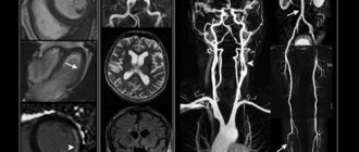
How to do an MRI of the spine: photo
The photo shows the results of an MRI examination of the spinal column. A large number of sections are shown, allowing for detailed examination.
Advantages of magnetic scanning:
- High level of accuracy and information content;
- The method is non-invasive (there is no introduction of instruments and devices into the body);
- The appearance of pain is impossible;
- Checking your back makes it possible to detect pathology in the early stages of development, and there is a chance of quickly eliminating the disease.
The technique allows you to obtain complete information about the state of the departments of the area under study:
- The spinal canal, its nerve fibers;
- Intervertebral discs;
- Vascular channels.
With the help of MRI diagnostics, it is possible to determine the speed parameters of the movement of cerebrospinal fluid and examine the cranium (required procedures for dangerous injuries).
A nuclear magnetic resonance session is in great demand among specialists involved in the treatment of spinal diseases, oncologists, and neurologists.
The study must be completed:
- Preoperative examination;
- If the structure of areas of the spinal column is disrupted;
- If there are anomalies of a degenerative-dystrophic nature (damage to articular cartilage, hernia, vertebral deformity);
- Determination of sources of chronic pain syndromes;
- In case of abnormal phenomena;
- After surgical interventions (to track recovery processes).
Spinal cord tomography is done:
- For post-traumatic consequences;
- There is an assumption of the presence of cancerous formations, secondary tumors;
- Study of cerebrospinal fluid, detection of cystic inclusions.
During diseases accompanied by destruction of the myelin sheath of neurons:
- Acute polyradiculoneuritis;
- Multiple sclerosis.
The picture shows scanning with verticalization. The method helps to visualize the displacement of the vertebrae under axial loads.
Indications for MRI examination
The procedure is always prescribed by a doctor. He should check the medical history, listen to the patient's current complaints and evaluate the treatment history.
Based on this, a decision will be made whether it is worth booking an MRI or whether any alternative means can be used.
There are several reasons to use this type of diagnostic test:
- Suspicion of various types of injuries, damage to bones, ligaments and soft tissues.
- Problems with the pelvic organs - frequent urination, foreign inclusions in the urine, menstruation problems and much more.
- Suspicions of problems in fetal development, the likelihood of hereditary pathologies.
- Diseases of the cardiovascular system, manifesting themselves in arrhythmia, tachycardia, shortness of breath and other symptoms. If there are signs of impaired blood flow in the internal organs, it is also worth signing up for a test using magnetic resonance.
- Diseases of the endocrine system - from incorrect functioning of the thyroid gland to the appearance of neoplasms with different localizations. They can manifest themselves in increased fatigue, hormonal instability and other symptoms.
And this is only part of the indications for use. The technique is also actively used in pediatrics.
Unlike X-rays, this method helps to give more accurate readings about the condition of a growing organism without radiation harmful to the body.
How to make a video MRI of the brain
Depending on the indications, with an MRI of the head, doctors check:
- Eye orbits;
- Vascular channels;
- Nerve fibers of the cranium;
- Brain;
- Central organ of the endocrine system;
- Paranasal cavities;
- The joint connecting the mandible to the temporal bone.
In the video you can better understand how tomography is performed. The sound inside the device indicates vibration of the metal spirals caused by electromagnetic pulses.
Diagnostics helps to study the state of the brain and its parts. The use of a contrast agent makes it possible to more accurately display the size, contours of neoplasms, and their locations.