Computed tomography of the throat is one of the new ways to examine bone structures and soft tissues in this area. Diagnostics provides an opportunity to identify existing pathological conditions and structural features of the tissues of the larynx. Through scanning, you can obtain highly accurate images of the part of the body being examined, and therefore making the correct diagnosis will not cause difficulties for a specialist.
What does a CT scan of the larynx reveal?
Using CT images, specialists have the opportunity to visualize a number of diseases and pathological conditions:
- neoplasms of soft and bone tissues of the larynx with the ability to assess their size and location;
- presence of esophageal diverticula;
- places of destruction of cartilage in the larynx;
- observe changes in the structure of the walls of the larynx, which may be a sign of a developing tumor;
- identify the presence of foreign bodies in the area being examined;
- determine the severity of injuries received;
- assess the condition of the lymph nodes;
- identify existing inflammatory processes in the cavity being examined;
- diagnose various developmental pathologies;
- examine the vascular network of the larynx.
Computed tomography scan of the larynx is normal
At the level of the hyoid bone, the entrance to the larynx is visible, which is limited on the sides by the lumens of the pyriform sinuses. Outside the vertebral body on both sides are the internal jugular vein and the common carotid artery. Anterior to the air column of the larynx, the following anatomical formations are determined:
- Epiglottis;
- Preepiglottic space;
- Body of the hyoid bone.
Below are the vocal and ventricular folds, the anterior commissure, the cricoid cartilage, and the plates of the thyroid cartilage. Normally, the vocal folds have a uniform structure, their free edges are symmetrical. Tomograms of the subglottic space on both sides of the larynx show the arch of the cricoid cartilage and the lower horns of the thyroid cartilage.
On a computed tomogram, the tracheal gap appears as a thin line. The anterior and lateral walls of the larynx on a CT image are represented by dense cartilaginous rings, the posterior - by dense elastic fibers. From the lower edge of the seventh cervical vertebra to the level of the upper edge of the fifth thoracic vertebra, the trachea has the clearest image.
The normal intrathoracic portion of the trachea on CT scans varies from person to person and at different levels. Typically round or oval in shape, but can be square or horseshoe shaped. The diameter of the trachea is normally from 15 to 18 mm, in adults it does not exceed 22 mm. The average diameter of the trachea in men is 19.5 mm, in women – 17.5. In patients over 40 years of age, CT images show calcification of the tracheal cartilage.

CT scan of the throat and larynx using a contrast agent
A CT scan of the larynx, performed without contrast enhancement, allows one to obtain information about the condition of the bone tissue and the existing pathological process in the hyoid bone and in the cervical spine. Using this diagnostic method, the cartilage of the larynx and the soft tissues of the neck are practically not distinguished, which is a significant disadvantage of this scanning method.
Therefore, in order to improve the quality of images when performing a CT scan of the throat, the doctor may prescribe a CT scan of the throat and larynx using a contrast agent.
Contrast is most often injected into the patient's body intravenously, after which it immediately spreads along with the bloodstream to all tissues and organs. Through the use of a contrast agent, it is possible to examine anatomical formations and organs in great detail. They acquire detail and the clearest outlines.
Contrast can also be introduced into the patient gradually throughout the entire scan. Exactly how the contrast will be administered is always decided on an individual basis, based on the purposes of the examination and the intended disease.
Regardless of how the contrast enters the body, the following discomfort cannot be ruled out:
- the appearance of a metallic taste in the mouth;
- feeling of warmth in the body;
- feeling of rising heat.
Such symptoms are considered not dangerous. They represent a normal reaction of the body to the injected drug, so there is no need to stop the procedure.
Dangerous signs that may indicate the appearance of intolerance to contrast during a CT scan of the throat are:
- the appearance of severe swelling of the face;
- sore throat;
- dizziness;
- nausea, vomiting;
- a skin rash and severe itching;
- decrease in blood pressure.
The presence of at least one such symptom becomes a compelling reason to stop administering the drug and provide first aid to the patient. Although such cases are extremely rare. Ideally, each patient should undergo a sensitivity test before using contrast. If, based on the test results, the doctor has no doubts about the safety of the contrast agent for the patient, then you can safely proceed to the next stage of the examination.
Features of the technique
Computed tomography, which is simply abbreviated as CT, allows you to draw a conclusion about the health of the larynx, as well as evaluate the blood and lymphatic vessels located in the scanning area.
Content:
- Features of the technique
- Indications for use
- Contrast enhancement
- Contraindications for examination
- Preparatory stage
Thanks to the constant improvement of the diagnostic system, with the advent of spiral CT, it became possible to obtain detailed visualization of the problem area. With multislice CT, it is possible to collect a larger number of images from different angles, which increases the final quality of the projection.
The second option is designed to reduce testing time, as well as reduce the radiation exposure to the body by an order of magnitude, which is especially important for elderly patients. But, regardless of how many beams of rays are used, the principle of analysis remains identical. The scanner with detectors will rotate around the medical couch with the patient, allowing a three-dimensional image of the organ to be formed in the shortest possible time.
Most often, when examining this area, the doctor additionally insists on including a contrast stage. It is not always necessary, but if the doctor recommends undergoing it, then you should not refuse, because the results when using contrast agents are more informative.
The contrast mechanism is based on the introduction of a special substance into the body. Most often this is an intravenous route. Due to the difference in contrast accumulation, it is possible to detect even the smallest deviations, which is more typical of oncological tumors.
Indications for CT scanning
Each patient can receive a referral for a CT scan of the throat and larynx from a doctor in the following cases:
- in case of previous neck injuries, as a result of which there is a high probability of injury to internal organs;
- when foreign bodies get into the throat;
- with existing pathologies of the development of the throat and larynx;
- with progressive inflammatory diseases;
- for pathologies of the vascular network of the neck;
- for the purpose of precise location of the pathological process before surgery;
- to assess the effectiveness of treatment after surgery.
Doctors prescribe a CT scan of the larynx only in cases where another diagnostic method carried out in the future will not provide sufficient information and will complicate diagnostic measures.
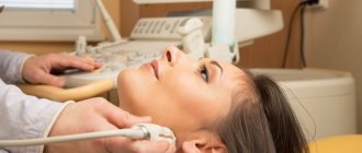
As a rule, the patient is initially referred to undergo ultrasound diagnostics or x-rays, and only if these examination methods do not lead to results, the patient will be sent to a CT scan of the throat and larynx.
Contraindications for diagnostics
CT scan of the throat and larynx has some contraindications for the following patients:
- women expecting the birth of a baby;
- patients with excess body weight;
- patients with mental illness.
CT scans are performed with extreme caution:
- children under 12 years of age;
- women breastfeeding babies;
- patients with kidney problems;
- patients with multiple myeloma.
Carrying out computed tomography using contrast is unacceptable:
- during pregnancy and breastfeeding;
- patients with disorders of the liver and kidneys;
- diabetics;
- patients with an allergic reaction to contrast.
Contraindications for examination
Despite the fact that the technique is relatively safe, there are certain groups of patients who are at risk. We are talking about pregnant women at any stage of pregnancy, since even a reduced dose of X-ray waves can significantly harm the development of the fetus. Radiation acts on the baby according to an identical principle during breastfeeding, but here the risks are slightly less.
If the benefit from the study significantly outweighs the harm, then during the lactation period women are still prescribed a CT scan, but after the manipulation they will need to refrain from breastfeeding for at least a few days.
You should also carefully approach the choice of diagnostics with the presence of iodine. To do this, an additional test for an allergic reaction is performed.
Other relative contraindications include:
- kidney diseases;
- severe cardiovascular diseases;
- age under 16 years;
- thyroid disease with overproduction of hormones;
- severe pain syndrome;
- hyperkinesis;
- mental disorders.
But it’s not for nothing that they have the clarification “relative”. Sometimes the doctor gives a referral for a CT scan even to people at risk if there is no alternative.
How to properly prepare for a CT scan of the larynx
For those patients for whom CT of the throat and larynx is planned to be performed in the native mode, there is no need to observe any strict restrictions in their usual lifestyle or follow a special diet.
However, those patients who are indicated to undergo a scan using a contrast agent will have to comply with some restrictions. So, before undergoing a CT scan of the throat, it is prohibited to eat any food 6 hours before the start of the scan. However, drinking water in small volumes is allowed.
All patients, regardless of how the examination will be carried out: with or without contrast, must remove absolutely all jewelry and objects with metal parts before entering the office. You will also have to leave your mobile phone, watch, bank cards outside the office - all these items can cause poor-quality photographs.
Contraindications for diagnosis
Despite the safety and information content of the technique, there are still contraindications to its use, including:
- Children under 14 years of age;
- Pregnant women;
- Myeloma patients;
- Heart, kidney and liver failure (when performing a procedure with contrast);
- Allergy to any of the components included in the contrast agent;
- Claustrophobia;
- People who are unable to independently control their body movements (usually under anesthesia);
- The patient's weight is over 150 kg.
Progress of CT scan of the larynx
To conduct a CT scan of the throat and larynx, the patient is asked to take a horizontal position on a sliding table, where he will remain for the duration of the study.
His body is fastened with belts for secure fixation, after which the table slides inside the device. All that is required of the patient during a CT scan of the throat is to remain completely calm and not make any movements. The tomograph scans, resulting in a series of images. As a rule, the entire examination process takes no more than 15-20 minutes.
During the scan, the patient is alone in the room. Medical staff monitors the examination progress through a special window from an adjacent office. The patient is not left for a minute without the supervision of doctors. Both the patient and the doctors have the opportunity to communicate through the use of a microphone, so if there are any changes in well-being, the patient will always be able to inform the staff and not be afraid that he is isolated from the outside world.
The latest generation of tomographs meets all safety requirements, so they do not pose any significant harm to the health of patients. Although CT is based on X-ray radiation, it has a very low intensity and, when compared with a classic X-ray examination, it is quite insignificant.
However, due to the fact that X-rays are used during a CT scan of the larynx, this procedure cannot be called absolutely harmless, and it has certain time restrictions for repeated scanning.
Thus, it is unacceptable for women who are expecting the birth of a baby to undergo a CT scan, regardless of the stage of pregnancy, as well as for patients who have already undergone this type of examination in the recent past.
A CT scan of the throat cannot be performed an unlimited number of times, since the radiation dose cannot be exceeded. And only in exceptional cases, if there is strong evidence for this, specialists can prescribe an additional CT examination.
Contrast enhancement
In most standard cases, classical computed tomography is sufficient. But if there is a suspicion of cancer, then you cannot do without contrast. This manipulation will be especially important when looking for a disease at an early stage of development in order to suppress it in the bud with minimal losses.
In such situations, a CT scan is sent directly from the oncology clinic to help quickly detect possible:
- primary tumor process of cervical localization up to the vocal cords;
- secondary oncology of metastatic type and extent of damage;
- tumors that degenerate into cancer due to malignancy;
- changes in the growth of a cancerous tumor.
Even ordinary papillomas, adenomas, polyps and other benign neoplasms are also diagnosed using this diagnostic procedure.
Also, patients at the intermediate stage of treatment in an oncology hospital will also have to undergo a CT scan again.
This is necessary to track the dynamics of recovery and be able to adjust the course of treatment if necessary.
The secret to the increased clarity of images displayed on the monitor screen is special iodine-based substances. Due to the ability to accumulate in the affected tissues, the contrast agent colors these areas especially brightly.
For the convenience of conducting the study, the drug is administered through a vein, after which a spiral test format is first performed, and then a contrast test.
Alternative diagnostic methods
Alternative diagnostic methods include ultrasound and magnetic resonance imaging. However, CT of the throat has a clear advantage - this method is the most informative and fastest. In addition, it allows you to identify even minor pathological processes and evaluate the structures of internal organs.
If we talk about which type of diagnosis is better: MRI or CT, then it will be quite difficult to answer this question unambiguously. Both research methods serve completely different purposes, although many of the data obtained may be duplicated.
A CT scan of the throat is performed using X-rays, so it allows you to examine dense formations and hollow organs. Magnetic resonance imaging is the best way to identify soft tissue conditions and is widely used to evaluate injuries to the neck and ligaments.
If there is a need to scan patients in an emergency situation, the choice of one or more examination methods is at the discretion of the doctors, based on the nature of the pathological condition.
Where to get a CT scan of the throat and larynx
Regardless of the exact reasons for which the patient was prescribed a CT scan of the throat, a fundamentally important point is to find a suitable clinic. She must not only satisfy the patient with her pricing policy, but also perform the examination at the highest level.
Unfortunately, sometimes it is not so easy to make the right choice: some medical centers do not have modern equipment, and tomography performed on old scanners does not provide a complete picture of the existing pathological process. In other clinics, the medical staff does not have sufficient experience, so there is a high probability of making an incorrect diagnosis.
We are pleased to offer our services to patients: on our website, every visitor can see for free a large list of medical clinics, as well as detailed information on each of them.
After selecting the required medical center, you can call the phone number listed on the website to make a preliminary appointment. We provide a discount on CT scanning to everyone who turns to us for help.
By contacting us for help, you can easily decide on the choice of the necessary medical center from a huge list, and also receive a guarantee of the lowest price.