MRI of the vessels of the brain and neck is prescribed to patients to identify existing pathological processes. Tomography is rightfully considered the most highly accurate type of examination. Diagnosis of soft tissue disease through other measures is very problematic.
Thus, ultrasound examination is considered a low-informative procedure, which is used for this purpose in rare cases. For example, when for one reason or another it is not possible to undergo an MRI of the vessels and arteries of the neck or brain, and it is advisable to perform a computed tomography scan only to identify tumors. In addition, CT scanning is associated with radiation exposure to the patient’s body, so scanning cannot be called completely safe and cannot be carried out without compelling reasons.
The main advantages of MRI diagnostics of brain and neck vessels
The high accuracy of MRI of brain and neck vessels is far from the only advantage of scanning. This may also include:
- absolute safety for the health of the subject;
- ease of examination;
- a small number of prohibitions and contraindications for undergoing scanning;
- a real possibility of identifying a pathological process at the very initial stage of its occurrence;
- Possibility of scanning an unlimited number of times.
As a rule, an examination of the vascular system of the brain is carried out together with an MRI of the vessels and arteries of the neck.
Carrying out two types of diagnostics simultaneously is determined by the fact that normal blood flow in the brain can only be ensured if there are no disturbances in blood circulation in the neck. For example, improper blood circulation as a result of mechanical injury in the neck area in most cases leads to oxygen starvation of the brain, which can cause the death of the victim. This leads to the logical conclusion that it is not advisable to conduct exclusively magnetic resonance imaging of brain vessels.
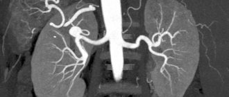
The scan allows specialists to draw conclusions based on 3D images of blood supply in a particular part of the brain.
MRI of cerebral vessels is a unique diagnostic method that detects diseases of the veins and arteries, so no type of examination can be compared with tomography due to its significantly low information content.
Due to layer-by-layer scanning and high-quality images, the likelihood of error during diagnosis is practically reduced to zero.
Main types of headaches
Neurologists determine the types of cranialgia by clinical symptoms:
- The most common type is tension pain. Characterized by a feeling of circular soreness along the skull, inside the eye sockets;
- Cluster pain syndrome is accompanied by pulsation of a separate area of the head;
- Tumor pain occurs in the morning, is accompanied by nausea and vomiting, and can gradually progress;
- Intracranial hemorrhage leads to pain in a certain part of the skull, characterized by secondary symptoms (vision loss, unsteadiness of gait, hearing loss, hallucinations);
- Migraine is long-term pain on one side, intensifying against the background of photophobia and loud noises. Often before an attack there are precursors - visual photophobia (rings), auditory hallucinations;
- Temporal arteritis - characterized by local pain in the area of the temporal bone (lateral parts of the skull).
Treatment depends on the type of disease. The combination of several forms at the same time complicates therapeutic tactics. Magnetic resonance scanning makes it possible to study the morphology of the cerebral parenchyma and verify changes in the arteries.
Indications for MRI of head and neck vessels
As a rule, such an examination is not prescribed by specialists for no apparent reason. Reasons for prescribing a scan may include:
- ischemic disease;
- very frequent headaches;
- the appearance of dizziness;
- sensation of ringing and noise in the ears;
- vegetative-vascular dystonia.
After the scan, the doctor evaluates the resulting images, where it is possible to clearly visualize the blood supply to various areas of the brain and neck.
Classification of hypertension
Primary and secondary hypertension are classified into several forms of the disease. In the first case, there are:
- Hyperadrenergic form, which is more often observed in young patients. Its symptoms: rapid change in facial skin color, throbbing headaches, anxiety for no reason.
- Normo- and hyporenin form. It is detected mainly in elderly people. The cause of the pathology is the retention of sodium salts in the body, which changes the volume of circulating blood. Signs of the disease are swelling of the face, swelling of the limbs.
- The hyperrenin form develops in individuals with rapidly progressive hypertension and is diagnosed in young men. The pathology is characterized by an increase in blood pressure up to 230 mmHg, accompanied by dizziness, nausea, and kidney pain.
Secondary hypertension is also classified into several forms:
- Renal, developing due to diseases of the renal system (pyelonephritis, polycystic disease, nephropathy, tumors of various etiologies).
- Endocrine, associated with increased or weakened thyroid function, Cushing's syndrome, tumors, hypothalamic syndrome.
- Cardiovascular, caused by heart defects.
- Neurogenic, occurring against the background of encephalitis, atherosclerosis of the arteries and brain tumors
- Drug-induced, which develops with long-term use of drugs that increase blood pressure.
What does the scan reveal?
It is advisable to conduct MRI of the vessels and arteries of the neck to identify many diseases and pathological processes, which include:
- cerebrovascular accident due to the presence of atherosclerotic plaques on the inner walls of blood vessels;
- received injuries to the head and neck;
- brain infections of any etiology;
- life-threatening conditions due to brain damage (for example, cerebral infarction);
- any neoplasms;
- pathological structure of the vascular system of the brain;
- multiple sclerosis.
Brief characteristics of cerebral pain syndrome
Tension pain is provoked by pathology of the cervical spine, tension in the muscles of the face and eye sockets. The duration of the syndrome is determined by the intensity of stress. The aggravation is observed in the evening.
The causes of migraine are not clear, but it was possible to identify a pathogenetic mechanism - dilation of cerebral vessels with increased activity of certain areas of the cerebral cortex. They disappear after rest and elimination of the provoking factors of the disease.
Cluster pain is difficult to treat. Localized near the eyes. Combined with redness, tearing, runny nose. The average duration is about 15 minutes.
As an oncological tumor grows, it leads to compression of cerebral structures. Neurological signs of nosology are varied and depend on the location of the cancer.
With intracranial hemorrhage, a person often loses consciousness. Excessive bleeding can lead to death without timely medical attention. Arterial rupture occurs after traumatic brain injury or thinning of the vascular wall.
Inflammation of the temporal arteries (arteritis) is caused by viral and bacterial infections, trauma, and hypothermia. Steroid medications are used to treat the disease. The nosology is combined with clouding of the lens and loss of vision.
Contraindications for examination
Despite the fact that MRI of the head and neck vessels is an absolutely safe diagnosis, there are still some contraindications for its use. If we look at it as a whole, they are not at all different from those contraindications that apply to scanning other parts of the body.
First of all, MRI of cerebral vessels is unacceptable for people with metal objects in their body. Any implants, stimulators, prostheses act as contraindications. The exception is products made of titanium. After the doctor is aware of the presence or absence of such objects in the body, the patient will be allowed to undergo the examination or will be refused. In this case, the patient must provide the specialist with appropriate documentary evidence of his words. The type of material from which a particular implant is made is recorded in a special certificate issued after the surgical intervention.
Another contraindication to MRI of the vessels of the head and neck is scanning using contrast for women carrying a child. The chemical substance, despite its relative safety for an adult, can cause irreparable harm to a developing fetus, which is why such a ban is due.
The use of contrast is unacceptable regardless of the duration of pregnancy, as well as during the lactation period. At the same time, MRI of the vessels and arteries of the neck without contrast enhancement is quite acceptable for pregnant women in the 2nd and 3rd trimesters, when the fetus has developed all vital organs and systems.
MRI for hypertension
The main cause of hypertension is vascular atherosclerosis. Scientists have identified the dependence of hypertension on hereditary factors, age characteristics and the presence of chronic diseases in humans.
Among the risk factors that provoke high blood pressure are:
- stressful situations and emotional turmoil;
- diabetes;
- disruption of the adrenal glands;
- malfunction of the thyroid gland;
- passive lifestyle and bad habits;
- violation of fat metabolism in the body.
Hypertension is more often diagnosed in older women. People who consume excessive amounts of salt are considered at risk.
There are several types of hypertension:
- hyperkinetic with increased hemodynamic shock;
- hyperkinetic with a relative increase in peripheral blood vessels;
- normal kinetic with increased peripheral resistance.
Limitations for MRI of brain and neck vessels
It is not always possible to conduct tomography for people with mental illness, patients with uncontrolled movements of the limbs, as well as people with a fear of being in a confined space. Often, scanning of such patients is carried out in a state of medicated sleep, since otherwise it is impossible to carry out a diagnosis.
It is especially difficult to perform MRI of cerebral vessels in patients on mechanical ventilation. Not every medical institution has specialists who can take on such responsibility.
Very often, tomography is performed under anesthesia and in young children. It is very difficult for children to remain immobilized throughout the entire examination period, and under anesthesia, the scanning is very calm for both them and the doctors. Another advantage of using anesthesia is that young patients will not experience any stress and will not have negative memories.
Preparation for MRI of brain and neck vessels
Many patients before undergoing MRI of cerebral vessels are worried that they will have to comply with some strict restrictions before scanning. Such speculation is completely unfounded. There is no need to carry out any preparatory measures on the eve of the diagnosis, which is a huge advantage of magnetic resonance imaging. As discussed above, the most important condition is that the patient should not have any metal objects in his body, and it is also unacceptable to undergo scanning in clothes that have rivets, buttons, zippers and other metal parts. It is recommended to change your clothes to looser or hospital clothes.
Patients who have been scheduled to undergo an MRI of cerebral vessels using contrast should not eat any food on the day of the examination. The scanning itself takes place very quickly (usually it does not take more than 15 minutes), so no unpleasant sensations should arise during this time.
Where is the diagnosis carried out?
You can sign up for a test at any municipal or private diagnostic center. However, not every district clinic contains a tomography room due to the complexity and high cost of its maintenance. You can find the nearest medical institution that provides MRI appointments on the unified city service mrt-v-spb.
Go to the main page of the site, mark the name of the service you are looking for, and study the list of clinics that provide diagnostics that opens. The centers are graded according to ratings and prices for services. Read reviews, compare offers and choose the best ones. Sign up for the procedure through the portal by calling the phone number indicated at the top of the page and receive 1000 rubles for any type of scan.
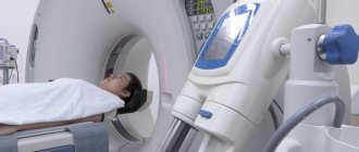
How is MRI of the vessels of the brain and neck performed?
Diagnosis is carried out using a special device - a magnetic resonance imaging scanner, which looks like a large tunnel from the outside. Before the scan begins, the patient is explained how to behave correctly inside the device, namely:
- It is prohibited to bring any gadgets with you;
- it is unacceptable to make any, even the most insignificant movements;
- You cannot make movements with your mouth (for example, try to speak).
Of course, in exceptional cases, when the patient feels significant changes in well-being during an MRI of the vessels and arteries of the neck or brain (which happens extremely rarely and is most likely caused by fear of undergoing diagnostics), he will always be able to contact the medical staff using a speaker and report changes in your health. If there are compelling reasons, the doctor may decide to interrupt the procedure.
The patient is also informed that there is no need to be afraid of the characteristic sounds of a working tomograph - this is a completely natural phenomenon. The doctor will ask how you are feeling and suggest taking sedatives if necessary.
If MRI of brain vessels needs to be performed using contrast, the doctor will definitely ask about the presence of allergic manifestations to medications.
The scanning takes place without the use of radiation exposure, which means it is completely harmless to the patient. Due to the fairly short examination time, patients in most cases tolerate it well, because the likelihood of panic attacks during an MRI of the vessels and arteries of the neck or brain is minimized.
Decoding the received data
As soon as the examination is completed, the patient will be able to leave the office to await the conclusion. Based on the images obtained, the radiologist will make a diagnosis and write a report. It can be issued both in paper and electronic form on any digital medium. The patient must show the obtained images and the report to the doctor who has been prescribed a referral for magnetic resonance imaging.
In the event that the tomography was carried out on your own initiative, you need to contact the specialist who is involved in the treatment of the identified pathology. It is important to remember that the earlier the correct treatment is started, the higher the likelihood that it will be successful and will not take long.
Potential Risks
Carrying out magnetic resonance imaging does not involve any risks for the patient. Due to the fact that the subject’s body is securely fixed with special straps, throughout the entire scanning period the likelihood of any injury is reduced to zero.
However, the possibility of unpleasant sensations due to being inside the tomograph cannot be completely excluded. The patient may experience increased heart rate, dizziness, and a feeling of tightness in the sternum, which has a psychosomatic cause. Therefore, you should not ignore the doctor’s suggestion to take sedatives before starting the examination.