Leukopenia
The diagnosis of “leukopenia” means a low level of white blood cells - leukocytes, which are an important element of the immune system (doctors call them immunocompetent cells).
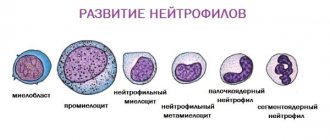
In adults, the limit is considered to be 4000 leukocytes per 1 microliter of blood. For children, the threshold value is calculated individually, taking into account age. Experts distinguish several types of leukopenia depending on which white blood cells are insufficient (lymphocytes, neutrophils, monocytes, etc.). Most often, a deficiency of neutrophils is diagnosed, which are responsible for protecting the body from bacterial and fungal infections; in this case, a diagnosis of neutropenia is made. In second place in the frequency of cases is a lack of lymphocytes, which provide protection against viral diseases.
Leukopenia in a child: causes
A low white blood cell count usually means that the body is not producing enough white blood cells. It is important to remember that this can increase the risk of developing all kinds of infections.
Common reasons that cause low white blood cell count are as follows:
- Treatment of cancer through radiation therapy.
- Taking antipsychotic drugs.
- Taking medications to reduce thyroid activity.
- Development of infectious diseases such as HIV or hepatitis.
- Autoimmune disorders such as rheumatoid arthritis.
Certain populations of Afro-Caribbean and Middle Eastern descent are often more prone to low white blood cell counts. Also, sometimes a low number of white blood cells in the blood is inherited. Such cases are considered normal and do not increase the risk of infection, so drug correction is not required.
Video: Clinical blood test - School of Dr. Komarovsky
Reasons for the development of leukopenia
A decrease in the number of white blood cells in the bloodstream can be due to three main reasons:
- Disruption of the process of formation of leukocytes. The latter are produced in the thymus gland, lymph nodes, spleen, tonsils, intestinal lymphoid nodules and bone marrow tissue - problems in the functioning of any of these organs can lead to disruptions in the production of blood cells.
- Destruction of leukocytes.
- Impaired circulation of leukocytes in body tissues.
The listed disorders can be caused by hereditary factors. These are congenital pathologies that impair the functions of the bone marrow, such as Kostman's syndrome - in such cases they speak of primary leukopenia, which is quite rare. A more common option is secondary (acquired) leukopenia. One of the most common factors causing the development of leukopenia is radiation or chemotherapy, which disrupts the production of mature leukocytes. However, there are other reasons:
- Autoimmune disorders accompanied by the destruction of bone marrow cells and/or leukocytes.
- Blood diseases (acute leukemia, anemia).
- Viral infections that deplete leukocyte reserves (the body does not have time to reproduce new blood cells).
- Taking a number of medications. These include antibiotics, cytostatics, anti-inflammatory drugs (such as analgin), as well as anticonvulsants, neuroleptics, etc.
- The effect of toxic substances such as carbon monoxide, mercury, lead.
- The effect of negative physical factors, for example, ionizing radiation.
Leukopenia often accompanies the following diseases:
- HIV infection;
- tuberculosis;
- systemic vasculitis;
- joint diseases (rheumatoid arthritis, etc.);
- inflammatory bowel diseases (Crohn's disease, etc.);
- multiple sclerosis;
- endocrine disorders.
A decrease in the number of leukocytes can also be observed after a blood transfusion and during protein starvation (malnutrition, lack of vitamins).
In children, the most common causes of secondary leukopenia are infectious diseases such as influenza, measles, and mumps.
Medical Internet conferences
Early diagnosis of oncohematological diseases in children is extremely difficult due to the nonspecificity of primary symptoms, which are often hidden under the “masks” of other diseases [2, 27]. Oncological alertness and knowledge of probable symptoms will allow the practitioner to diagnose this pathology earlier and, therefore, significantly improve the prognosis for the patient [1, 3, 4, 5].
In most children, acute lymphoblastic leukemia (ALL) manifests rapidly and is characterized by clinical polymorphism. The preleukemia stage does not have characteristic clinical symptoms [6, 7], and the analysis of the symptoms of the initial period is carried out retrospectively [7], and therefore the diagnosis is not made on time. Despite the progress of laboratory diagnostics, it is difficult to detect leukemia in children in the early stages, because For characteristic changes in the hemogram to appear, the tumor must reach a critical mass [4], and the manifestation of ALL is associated with blast infiltration of various organs with disruption of their function [8]. Clinical symptoms in this case are ahead of laboratory changes, and the child is not hospitalized in a specialized hematology hospital [55].
ALL occurs predominantly in children aged 0–15 years, with a peak incidence observed at the age of 2–5 years [54]. A more favorable prognosis and effect of therapy is observed in children from 1 to 10 years of age, while in children younger than 1 year and older than 10 years, ALL has an unfavorable course and is difficult to treat [46].
Gender differences:
Studies aimed at studying gender differences in ALL showed that in B-ALL the gender distribution was the same: boys - 51%, girls - 49%, while the T-variant was found predominantly in boys (90%).
Constitutional features. There is data in the literature on the connection between the onset and course of the initial period of ALL with the constitutional type of the child (asthenoid, thoracic-muscular, digestive): the most severe course is observed in children with a digestive body type [55].
Predisposing factors. When diagnosing ALL, the doctor must take into account a wide range of predisposing factors that can lead to the development of ALL: socio-economic status of the child’s family, chromosomal abnormalities, Down syndrome, neurofibromatosis, variable immunodeficiency, Ig A deficiency, Fanconi anemia, Shwachman syndrome, congenital X- linked agammaglobulinemia [56].
In the clinical picture of acute lymphoblastic leukemia, 5 main syndromes are most common (Table 1):
- Intoxication syndrome. Most often, patients are concerned about prolonged fever of unknown origin, intoxication of varying degrees, weakness, fever, malaise, and weight loss. Fever may also be associated with the presence of a bacterial, viral, fungal or (less commonly) protozoal infection, especially in children with neutropenia [7].
- Osteoarticular syndrome. Manifestations from the musculoskeletal system are detected mainly in children with B-cell ALL, which is characterized by less involvement of extramedullary organs and unclear changes in the peripheral blood [18]. Osteoarticular syndrome is often the first manifestation of ALL in children [9, 10]. Studies have shown that in patients with ALL, osteoarticular syndrome was recorded in 54% of cases, with a higher frequency in children from 1 to 9 years of age. The presence of joint pain was noted in 16.2% of children with ALL, arthritis in 26.6%, and changes in gait in 32.8%. Large joints were most often affected: knees - 10.6%, ankles - 9.4%, elbows - 4.4%, shoulders - 3.6% [11]. Upon examination, swelling of the joints, the presence of effusion, and dysfunction of the affected limb were revealed [12]. Patients often complained of frequent and multiple fractures, which may be due to osteoporosis [13]. Clarifying diagnosis is also necessary if recurrent multifocal osteomyelitis is suspected [14]. Acute lymphoblastic leukemia in rare cases manifests itself as osteoarthritis, the incorrect interpretation of which can lead to errors in differential diagnosis with juvenile rheumatoid arthritis (JRA). Children with ALL have asymmetric oligoarthritis, which is manifested by fever, pallor, arthritis, night pain, and bone pain [19]. Damage to the skeletal system at the onset of ALL is characterized by polymorphism of radiological manifestations. In some cases they are uninformative (only a slight periosteal reaction is revealed) [15], in other cases osteolysis, osteopenia, osteosclerosis, pathological fractures, periosteal reactions and mixed lesions in the form of lysis-sclerosis were detected [16]. An important symptom is the discrepancy between clinical symptoms and radiological data. The combination of hyperthermia, intoxication and articular syndromes can mimic septic arthritis. However, the lack of an adequate response to antibacterial therapy allows one to suspect ALL. The pathological process in this case develops as a result of metaphyseal sclerosis [3]. Intractable pain and joint syndromes characteristic of juvenile idiopathic arthritis must be considered from the standpoint of differential diagnosis with ALL [16]. Similar clinical symptoms in combination with pancytopenia are characteristic of necrosis of the bone marrow substance [17]. These data highlight the importance of including ALL in the differential diagnosis of musculoskeletal disorders even in the presence of apparently normal peripheral blood findings.
- Lymphoproliferative syndrome. Enlarged lymph nodes are associated with leukemic tissue infiltration [57]. The most often enlarged cervical, inguinal, axillary lymph nodes, which have a dense elastic consistency, are painless on palpation, are not fused with the surrounding tissue, are mobile, the skin over them is not changed. Some children may experience Mikulicz's symptom complex (simultaneous enlargement of lymph nodes in the submandibular, parotid and periorbital region). There may be a significant increase in the lymph nodes of the mediastinum, up to compression of the superior vena cava (shortness of breath, cyanosis, puffiness of the face, pasty eyelids, bulging of the jugular veins). Along with lymphadenopathy, in 75% of cases hepatosplenomegaly is detected, which is not accompanied by pain [2, 6].
- Anemic syndrome is characterized by increasing pallor with worsening general condition, dizziness and headache, shortness of breath, tachycardia [2].
- Hemorrhagic syndrome is associated with both thrombocytopenia and intravascular thrombosis (especially with hyperleukocytosis) [46]. Patients may complain of polymorphic hemorrhages (from petechiae to large hemorrhages) on the skin and subcutaneous tissue of various locations. Bleeding from the mucous membranes (nasal, gingival, gastrointestinal, renal, uterine) is observed [2, 58].
, clinical manifestations from other organs may be observed, and in some cases predominate [27] (Table 1).
- Lesions of the mucous membranes. One of the early symptoms of ALL in children is lesions of the oral mucosa in the form of erythema, ulcers and swelling of the lips, tongue, palate and gums [21]. Often the diagnosis is accompanied by difficulties in differential diagnosis with surgical pathology of the face. Thus, a case of ALL was described in a girl with swelling of the nasolabial area, which was regarded as phlegmon. The changes described above were associated with infiltration of the mucosa and soft tissues by blast cells [22].
- Skin manifestations. ALL in children in some cases manifests itself in the form of various skin manifestations, which are usually based on a leukemic infiltrate with damage to the skin and subcutaneous tissues. Localization can be different: the head area is affected in the form of small nodules [23], or other areas of the skin [24]. A case of skin lesions in the external auditory canal is described, in which the child was initially diagnosed with diffuse external otitis media [25].
- Eye lesions. Ophthalmological manifestations in patients suffering from acute lymphoblastic leukemia may also be its first manifestation [26, 27]. Complaints and clinical manifestations differ in polymorphism [28]. The most common sign of eye damage in ALL is hemorrhages in various parts of the eyeball: retina, subconjunctival hemorrhages [29]. When examining the fundus, swelling of the optic nerve nipple and vascular infiltration may be detected.
- Abdominal syndrome. Abdominal syndrome manifests itself in the form of abdominal pain of unknown etiology, alternating diarrhea and constipation, which is associated with lymphoid infiltration of parenchymal organs and the intestinal wall. In rare cases, ALL can be hidden under the “mask” of acute pancreatitis, which is difficult to treat [30], as well as typhlitis [31], which requires additional differential diagnosis.
- Damage to the genitourinary system. Clinical manifestations of kidney disease caused by malignant infiltration may be the primary manifestation of the disease in patients with ALL and are represented by acute renal failure [32, 33], renal arterial hypertension [34], palpable mass syndrome and bilateral nephromegaly, which must be differentiated from Wilms tumor [ 35, 36]. A feature of the clinical picture of acute renal failure in such cases is the absence of oliguria against the background of severe nephromegaly [37, 38]. In addition, there is evidence of the primary manifestation of ALL in the form of nocturnal enuresis in a child [39].
- Neurological symptoms. Neurological symptoms are not only a sign of the development of neuroleukemia in children with ALL, but may also be the only early clinical manifestation of the disease. The clinical picture is predominantly associated with damage to the cranial nerves (CN). In one case, with damage to the motor branch of the trigeminal nerve in a child, the only symptom was trismus of the masticatory muscles [40], and in another, with damage to the sensory branch, sensory neuropathy (Numb chin syndrome) was noted [41]. The primary manifestation of ALL in a number of cases was an isolated lesion of the abducens nerve with corresponding oculomotor symptoms [42, 43]. In addition to lesions of the cranial nerve, manifestations of neurodegeneration (Louis-Bar ataxia-telangiectasia) with corresponding immune and hematological disorders can also be observed in the clinic [44]. ALL can debut as a paraneoplastic neurological syndrome (PNS), manifested by acute muscle weakness of the proximal muscles of the upper and lower extremities [20].
- Lung damage. Respiratory system disorders may be associated with enlarged mediastinal lymph nodes, characteristic of T-cell leukemia, leading to the development of superior vena cava syndrome or respiratory failure. There may be leukemic infiltration of the lung tissue and/or hemorrhages into it. It is sometimes difficult to differentiate these complications from an infectious process [45].
- Disorders of mineral metabolism. Disorders of mineral metabolism that develop in ALL are characterized by hypercalcemia, which can be the first symptom of the disease [50, 51] or its complication [52]. In patients with hypercalcemia, complaints from the gastrointestinal tract and osteoarticular system prevail [53].
- Infectious diseases. In some cases, the onset of ALL can occur under the “mask” of some infectious diseases, for example, acute respiratory viral infection, infectious mononucleosis, tonsillitis, whooping cough, parawhooping cough, pneumonia, mumps [59, 60, 61, 62].
Hematological changes. The most important diagnostic test to suggest ALL is a complete clinical blood test with a leukocyte count.
Hematological changes in ALL do not have clear patterns, and often their severity may not correspond to clinical symptoms; changes in blood tests characteristic of ALL may be absent, which indicates a comprehensive assessment of clinical and laboratory data.
Most often, a hemogram reveals pronounced leukocytosis, while the number of leukocytes varies widely and depends on the type of ALL. Thus, the average number of leukocytes in children with the B-variant ALL was 37.1 ± 12.2*109/l, and in the T-variant ALL – 123.3 ± 36.5*109/l [63]. It is noted that forms of ALL with hyperleukocytosis have a more unfavorable prognosis [46].
Along with leukocytosis, blastemia is observed up to 80-90% [27], and a small percentage of blast cells does not provide grounds for making a diagnosis of ALL, while their absence does not allow it to be completely excluded [47, 64].
In rare cases, persistent unexplained hypereosinophilia (up to 80%) is observed at the onset of the disease, which can be combined with skin manifestations [48], and in 25-30% of cases - lymphocytosis. The cytopenic variant of ALL is characterized by leukopenia and absolute neutropenia [4]. An increase in the leukocyte pool leads to inhibition of other hematopoietic germs, which is accompanied by a decrease in the number of red blood cells and hemoglobin, as well as thrombocytopenia [27, 46, 65, 66].
A number of studies contain data on the development of metabolic lactic acidosis in the initial period of ALL [49].
Thus, the primary clinical manifestations of acute lymphoblastic leukemia in children are characterized by pronounced polymorphism, which requires doctors of all specialties to be oncological vigilant, a comprehensive assessment of the anamnesis, consistent analysis of clinical data from all body systems, and the use of an adequate set of laboratory and instrumental studies. In doubtful cases, the child should be referred for consultation to a pediatric hematologist-oncologist.
Symptoms
Leukopenia is often discovered by chance, for example, during preventive medical examinations, since a person may not notice any disorders for a long time. The first signs of leukopenia may be:
- headache;
- dizziness;
- feeling of weakness;
- cardiopalmus;
- bleeding gums, mouth ulcers;
- hoarse voice.
As the pathology progresses, the following are observed:
- increased body temperature;
- fever, chills;
- enlarged tonsils and spleen;
- exhaustion of the body.
High susceptibility to infectious diseases is another symptom that may indicate leukopenia.
Depending on the number of leukocytes, three stages of pathology are distinguished: first (less than 1.5 g/l), second (1 g/l) and third (0.5 g/l). At the third, most severe stage, called agranulocytosis, thrombocytopenia may develop, a pathology characterized by a decrease in the number of platelets, which threatens bleeding and hemorrhage.
Agranulocytosis is a severe pathology in which the number of leukocytes and granulocytes sharply decreases. Since granulocytes provide antimicrobial protection to the body, a critical decrease in their number can lead to serious infectious complications, including death. A patient with this diagnosis is prescribed hospitalization and emergency treatment in the hematology department, with the obligatory creation of aseptic conditions.
Acute leukopenia can last up to three months; for longer periods, a diagnosis of chronic leukopenia is made.
Leukopenia in children: symptoms
Symptoms depend on the type of quantitative change in white blood cells in the child. Common signs of a condition where there are too few white blood cells or they are not functioning properly include the following:
- Frequent infectious diseases, mainly affecting the lungs, ears or paranasal sinuses.
- Abscesses of the skin.
- Ulcers in the mouth.
- Invasive fungal diseases.
- Severe damage to teeth and gums (periodontal disease).
Brief clinical characteristics:
- Ear infections cause pain in the affected ear.
- Sinus infections can cause headaches, difficulty breathing, or coughing.
- Pneumonia is an inflammation of the lungs that can cause chills, cough, shortness of breath, and muscle pain.
- A bladder infection often causes fever, nausea, or pain when urinating.
Some disorders may present with unique symptoms, such as:
- Delayed wound healing due to deficiency of leukocyte adhesion.
- Chronic, sometimes fatty, diarrhea, which is characteristic of Shwachman-Diamond syndrome.
Less typical signs include weakness, fatigue, dizziness, chills and trembling, but their appearance should not be ignored. If your child has even the usual symptoms of a cold, you should contact your pediatrician. If necessary, the doctor will refer you to a hematologist for further research, and if necessary, prescribe medications.
Leukocyte adhesion deficiency
Diagnostics
Clinical tests of blood elements help make an accurate diagnosis. A general practitioner can prescribe the necessary tests. To determine the level of leukocytes, it is necessary to donate venous or capillary blood in the morning on an empty stomach.
First of all, hemograms are carried out to count the number of different types of blood cells (erythrocytes, leukocytes, etc.) and determine their percentage. To determine the type of leukopenia and the cause of its occurrence, cytological studies are prescribed. Comprehensive diagnostics also includes biochemical and immunological blood tests, as well as a blood test for hepatitis. In each case, a complete list of necessary laboratory tests is established taking into account the medical history of the individual patient.
Treatment
The regimen and drugs for the treatment of leukopenia are selected depending on which blood cell levels have dropped below normal and what caused this. As a rule, the patient is prescribed combination therapy, which involves taking medications, a special diet and lifestyle changes.
Standard treatment includes taking the following:
- improving metabolism;
- increasing the number of neutrophil granulocytes;
- stimulating the activity of bone marrow stem cells and other cells involved in the production of leukocytes.
Since leukopenia most often acts as a secondary pathology, it is first of all important to eliminate the root cause:
- If leukopenia is caused by taking medications, a decision may be made to urgently discontinue or replace them.
- If a deficiency of blood cells is a consequence of any disease, you must first begin to treat it. So, to achieve remission in autoimmune disorders, immunosuppressants and glucocorticosteroids are used. In the case of oncohematological pathology, chemotherapy drugs are prescribed. Patients with bacterial infections are prescribed antibiotics, etc.
- In the case of genetic abnormalities, as well as patients taking chemotherapy, they are additionally prescribed growth factors (proteins) that stimulate the production of leukocytes.
In addition to drug treatment, you need:
- Normalize your diet by adding foods rich in vitamins and minerals, in particular vegetables and fruits, to your menu. If leukopenia is diagnosed, the doctor may additionally prescribe vitamins B6, B2, B12, as well as folic acid, etc.
- Provide adequate rest.
- Create aseptic conditions. It is important to observe sanitary and hygienic standards and avoid injuries (cuts, scratches) through which infection can enter the body.