When can you suspect dysplasia?
There are many options for defining dysplasia, but their accuracy is questionable:
- The most popular method, which, perhaps, any mother has heard of, is based on the asymmetry of skin folds. Each person is an individual, so the development of subcutaneous tissue is not an indicator at all.
- Different leg lengths. It is rare to suspect dysplasia based on this criterion. Few people are able to see the difference with the naked eye, because a lot depends on the baby’s positioning.
- Limitation of leg mobility. This is noticed in children older than one year, but especially vigilant grandparents can pay attention earlier. The reason is a small diaper, an uncomfortable mattress and a number of other nuances. This method is not reliable.
The “gold standard” for diagnosis is considered to be radiographic examination, which dots all the i’s. It must be taken at three months for all children, without exception, for timely diagnosis of the disease.
Ultrasound examination is considered a more modern technique. It is often used in hospitals, but this is just a waste of money and time, because the data obtained does not show an accurate and complete picture of the pathology.
Requirements for radiographs
In order for the resulting images to serve as evidence of the absence of joint problems, they must meet several basic requirements:
- show clear outlines of bones and their architecture, including the superior edge of the acetabulum;
- display the shadow of the pelvis and knee joints, and so that the images of the left and right iliac crest are at the same level, and the shadows of the knee cups are located opposite the block.
If the photographs are taken in violation of the above standards, an international certificate will not be issued for them. You will have to pay money again for re-diagnosis.
Did you know? Dog nose prints are just as unique as human fingerprints. No two pets have the same patterns.
Types of dysplasia
The dysplasias themselves are divided into several types: A mature joint is included in categories 1a and 1b and does not require treatment.
| View | Type | Description |
| Immature joint | 2a | Pre-dislocation of the hip occurs when the joints are immature. Up to 3 months this is the norm for most children. There are no serious violations, but observation is necessary. Types 2b and 2c are subject to correction. |
| 2b | ||
| 2v | ||
| Subluxation | 3a | With subluxation of the hip joint, x-rays show displacement of the head upward and to the side. She is still in contact with the socket, but treatment is necessary. |
| 3b | ||
| Dislocation | 4 | The head of the femur is outside the socket. Surgery required |
A mature joint is included in categories 1a and 1b and does not require treatment.
For hip dysplasia type 2a, treatment is not carried out until 3 months of age, as this is considered normal.
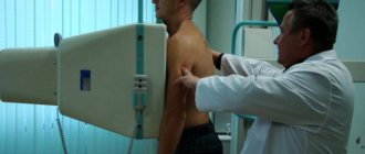
Preparing the child and technique of the procedure
There is no need to carry out any specific preparatory measures before taking x-rays of the hip joints. For very young patients, you need to follow a normal sleep and feeding schedule. For children older than a year or a year and a half, you can clearly and generally explain what will happen during an x-ray, tell them that the upcoming procedure is painless and there is no need to be afraid of it.
The child lies on his back during the x-ray. In this case, the patient’s legs should be straight and straight and should not be bent. In newborn babies, the legs move closer to the body and move slightly inward.
If the child’s pelvis is tightly pressed by the cassette to the plane of the couch or table, his movements will not interfere with the recording of the image. The genitals are covered with a protective lead apron to prevent exposure to X-ray radiation.
If there are no contraindications to the use of anesthesia, the procedure can be performed with the young patient under general anesthesia to ensure the accuracy and quality of the images.
Why is hip dysplasia dangerous?
Dysplasia is a pathology. It is a mistake to think that it does not have serious consequences. With the disease, the entire hip joint is formed incorrectly, and it is one of the largest and most important in the entire body. It ensures an upright posture and normal gait.
With dysplasia, the head of the femur is positioned incorrectly in relation to the acetabulum. As a result, gait is disrupted:
- may become a “duck”;
- there is excessive “swaying of the hips”, the gluteal muscles are “higher”;
- with unequal limb lengths, lameness develops.
Cartilage tissue is formed gradually; with dysplasia, this process is disrupted. The pelvic bone also undergoes changes. As a result, the joint loses some of its functions, which can lead to disability.
Pathology affects the entire musculoskeletal system. The load on him increases, the person constantly gets tired, and muscle weakness develops.
In nine out of ten clinical cases, patients who do not receive the necessary treatment in a timely manner develop a number of complications:
- coxarthrosis;
- impaired motor function of the leg;
- cosmetic deformities;
- neoarthrosis (formation of a new pathological joint).
Particularly dangerous is coxarthrosis - this is arthrosis of the hip joint. The disease destroys cartilage tissue and adjacent bones. As a result of coxarthrosis, the patient cannot walk normally and experiences constant pain.
Effective methods of treating the cervix. Radio wave surgery. Photodynamic therapy.
To treat cervical dysplasia, surgical treatment methods are used, i.e. removal of altered areas of the cervix with electric, laser or radiosurgical scalpels, the latter is most preferable, since it practically does not cause damage to the deep structures of the cervix and, as a result, there is no scar after exposure.
Radio wave surgical treatment of dysplasia (Surgitron device (USA)) is the “gold standard” for the treatment of cervical diseases - it is an atraumatic method of incision and coagulation of soft tissue using high-frequency waves. The dissecting effect of a radio wave is achieved due to the heat generated by the electrical resistance of the cervical tissues to the penetration of directed high-frequency waves into them. Moreover, pathologically altered and dying cells have the greatest electrical resistance, so they are the first to be destroyed. In this case, there is no direct contact of the electrode with the cells, and the electrode itself does not heat up. The high frequency energy is concentrated at the tip of the "active" or "surgical" electrode and causes a burst of intracellular molecular energy that heats the tissue and essentially vaporizes the cells.
As a result of radio wave exposure, tissue destruction is several times less than when using any other electrosurgical equipment or surgical laser.
The use of a radio wave scalpel for excision of a pathological focus is not accompanied by damage to healthy tissue, there are no postoperative consequences such as swelling, inflammation, the risk of severe cicatricial deformation of the cervix is minimized, and therefore the method can be used in the treatment of nulliparous women.
Very important! With radio wave surgery, coagulation and incision are performed without the tissue destruction caused by the use of electrosurgical low-frequency devices, such as electrocoagulation or diathermocoagulation. The advantages of the radio wave surgical method for treating cervical diseases are: controlled minimal damage to healthy tissue; minimal pain during surgery; no bleeding; fast healing; Possibility of use in nulliparous women.
IMPORTANT: With proper radio wave surgery using the Surgitron device, unchanged areas of tissue are practically not damaged and the normal anatomy and physiology of the cervical canal is preserved, which has the most beneficial effect on subsequent pregnancy and childbirth.
For young women who want to realize their reproductive plans in the future, maintaining the normal structure of the cervix is extremely important. For such patients, an even more ideal method of treating cervical diseases is photodynamic therapy. With this method of treatment, the integrity of the cervix is not compromised. A drug is introduced into the body - a photosensitizer, which accumulates only in pathological cells of the cervix, then, under the influence of laser energy, the cells that have accumulated the sensitizer are destroyed. The clinical effect in most cases is achieved after a single procedure. The effectiveness of the technique is so high that it is also used in the early stages of cervical cancer (in situ and stage IIB1).
Treatment of hip dysplasia
The most important thing in treatment is timely diagnosis. Ladisten uses only modern equipment to determine the exact angle of displacement and the degree of pathology. At the initial stages, treatment of dysplasia is possible with the help of special orthopedic structures: Pavlik stirrups, Gnevkovsky apparatus, Freik pillow, etc. This is done up to twelve months. Constantly wearing the structures is an integral part of treatment. If there are no positive results by one year, surgical intervention is recommended.
There are many articles on the Internet about the treatment of dysplasia: from wide swaddling to invasive procedures. Remember, at the slightest suspicion of a congenital dislocation, rush to consult a professional, and do not practice traditional medicine. This may make the situation worse.
There is no time to waste with this pathology. An experienced orthopedic doctor will accurately and accurately diagnose the disease and prescribe the correct treatment.
Advantages of contacting SM-Clinic in St. Petersburg
We employ some of the best specialists in the Northern capital. The examination is carried out using high-quality Siemens Sireskop equipment. This device is specially designed to allow radiography of the youngest children, and it also minimizes the radiation dose - depending on the area of study, it is 0.03-0.1 mSv.
There are no queues in our medical center, patients feel comfortable and calm. Call to find out prices for the services you are interested in or to sign up for a procedure. Registration is also possible through the website.
Treatment of dysplasia in children
In most cases, the diagnosis of hip dysplasia is made in the first year of life. Up to 3-4 months this is a natural condition; it goes away on its own in 80% of cases. But 20% of the pathology always remains, so the baby is observed by an orthopedist. Inspection is required at 3 months, 6 months, and a year. If there is pathology, you need to see a doctor more often. Timely measures give a positive treatment result almost immediately.
How to treat dysplasia in children:
- massages, gymnastics, physiotherapy and wearing stirrups are recommended for infants. As a rule, this therapy is sufficient. Time plays a major role: the sooner you put on the stirrups and do the procedures, the more favorable the outcome. Orthopedists try to do this as early as 4-6 months, before the child begins to sit and walk. It is important not to put the baby on his feet during this period. Parents can provide wide swaddling on their own;
- if the situation does not improve in the first year, the baby is constantly monitored. Conservative treatment can still be continued for several months. But this significantly delays development, because the child should already be walking. A positive result is achieved by surgery to reduce the dislocation or osteotomy;
- Treating hip dysplasia in adolescents is difficult. Conservative methods no longer help. The only way out is surgery. In the case of developed coxarthrosis, it is also eliminated in parallel.
If your child suffered from dysplasia at an early age, continue to see your orthopedist every year. This will make it possible to notice violations in time.
Diagnosis of cervical dysplasia
In addition to the mandatory cytological examination of smears from the cervix, an extended colposcopy or examination of the cervix under high magnification is carried out using specific tests and determination of HPV. In cases of detection of changes according to cytological examination or the presence of atypical visual changes in the cervix according to extended colposcopy, a more in-depth diagnosis is carried out, including histological examination of cervical biopsies and scrapings of the cervical canal mucosa and cervicoscopy.
List of examinations necessary for diagnosing and adopting the correct treatment tactics for cervical dysplasia:
- Extended colposcopy;
- Histological examination of cervical biopsies;
- Histological examination of scrapings from the cervix;
- Smear on vaginal flora;
- Quantitative determination of HPV;
- Ultrasound of the pelvic organs;
- Therapist's conclusion about the absence of contraindications.
Additionally from examinations, according to indications:
- Cervicoscopy;
- Extended examination for urogenital infections;
- MRI of the pelvic organs;
- Immunohistochemical examination of biopsy specimens and scrapings of the cervix;
- Blood test for immuno-interferon status;
- Histological examination of aspirate from the uterine cavity;
- Hysteroscopy and separate diagnostic curettage of the uterus;
- Special examinations in the presence of concomitant pathology.
Can hip dysplasia be cured with massage?
Massage plays an important role in the treatment of dysplasia, but not the leading one. The purpose of the massage is to normalize muscle tone, eliminate contracture of the adductor muscles, create range of motion in the joint, improve trophic processes, and create conditions for the normal position of the femoral head.
It is impossible to cure dysplasia with massage alone. There are main factors that influence the result:
- Patient's age. Dysplasia in infants from 3 months to a year can be cured with massage. But it needs to be done together with gymnastics. At an older age, massage does not help.
- Degree of dysplasia. For dysplasia types 2b and 2c, as well as congenital hip dislocation, massage is not the main treatment. But it can be used in combination with physiotherapy, stirrups and splints. If the patient has undergone surgery, then massage is necessary during the recovery stage. It significantly speeds up this process.
Consider this important point! When performing a massage, you do not just make movements, but feel the anatomical location of all structures, feel the joint, calculate the force of pressure and the angle of rotation. Can a person without special qualifications do this? Therefore, you should not resort to massage yourself. An incorrectly performed procedure can only cause harm.
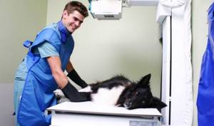
How is the procedure performed?
An X-ray examination to determine the tendency to develop dysplasia consists of two main stages: preparation and the procedure itself. Before the x-ray, the doctor must examine the dog and identify it by its mark or chip. It is not recommended to feed the animal 8–10 hours before the start, and the day before this it is worth minimizing physical activity and ensuring that the pet’s emotional state remains stable. When visiting a veterinary clinic, you should have the dog's pedigree and a muzzle with you if it may behave aggressively.
The reliability of the research results directly depends on the calmness and maximum relaxation of the dog, so a sedative is administered through an intravenous catheter. After this, the pet is placed on its back with its limbs extended parallel and a photo of the right and left sides is taken.
Important! During the shooting, the x-ray image must contain an identification number stamped using an x-ray stencil. The pedigree number, dog breed, and date of the procedure are also noted.
X-rays of the elbow joints are taken in two positions: the mediolateral projection (the joint bent at an angle of 45 degrees) and the craniocaudal projection, when the dog lies with its paws extended parallel to the front. The examination of the right and left joints is performed alternately.
After a short period of relaxation in the relaxation room, the animal quickly recovers and after 1-2 hours is able to leave the clinic on its own. The doctor can only fill out all the accompanying documents for the RKF and burn the digital images onto a disk for transfer to the owner.