directions
Almost every person has experienced pain in the lumbar region. A sedentary lifestyle, especially among office workers, drivers and other professionals who spend a lot of time in a sitting position, excessive physical activity, improper exercise and other negative factors contribute to the occurrence of pain in the lumbar region, but are not the exhaustive causes of ailments. If discomfort in the lumbar region begins to torment you, the pain becomes severe and regular and lasts more than 3 months - this is a clear signal to seek help from a doctor. After all, very often back pain, especially in the lumbar region, can indicate problems not only of the spine, but also of the kidneys and other organs located nearby. Only a doctor is competent to identify the cause of the disease, make a correct diagnosis, and prescribe effective treatment. Most often, in case of prolonged pain in the lumbar spine, you will be offered an X-ray examination of the lower back.
Our clinics in St. Petersburg
Structural subdivision of Polikarpov Alley Polikarpov 6k2 Primorsky district
- Pionerskaya
- Specific
- Commandant's
Structural subdivision of Zhukov Marshal Zhukov Ave. 28k2 Kirovsky district
- Avtovo
- Avenue of Veterans
- Leninsky Prospekt
Structural subdivision Devyatkino Okhtinskaya alley 18 Vsevolozhsk district
- Devyatkino
- Civil Prospect
- Academic
For detailed information and to make an appointment, you can call +7 (812) 640-55-25
Make an appointment
We draw your attention to the schedule of technological breaks in the CT and X-ray rooms.
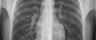
X-ray of the lumbar spine today, when X-ray diagnostic equipment is available in almost every clinic and medical center, is one of the most popular, quick and simple methods of diagnostic research.
If you are looking for a clinic in St. Petersburg with good and safe X-ray equipment, then in the Medicenter network of multidisciplinary clinics you can do an X-ray examination of the lumbar spine. The trauma department of the Medicenter accepts patients seven days a week and on holidays. The department is equipped with high-tech X-ray equipment made in Italy, Clinomat, which allows you to quickly, efficiently and effectively take X-rays of various areas of the body, and study the results obtained in various processing modes. Qualified doctors - surgeons, traumatologists, neurologists, orthopedists and other specialists at the Medical Center will provide you with first aid for injuries, conduct a thorough examination, conduct the necessary examinations and prescribe a course of treatment and recovery.
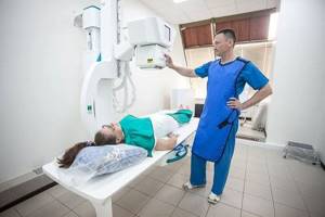
How is the procedure done?
X-rays of the lumbosacral spine may be performed on an outpatient basis or as part of a hospital stay. Procedures may vary depending on the patient's condition.
Typically, X-ray examinations of the spine follow the following process:
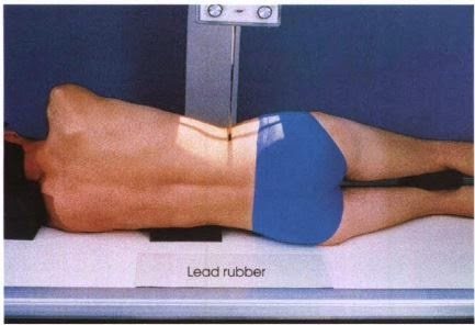
- The person will be asked to remove any clothing, jewelry, hair clips, glasses, hearing aids, or other metal objects that may interfere with the procedure.
- The patient is positioned on an x-ray table, which carefully positions the part of the spine to be x-rayed between the x-ray machine and an x-ray film cassette or digital media. Your doctor may also require that x-rays be taken from a standing position.
- Parts of the body that do not need to be examined may be covered with a lead apron (screen) to avoid exposure to X-rays.
- The radiologic technologist will ask the patient to remain still in a certain position for several minutes during the X-ray exposure.
- If x-rays are used to determine injury, special attention will be given to preventing further injury.
- The X-ray beam will be focused on the area to be photographed.
- An x-ray technician stands behind a protective window during an examination.
Although the X-ray procedure itself does not cause pain, manipulation of the body part being examined may cause some discomfort or pain. This is especially true if there has been a recent injury or an invasive procedure such as surgery.
Indications for X-ray examination of the lumbar spine
Of course, not all patient complaints of lower back pain require an x-ray of the lumbar spine. But there are symptoms and suspicions of certain ailments for which an X-ray of the lower back is indicated:
- prolonged pain in the back, lower back, limbs;
- hernia formations;
- neoplasms, protrusions;
- tumor processes;
- feeling of numbness in the limbs;
- Schmorl's hernia;
- injuries of various types;
- birth injuries;
- congenital anomalies;
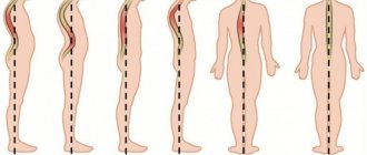
- curvature of the spine;
- movement disorders;
- preoperative control;
- monitoring the effectiveness of treatment, etc.
Purpose of X-ray
- Essentially, x-rays of the lumbosacral spine are used to evaluate back injuries as well as low back pain.
- This method is also used to identify problems in the lower back if there is weakness and persistent numbness.
- X-rays of the lumbosacral spine are used to detect other back injuries such as bone spurs, spinal deformities, fractures, spinal dislocations, osteoporosis, and slipped discs.
- Another goal of the study is to identify the cause of low back pain and numbness.
Preparation for x-ray of the lumbar spine
In order for the examination to be effective and the x-ray to be accurate, it is necessary to follow certain recommendations before taking the x-ray. A few days before an x-ray of the lumbar spine, it is important to follow a diet. Foods and drinks that cause increased gas formation (some vegetables, fruits, bread, carbonated drinks, etc.) should be excluded from the diet. The evening before the examination, you need to do a cleansing enema and repeat it in the morning before x-ray of the lumbar region.
What do abnormal results mean?
An X-ray of the lumbosacral spine may show:
- Abnormal curvatures of the spine
- Abnormal wear and tear on the cartilage and bones of the lower spine, such as bone spikes and narrowing of the joints between the vertebrae.
- Cancer
- Fractures
- Signs of thinning bones (osteoporosis)
- Spondylolisthesis
Although some of these findings may be visible on x-rays, they are not always the cause of back pain.
3. How to prepare and how is the research carried out?
How to prepare for a spine x-ray?
Tell your doctor if you may or may be pregnant. X-rays are a type of radiation, so your doctor may advise against taking an X-ray unless it is absolutely necessary. You may need to remove all jewelry and other iron objects just before the x-ray.
How is a spine x-ray performed?
X-rays of the spine are taken and interpreted by special doctors (radiologists), although many other doctors can also do this.
During the procedure, you will need to remove any clothing that might interfere with the X-rays and lie on your back. If you have had a neck injury and are wearing a collar, your doctor may examine you to make sure the collar can be removed. Typically 3 to 5 x-rays are taken, and you will need to try to remain calm to avoid blurring the pictures.
An X-ray of the spine takes about 15 minutes.
About our clinic Chistye Prudy metro station Medintercom page!
Risks
- The patient has the right to find out from the doctor about the amount of radiation that will be received during the study and the possible risks. The patient needs to record data on previous radiological studies, since X-ray radiation has a cumulative effect.
- If the patient is pregnant or suspects that she is pregnant, she should inform her health care provider due to the risk of birth defects in the baby. Therefore, it is better to replace x-rays with other diagnostic methods, such as MRI.
- Depending on the specific disease, there may be other risks.
X-ray of the spine
01/01/2020 One of the conservative methods for diagnosing injuries and diseases of the spine is x-ray.
This is the easiest and most inexpensive way to identify many problems associated with spinal deformity. Depending on the extent of the lesion and the location of the injury, there are several options for conducting such an examination.
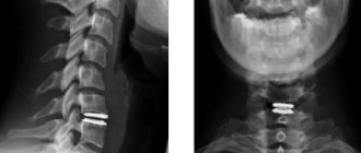
X-ray of the cervical spine
Indications for x-rays of the cervical spine are headaches or short-term dizziness during a sharp tilt of the head or turn of the neck.
Pictures are taken in two projections. In frequent cases, in order to take an X-ray of the cervical spine, the examination is carried out through an open mouth. Afterwards, the doctor analyzes the images and determines the severity of the disease. X-rays of the cervical spine do not require special preparation.
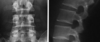
X-ray of the lumbar spine
For x-rays of the lumbar spine, preparation is necessary. How to prepare for a spine x-ray? Two days before the examination, you need to exclude from your diet those foods that provoke the formation of gases in the intestines, since such effects can distort the image. On the day before the examination, you should take medications to relieve flatulence and also skip dinner. An X-ray of the lumbar spine is performed on an empty stomach, having previously cleansed the intestines with an enema. This is the only way the picture will be as accurate and easy to read as possible. X-rays of the lumbosacral spine are also performed in the same mode.
X-ray of the thoracic spine
Pain in the chest or abdomen may be an indication for an x-ray of the thoracic spine. This examination is carried out without preparation. For higher information content and diagnostic accuracy, the image is taken in several projections. The results are analyzed by a radiologist. Then a neurologist or orthopedic traumatologist prescribes treatment if necessary.
X-ray of the spine is effective:
- in the diagnosis of intervertebral hernias
- when vertebral displacement is detected
- in the diagnosis of vertebral compression fractures
- when identifying tumors of various origins
- upon detection of infectious diseases (spinal tuberculosis)
- to identify spinal curvatures
- to confirm congenital pathologies of the spine (torticollis, sacralization)
How are spinal x-rays done?
In the X-ray room, you will be asked to remove your waist-length clothing and body jewelry. An x-ray of the spine will be informative if you followed all the rules for preparing for the examination and also carefully listened to all the commands of the doctor who performed the x-ray. It may ask you to turn several times, depending on the number of shots needed in different projections.
The frequency of the procedure is calculated by the radiologist depending on the severity of the disease and the radiation dose received. It is worth noting that modern X-ray machines are equipped with a program that significantly reduces the radiation dose per procedure. This allows examinations to be carried out more often and without much risk. But after the x-ray procedure, it would still be a good idea to ask the doctor to write down the received radiation dose in your card to calculate the possibility of further x-ray examinations.
X-ray is an inexpensive and effective method for diagnosing the condition of your spine, and therefore the condition of the entire body!
At the Ekaterinburg Medical Center, x-rays are performed on a universal digital radiography system AXIOM Aristos MX from Siemens, which makes it possible to obtain:
- minimum safe radiation dose
- high quality x-ray images
- digital recording
- low cost of x-ray
The X-ray room is located in the branch on Shevchenko, 9.
The cost of X-rays can be found in the Services and prices section
Make an appointment for an x-ray in Yekaterinburg by phone: 8 (343) 379-07-70 or through the clinic’s website.
There are contraindications. Read the instructions or consult a specialist.
Before the procedure
- The attending physician should explain the procedure to the patient and answer any questions they may have about the procedure.
- As a rule, no preliminary preparation is required for an X-ray of the spine.
- It is necessary to inform the radiologist if there is or is suspected pregnancy
- It is important to notify the radiologist of a recent barium study as this may interfere with optimal image quality.
- Depending on your health, your doctor may recommend certain preparations.
Contraindications to undergoing the study
Like any other study, X-ray diagnostics has certain contraindications. It is highly not recommended for women to have X-rays during pregnancy, since irradiation of the fetus can lead to serious pathologies in development, fetal death, and premature birth.
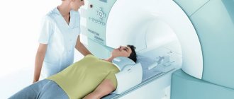
Women during pregnancy (if urgently needed) are recommended to undergo an MRI - this method is both safe and more informative.
Women of childbearing age are recommended to undergo X-ray diagnostics of this part of the spine in the first two weeks after the end of menstruation - in order to avoid a situation where the woman does not yet know about pregnancy, and thereby avoid possible exposure of the fetus.
Among the contraindications, it is also worth mentioning the recent presence of X-ray diagnostics using barium suspension.
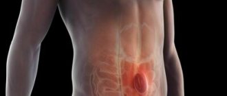
When should an x-ray of the coccyx and sacrum be taken?
Doctors order x-rays after falls on the buttocks, especially in cases where it becomes painful for the patient to sit or stand up. Pain in the lower back some time after a fall may also indicate damage to the tailbone. The danger lies in the fact that many nerve endings are concentrated around this organ, damage to which can lead to serious problems of the internal organs. An important indication for radiography of the sacrococcygeal spine is suspicion of existing developmental anomalies, myeloma.
How to get tested and what you need for this
To conduct a CT scan, you will need to visit a doctor and get a referral for an examination. In the case of an MRI, a referral is not necessary; it is enough to have the results of a previous diagnosis and a doctor’s advisory opinion with you. They will be reviewed by a radiology doctor, who will recommend the required scope of examination and select the desired scanning mode.
You can sign up for an examination by calling the number listed on the official website. An experienced employee will inform you on all questions regarding the tomography procedure, and you can also check the prices for the examination with him. The average price for a CT scan of the spine is 2,700 rubles; an MRI of the spine will cost a little more - 3,300 rubles. The cost of the examination is slightly lower than in other hospitals in St. Petersburg, which is due to the current promotion.
How is the examination carried out?
X-rays of the sacral region are usually taken in direct and lateral projections. This is enough to visualize structures and examine their condition. To begin the diagnosis, the patient is asked to lie down on a special table. When performing an x-ray of the sacrum, a picture is also taken of part of the spine in the lumbar region
and
coccyx
. This allows you to determine whether there are problems with the vertebrae, their size and condition, since the sacrum often hurts due to diseases of the neighboring vertebrae. The sacral x-ray procedure itself takes very little time. You will need to spend about five minutes in the office, after which the patient will be asked to wait outside the office while the doctor draws up a report. An important advantage of the procedure is that it does not require surgical intervention in the body and does not cause any discomfort.