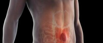
B-scan is a technique for recognizing the internal structures of the eyes using an ultrasound machine.
It is a non-invasive method and does not cause discomfort or pain during the procedure.
Therefore, all categories of patients easily tolerate the process. Using the technique, it is possible to recognize changes in the internal structure of the eyeball when it is impossible to examine the fundus using a slit lamp. It is recommended that the examination be carried out by the surgeon who will perform the operation so that he can make an accurate diagnosis.
What is a B-scan of the eye?
The technique is carried out using an ultrasound machine, which is brought to the patient’s closed eyes. First, the doctor applies a gel that eliminates the possibility of air appearing between the patient’s eyes and the sensor. The device sends ultrasonic waves into the eyeball, which are reflected and returned back. All wavelength data is displayed on the monitor screen. They are deciphered by an ophthalmologist after completion of the study.
Using a B-scan, the procedure is performed quickly, and it is possible to detect a large number of deviations in the normal structure of the eyeball.
How is an ultrasound examination of the eye performed?
The procedure is carried out in a sitting or lying position. It does not require any special preparation and its duration on average ranges from 15 to 30 minutes.
Depending on the data that the ophthalmologist needs to clarify, there are two types of research.
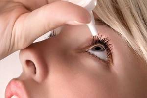
Diagnostics in A-mode is carried out by contact method. The patient is with his eyes open. Superficial anesthesia is performed. This is done for two purposes: achieving eye immobility and ensuring painless manipulation. The eyes remain open. A sterile probe is placed on the surface of the eyeball. The ultrasound machine's probe comes into contact with the eye and is moved slowly across the surface. Tear fluid acts as a natural contact medium.
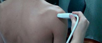
Diagnosis in B-mode is carried out with eyes closed. A hypoallergenic gel intended for ultrasound diagnostics is applied to the eyelids. A sensor is installed on the upper eyelid. The specialist moves the sensor across the eyelid, periodically indicating what actions the patient needs to perform with his eyes.
The results of the study are recorded in the protocol. The procedure takes about half an hour. The information received is deciphered by a qualified doctor specializing in this area.
Indications for eye ultrasound
B-scanning of the eyeballs is carried out to determine the following pathologies:
- cataract – clouding of the lens;
- glaucoma - increased secretion of fluid inside the eye chamber, which leads to enlargement and compression of surrounding elements;
- penetration of a foreign body into the internal structures of the eyeball;
- injury to the internal structure of the eyeball;
- the presence of malignant and benign tumors;
- decreased visual acuity, when a person sees well near, but poorly at a distance (myopia);
- disruption of the structure of the muscle around the lens or pupil;
- dystrophy, mechanical damage and other pathologies of the optic nerve;
- pathology of the vitreous body;
- diseases affecting the retina (atrophy, mechanical damage, detachment);
- decreased permeability of blood flow through the vessels of the eye microcirculation (due to penetration of a blood clot, atherosclerotic plaque, glucose conglomerate, vascular ischemia).
It is recommended to conduct an examination before surgery to identify the exact structure of the eyeball. The procedure is also carried out after completion of the operation to identify the patient’s tendency to recovery.
Ultrasound eye diagnostic techniques
In ophthalmological practice, several ultrasound diagnostic methods (scanning modes) are used, each of them has its own technical features, differs in diagnostic capabilities and is intended for specific medical cases.
Content:
- Ultrasound eye diagnostic techniques
- How is an ultrasound examination of the eye performed?
- Indications for ultrasound examination of the eyes
- Diagnosed pathologies
One-dimensional echography (A-mode) is an effective way to determine the size of the eye, internal structures and elements. It is used before surgery.
An anesthetic is instilled into the eye to relieve pain. The essence of the procedure is that the specialist moves the sensor over the eyeball. The information obtained is displayed on a computer monitor in the form of a graph, which shows the key parameters of the eyeball.
Examination in A-mode allows you to accurately measure the axial length of the eye, the depth of its anterior chamber, and lens parameters. These data are crucial in preparing for cataract extraction, for accurately calculating the optical power of an artificial lens, and in diagnosing refractive errors. Ultrasound diagnostics in A-mode also makes it possible to detect and evaluate intraocular tumors.
Two-dimensional echography (B-mode) is a scan used to assess the internal structure of the organ of vision. A two-dimensional picture of the structures of the eye is displayed on the computer screen. B-mode scanning has more advanced capabilities compared to A-mode. With its help, the monitor no longer produces a graph, but a two-dimensional image detailing the internal structures of the eyeball: lens, retina, eye muscles, etc.
B-mode examination allows one to highly accurately assess and differentiate intraocular changes: determine the nature of the pathology, its shape, and extent. Unlike the static A-mode, B-scan displays the intraocular picture in dynamics, which significantly expands its diagnostic potential.
Along with the A- and B-methods, other scanning modes are also used in ophthalmological diagnostics.
Combined method – a mode that combines the capabilities of A- and B-diagnostics. This technique combines the advantages of one- and two-dimensional echography.
Three-dimensional echography is a highly informative method that provides three-dimensional acoustic visualization of intraocular structures. The advanced technique of three-dimensional echography provides a three-dimensional image of the organ of vision. This is an effective method that opens up wide possibilities. The image is given in volume and broadcast in real time, which allows you to assess the general condition of the eye over time.
Color duplex scanning is a method that allows you to evaluate blood flow parameters in the orbital vessels. Studying the blood flow of the vessels of the eye makes it possible to diagnose vascular pathologies, which are sometimes the cause of visual impairment.
All ultrasound eye examination techniques are absolutely safe. Ultrasound waves do not have a negative effect on the structures of the eye.
Performing an ultrasound of the eye
The technique is carried out in several stages:
- the patient lies down on the couch and closes his eyes;
- the doctor applies a special gel developed for ultrasound techniques;
- a sensor is applied to the patent's eyes that produces ultrasonic waves;
- the device reads the indicators, transferring them to the screen monitor;
- After completing the study, the patient is given a dry cloth with which to wipe off the gel.
There are practically no contraindications to the ultrasound technique. Therefore, even a person with strong eye sensitivity can perform it. There are no side effects after completion of the procedure.
What is diagnostics
The best prevention of eye diseases is regular vision examination. Successful treatment of eye diseases depends on the timing of their detection.
Widely used in ophthalmology, methods of computer vision diagnostics, based on the achievements of modern science, allow for early diagnosis of acute and chronic diseases of the organ of vision even at the stage of reversible changes.
A complete vision diagnostics not only helps to make an accurate diagnosis, but also allows you to monitor and effectively manage the process of treating diseases. You can find out the cost of the examination in the “Prices” tab.
Decoding the result
There are normal indicators that the device sensor detects:
- the vitreous body and the internal structure of the lens should not be cloudy;
- the lens capsule is clear and clearly visible;
- the volume of the vitreous body should not exceed 4 mm;
- The normal length of the eyeball is 24-27 mm;
- the length of the optic nerve should not exceed the parameters of 2-2.5 mm;
- the cornea should not be distorted, damaged, or clouded.
If abnormalities are detected in one of the test results, it is recommended that a repeat diagnostic test be performed. After this, the doctor prescribes medication and surgical treatment.
Eye examination methods
Eye examination
includes accurate determination of the patient's visual acuity and refraction, measurement of intraocular pressure, examination of the eye under a microscope (biomicroscopy), pachymetry (measurement of corneal thickness), echobiometry (determining the length of the eye), ultrasound examination of the eye (B-scan), computed keratotopography and a thorough examination retina (fundus of the eye) with a wide pupil, determination of the level of tear production, detailed examination of the patient’s field of vision. If necessary, the scope of the examination can be expanded.
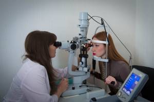
As a rule, the doctor begins the examination by obtaining information from you about your health status and family diseases. Then the doctor will most likely ask you to name the letters or numbers that he points to on a table of letters and numbers arranged in rows of gradually decreasing sizes. It allows you to determine the acuity of central vision.
If you wear contact lenses, your vision will be checked at this time. Additionally, your doctor will use a special instrument called a lensometer to check your glasses to confirm whether your prescription was written correctly.
If the chart test indicates that you need a prescription or contact lens prescription, your doctor will measure your eye's refractive error, or focusing, using instruments that contain a variety of contact lens strengths. To confirm the readings and find out which lenses provide the best vision, you will be given lenses of varying strengths to test.
Peripheral vision examination
External eye examination
Visual coordination test
Biomicroscopy
Dilated pupil examination
Fundus examination
Data from studies of natural and corrected visual acuity
using a slit lamp should be assessed using the symbols of the Snellen or Sivtsev tables. If the patient cannot distinguish large letters, then vision is assessed by determining the number of fingers. Then the patient's perception of finger movements and, finally, the ability to distinguish light from darkness are determined.
Determination of fields of view
performed using a contrast study, which can be used to estimate the approximate degree of visual field loss.
Study of pupil reaction to light
(indirect and involuntary) allows you to assess the state of the visual tract. The absence of a direct light reflex is observed with unilateral damage to the optic nerve and occlusion of the central retinal artery.
A patient with glaucoma has an arcuate scotoma (an isolated area in which vision is weakened or absent along the nerve fibers, along the edges of the optic nerve head). Central scotoma can be observed with optic neuritis. Bitemporal hemianopia/homonymous hemianopia (loss of the right or left halves of the visual fields) and quadrant hemianopia (loss of one quadrant of the visual field of one or both eyes) is observed in patients with neurological pathology.
Intraocular pressure
, as a rule, is measured using a non-contact tonometer. If necessary, intraocular pressure is measured using a Maklakov contact tonometer or Goldman tonometer. To exclude glaucoma, it is possible to conduct computer perimetry, that is, a study of visual fields.
Before any surgical intervention, a refractive examination
, which includes: determination of visual acuity without correction and with optimal correction, biomicroscopy, ophthalmoscopy, tonometry, refractometry (using an autorefractometer), computer topography of the cornea on a computer topograph, ultrasound biometry, ultrasound pachymetry. The data obtained during diagnosis is used by the surgeon when performing excimer laser correction.
Before refractive surgery, patients undergo pachymetry
a device for measuring the thickness of the cornea, which allows you to calculate the maximum permissible depth of laser exposure, which in cases of very high degrees of myopia determines how completely the correction can be carried out.
Any microsurgical or laser interventions are preceded by a complete comprehensive computer diagnostic vision examination
. The examination identifies the range of existing problems and determines treatment tactics.
Any disease of the visual organs requires a special approach to treatment, including at the stage of determining the diagnosis. Many ophthalmological diseases have similar symptoms, so even experienced professionals cannot make a diagnosis, much less determine the extent of the disease, without a thorough examination using specialized equipment. At an early stage, it is advisable to carry out diagnostics as planned. Below is brief information about the main methods for diagnosing eye diseases.
Visometry
– vision testing using special tables and trial lenses. Currently, their place is gradually being taken by more modern projectors and phoropters. This study is subjective, as it largely depends on the state of the visual organs and the body at the time of diagnosis. One of the types of viziometry is the selection of spectacle lenses.
Autorefractometry
- a study of refraction and the strength of curvature of the cornea of the eye, carried out using an automatic refractokeratometer. Using this device allows you to fully automate the process. All that is required of the patient is to look at a special mark, thereby keeping the head in a static position.
Keratometry
– measurement of the curvature of the anterior surface of the cornea. This test is used to calculate the optical power of an artificial lens, and also detects and measures astigmatism. Previously, keratometry was performed manually using a ruler, but in our center to conduct this study, a special device is used - a keratometer.
Tonometry
– measurement of intraocular pressure. Used to diagnose glaucoma and disorders of the optic nerve. The tonometry technique is simple: first, drops are injected into the patient’s eye to reduce the sensitivity of the cornea, and then an ophthalmic tonometer is applied to its surface. The device applies slight pressure to the cornea, and the result of the study is its reverse resistance.
Pachymetry
– measuring the thickness of the cornea at various points. The study is carried out using ultrasound. It is used to diagnose corneal dystrophy and is actively used before laser correction and to assess the condition of the eyes after corneal transplantation.
Biomicroscopy
– visual examination of the cornea, lens, iris and other parts of the eye by creating a contrast between illuminated and unlit areas. Allows you to assess the condition of optical media, blood vessels and optic nerves.
Ophthalmoscopy
– The most common examination using a slit lamp, during which the eyeball and adnexa are examined. It is carried out to diagnose diseases of the cornea, iris, lens and vitreous body.
Gonioscopy
- examination of the anterior chamber angle using a slit lamp and a gonioscope - a special device that is a system of mirrors. It is carried out to detect pathologies of the anterior chamber angle (tumors, foreign bodies, etc.)
Keratotopography
– examined eyes by scanning its surface, the main purpose of which is to determine the sphericity of the cornea. The scanning is carried out using a laser beam, and the results are a topographical “map”, on which areas of different heights are indicated in different colors.
Critical flicker fusion frequency
– a research method used to diagnose pathological processes in the visual system. FCSM allows you to determine the degree of eye fatigue based on the functional state of the cortical part of the visual analyzer and the central nervous system, as well as the degree of inertia of mental processes.
To the list of techniques
Indications for the study
Diagnosis using echobiometry is not always carried out; the following indications are required to prescribe it:
- myopia of moderate and high degree of development;
- cataract - clouding of the lens;
- glaucoma - increased intraocular pressure;
- visual impairment, in which a person cannot see small objects;
- farsightedness;
- suspected retinal detachment;
- disturbance in the vitreous region;
- malignant and benign neoplasms;
- congenital visual defects;
- disruption of blood supply to tissues;
- age-related changes;
- determination of the area of foreign body entry.
Data on the state of the internal structures of the eyes is accurate, so the likelihood of an erroneous diagnosis using this technique is minimal.
Echobiometry is carried out during treatment to identify its quality and progress.
Indications and contraindications for the procedure
An ophthalmologist prescribes ultrasound biometry of an organ after examining the patient and receiving test results. The manipulation is carried out with the aim of diagnosing diseases and establishing the dynamics of disease progression. It is also necessary to conduct research to test the treatment of myopia and other diseases of the organ.
Other indications for the procedure include: glaucoma, cataracts (complete or partial clouding of the lens), suspicion of benign and malignant neoplasms, pathological changes in the organ of vision, mechanical injuries to the eye, decreased transparency of optical media, mild inflammatory processes in the eyes.
Contraindications include:
- severe inflammation of the organ;
- purulent diseases of the studied area;
- lens diseases.
If the patient is not feeling well before the study, he should inform the attending physician. The doctor will take all necessary measures to protect the patient from adverse reactions and complications.
Evaluation of results
By assessing the condition of internal structures, damage to various fibers, pathologies of the ligamentous apparatus of the lens, and structural anomalies of various parts are revealed. Data are obtained from the density of the lens and the degree of transmission of the light beam through it.
The doctor can obtain informative data if it is impossible to check the condition of the fundus. This is possible in the case of a reduced effect from drops that disrupt the accommodation of the pupil.
Farsightedness and myopia are detected by changes in the length of the axis of the eye. Normally it is 23 mm. In people with farsightedness it is reduced, in people with myopia it is increased.
Echobiometry is a fairly informative study that can confirm or refute data obtained from other studies. It is carried out in clinical institutions equipped with specialized equipment. An appointment can be obtained from an ophthalmologist after a general examination.
The essence of the eye echobiometry method
The meaning of the method is the use of ultrasonic waves. Using a special device that uses ultrasonic waves, a specialist can measure various parameters of the eye - the thickness of the lens, the length of the eyeball, the depth of the anterior chamber.
The essence of this method is very simple. Ultrasound itself is used in various fields of medicine, both in diagnosis and in the process of carrying out a number of therapeutic procedures. Ultrasonic waves (echo signals) directed in a certain direction are reflected and thereby provide information (in centimeters or millimeters).
This procedure is also known as ultrasound scanning (A-scan and B-scan). Their main difference is that in the first case the procedure is carried out with the patient's eyes open. A-scanning is usually used when it is necessary to measure the curvature of the cornea and the length of the eye axis (which will help to correctly calculate the optical power of the intraocular lens). During a B-scan, the patient is asked to close their eyes. In this way, it is possible to scan the sclera and retina of the eye. In this case, the specialist places the sensor of the device at different angles.