Duration
15 minutes
The duration of the study itself is from 1 to 10 minutes.
After this, a decryption is carried out, and the person is immediately given the photographs. The patient is scheduled for 30 minutes. Soreness
No pain
The study is non-invasive and painless.
Preparation
Preparation required
Follow a diet 2-3 days before the test.
12 hours before the x-ray you need to stop eating solid food, and 2 hours before the examination you need to cleanse your intestines (about the same as when preparing for gastroscopy). Rehabilitation
Not required
After the study, patients feel well.
Work ability
Saved
After X-rays, patients feel well and their ability to work is not impaired.
Sports activities
No restrictions
The study is non-invasive, no restrictions.
Abdominal X-ray is a non-invasive, painless examination method. The procedure is performed without the use of a contrast agent and is 100% safe! X-ray allows you to see the overall picture of the condition of the abdominal organs.
If you are worried about bloating, pain, constipation, nausea, consult a specialist. Perhaps the cause will be determined using an x-ray.
In addition to radiography, there is also such a study as fluoroscopy. Fluoroscopy is a real-time study of organs, which allows you to evaluate their functioning over time. The main difference between radiography and fluoroscopy is that such observation is not carried out; one-time photographs are taken. However, in some cases, when the radiologist sees clear images with brightening, contrast is not needed.
Our patients can undergo both fluoroscopy with contrast agent and radiography. It is determined individually what kind of research needs to be carried out.
Traditional abdominal examination includes transillumination and serial plain radiography. Due to the anatomical features of the digestive system, it is impossible to recognize the disease only from photographs taken in standard projections.
The duration of the study itself is from 1 to 10 minutes. After this, a decryption is carried out, and the person is immediately given the photographs. The radiologist writes a description for the doctor who referred the patient for an x-ray.
A survey X-ray of the abdominal cavity allows you to determine what is causing the patient’s poor health: excess gases, fluid in the abdominal cavity (bleeding), foreign objects. In the images, the doctor can see damage that has arisen for various reasons: inflammation, injury, etc.
What is an abdominal x-ray?
An abdominal x-ray is a non-invasive, completely painless method of medical examination using x-rays. Modern X-rays allow you to project images of internal organs clearly and with the least harm to the body. An X-ray of the abdominal cavity shows the position of the internal organs, as well as their structure and tone. The study is carried out by a radiologist who monitors the correctness of the process. He also gives the patient a conclusion with the diagnostic results, which then must be shown to the doctor. Despite the high information content of modern x-ray research methods, a correct diagnosis requires a doctor’s examination, tests, etc. As a rule, x-rays are the final stage in drawing up a complete clinical picture.
Types of abdominal x-ray
The close arrangement of organs in the abdominal region causes difficulties during examination, which forced the improvement of diagnostic methods. Modern radiography of the abdominal cavity can be performed in two ways, each of which has indications and advantages.
Survey Study
This is a standard diagnostic method, the most accessible and simple. A plain X-ray of the abdomen shows what is causing the discomfort and discomfort: excess gas or fluid, bleeding, or foreign objects (such as kidney stones or small objects swallowed). Also, a survey X-ray of the abdominal organs allows you to see damage that has arisen for various reasons: from inflammation to injury.
Contrast study
Contrast x-ray of the abdominal organs is an improved research method that allows you to obtain a more detailed description of the condition of any organ. In this case, barium sulfate is used as a contrast agent, which stains the blood vessels from the inside and makes the pattern of the internal organs clearer.
Advantages of plain radiography

Unlike classical x-rays, survey radiography
is a more informative study.
A diagnostician evaluates the condition of not only the liver or kidneys, but all abdominal
. It is possible to evaluate the individual nuances of the location of organs and recognize existing pathological processes.
Computed tomography is based on the same principles as x-rays
: Using ionizing radiation, various areas of the human body are scanned.
The advantage of plain radiography
is that the method accurately visualizes bone tissue and is safer for health due to minimal radiation exposure to the body. In addition, X-ray diagnostics are cheaper and accessible to most patients.
Indications for the study
An abdominal x-ray is a serious examination that is prescribed by a doctor only if necessary. If there are certain indications and the results of other studies, radiography will help confirm or refute the suspected diagnosis.
Pain in the abdomen (acute abdomen syndrome) and lower back pain
Pain in the abdomen and lower back can be a symptom of many diseases: inflammation of the appendix, cholecystitis, acute pancreatitis, ectopic pregnancy, etc. An X-ray of the abdominal cavity, which shows the structure of the internal organs, allows us to identify an inflamed or damaged organ that is the cause of acute abdominal syndrome. Typically, an X-ray of the abdominal cavity for acute abdominal syndrome is prescribed in particularly difficult cases when making a diagnosis is difficult due to concomitant health problems.
Bloating
Typically, bloating occurs due to improper diet, however, if it is observed constantly, an x-ray of the abdominal cavity may be prescribed, which shows structural abnormalities of the organs. The cause of bloating can be inflammatory diseases of internal organs, as well as neoplasms and swelling. All these problems are clearly visible on a contrast radiograph.
Abdominal injuries
An X-ray of the abdominal cavity is indicated if internal injuries are suspected, since violation of the integrity of any organ can lead to disastrous consequences. Damage is indicated by free gas, which is visible on x-rays to varying degrees, depending on the organ. Plain radiography also allows you to see bleeding or hematomas.
Retroperitoneal abscess
A retroperitoneal abscess is the occurrence of purulent formations on internal organs. This disease can occur due to trauma, abdominal surgery, organ perforation, or the growth of metastases. The consequence of a retroperitoneal abscess can be sepsis and, as a consequence, death. An X-ray of the abdominal cavity can reveal the position of the abscess and its size. As a rule, it is prescribed after an ultrasound if necessary.
Acute intestinal obstruction
Since in case of acute intestinal obstruction, areas of the intestine with liquid and gas are visible in the horizontal position of the patient, an X-ray of the abdominal cavity is taken in a supine position. The shape and size of these areas determines which part of the intestine the obstruction is located in. The most effective in this case is a contrast X-ray of the abdominal cavity. Acute intestinal obstruction can be fatal if untreated, so correct diagnosis in this case is extremely important.
Intussusception
The cause of the development of acute intestinal obstruction can be intussusception - the introduction of one section of the intestine into another. Pathology can have consequences in the form of the development of inflammatory and tumor processes. The causes of intussusception are very diverse, and X-ray examination helps to understand them. For this purpose, a survey radiography of the abdominal cavity or a more informative one, contrast, is used. In a particularly difficult case, an additional computed tomography may be prescribed, which is also a type of radiation diagnostics.
What does the research reveal?
Presence of free gas during perforation of a hollow organ and free liquid
Violation of the integrity of the hollow organ is the reason for the presence of free gas. This phenomenon does not occur in a healthy person. The cause is often ulcerative lesions.
Perforation, or perforation, is the occurrence of a defect in an organ, resulting in a direct communication between its contents and the environment surrounding the organ.
Perforation can appear in any hollow organ, but most often it occurs in the stomach, duodenum and intestines.
The main reasons for perforation:
- destruction of the walls by an inflammatory process, necrosis or neoplasm (most often perforated ulcers of the stomach and duodenum);
- mechanical damage to the walls due to a gunshot or knife wound;
- violation of integrity during medical manipulation (uterine curettage, biopsy during gastroscopy);
- rupture of the wall due to increasing tension (when feces accumulate, for example).
The most striking symptom of perforation is pain. Besides:
- vomiting with the development of functional intestinal obstruction;
- bloating;
- severe pain at the time of perforation of an ulcer of the duodenum or stomach, occurring in the epigastric region and right hypochondrium, etc.
Malignant tumors can lead to the accumulation of fluid: cancer of the stomach, intestines, pancreas. Sometimes the cause is pathologies such as pancreatitis (inflammation of the pancreas), peritonitis (inflammation of the peritoneum). That is why it is important to pay attention to alarming symptoms in time and undergo an examination.
Peritonitis can be a manifestation of an acute abdomen, a phenomenon that is characterized by severe pain and pathological tension of the abdominal wall. An acute abdomen is observed in gastrointestinal pathologies such as perforated gastric ulcer, appendicitis and others. This condition requires hospitalization and surgery!
Intestinal obstruction
With intestinal obstruction, food is unable to pass through the intestinal tract due to mechanical obstructions or problems with the intestines.
Main symptoms:
- bloating;
- constipation;
- bursting pain in the abdomen;
- nausea.
A test for intestinal obstruction is done if you have bloating, constipation, or other symptoms. Thanks to the study, the diagnosis is confirmed or refuted.
Obstruction may be a consequence of the appearance of polyps, tumors, or impaired intestinal motility.
Early detection of signs of intestinal obstruction allows you to start treatment on time!
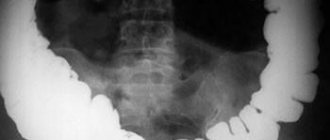
With the help of an intestinal examination, a specialist can detect liquids and gases that form “bowls” and “arches”. On an x-ray, the so-called “Kloiber cups” look like gas bubbles of a semicircular shape. The cup pattern is formed when the bowel loop contains more fluid and a small amount of gas; the arches are the opposite. The bowl can be transformed into an arch, and the arch into a bowl.
With intestinal obstruction, several Kloiber cups appear at once, which are located in the center of the abdominal cavity, in the area of the loops of the small intestine. On an x-ray, it appears as swollen areas of intestine filled with fluid and gas.
Examining the patient in an upright position gives a clearer picture than when examining a patient in a lateral position.
Also, a radiologist can detect intussusception , a type of intestinal obstruction when one section of the intestine penetrates into the lumen of another. But this pathology is typical for young children.
Historical reference
The German radiologist Kloiber in 1919 first published systematic radiological observations of acute intestinal obstruction. Unlike others, he did not use contrast agents.
Modern X-ray diagnosis of acute intestinal obstruction is based mainly on the study of the accumulation of gases and liquids in the intestines. They look like light spots on the film. With such a clear picture, the use of contrast is unnecessary.
Retroperitoneal abscess
An abscess usually occurs after surgery or after injury. The disease most often occurs in people 20-40 years old.
There may be no symptoms at first. And then intoxication occurs, characterized by nausea and chills. Unpleasant sensations arise first when walking, and then at rest.
Preparing for an abdominal x-ray
Plain radiography of the abdominal cavity does not require any preparation. However, for a contrast X-ray of the abdominal cavity, preparation is required, since if there are remains of food, liquids or gases inside the organs, their condition will be difficult to determine. 2-3 days before the test you will need to follow a diet, giving preference to foods that do not cause gas. 12 hours before the x-ray you will need to stop eating solid food; it is advisable to cleanse your intestines two hours before. Also, a few hours before the test, the patient is given a solution of barium sulfate to drink to stain the organs.
When is an abdominal x-ray required?
A survey X-ray of the abdominal cavity is performed in 35-45 minutes and does not require special preparation. The patient, without removing clothes, must fix the body in a comfortable position for the photograph.
The usefulness of a survey X-ray of the abdominal region: it allows you to quickly diagnose (start a treatment program) such dangerous pathologies as pancreatitis, intestinal volvulus, obstruction, stones in the bladder or kidneys, bleeding ulcers (stomach/intestines), appendicitis.
A survey x-ray allows you to determine the presence of inflammation, purulent foci, neoplasms, abscesses, gases or fluid in the abdominal cavity. In case of injury/mutilation, a plain X-ray helps diagnose bleeding and prescribe urgent abdominal surgery to save life.
Contrast x-rays are indispensable for studying hollow organs. It helps confirm/refute the presence of oncology, edema, bacterial lesions of the mucous membranes, etc.
How is an X-ray performed?
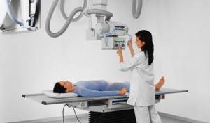
Immediately before the test, your doctor will ask you to remove items from your pockets that may interfere with the image being displayed on the screen. An x-ray of the abdominal cavity is taken in both vertical and horizontal positions. In some cases, it may be necessary to scan the body in two projections in order to better see the condition and structure of the internal organs. The patient takes a standing or lying position. In order for the pictures to be of high quality, it is necessary to remain still.
Procedure technique
Before diagnostic manipulation, the patient must remove chains and other metal jewelry from the area being examined. It is necessary to stand upright at the device and follow the instructions of the diagnostician; the device is first adjusted taking into account the patient’s height. The specialist makes the necessary settings and controls the process from a separate office.
When the body is exposed to ionizing radiation, a person must be motionless; only then can the most accurate images be obtained. If necessary, the diagnostician asks you to hold your breath for a couple of seconds. It may be necessary to take photographs in different body positions to obtain images of the abdominal
were in different projections.
In the supine position, radiography
is performed for the purpose of optimal viewing of through defects in the intestines or stomach. The finished radiographs are transferred to the patient or the attending physician.
Contraindications to X-rays
Contrast and plain radiography of the abdominal cavity is a relatively safe method of examination, but has contraindications. Radiation diagnostics is contraindicated for children under 15 years of age and pregnant women; obstacles may also arise if the patient’s condition is inadequate or severe, when it is impossible to remain still. In general, thanks to radioprotection methods, modern X-rays of the abdominal cavity
Even when carried out regularly, it is harmless to humans.