Diseases localized in the area of the skull and brain are, of course, most informative and safe for the patient to examine using magnetic resonance or computed tomography. However, not every medical institution has the appropriate equipment, and in some situations involving damage to the skull or brain, there is no time to transport the patient to a clinic where the necessary technical devices are available. The simplest, most common and accessible way to diagnose destructive processes in the bone or soft tissues of the head remains radiography. This procedure is also effective for examining such a part of the skull as the sella turcica.
What is the sella turcica and what organs surround it?
Content:
- What is the sella turcica and what organs surround it?
- Why is an x-ray of the sella turcica prescribed, what pathologies can it reveal?
- Indications for examination of the sella turcica
- What is “empty saddle” syndrome and why is it dangerous for humans?
- In what cases does a gynecologist refer for the procedure?
- Are there any contraindications for the procedure, and how to prepare for it?
- How is radiography of the sella turcica performed?
- Features of interpretation of survey results
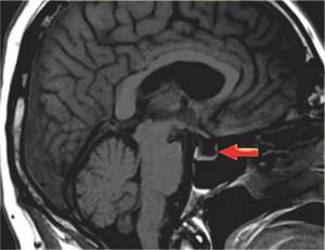
- Suspicion of a pituitary tumor
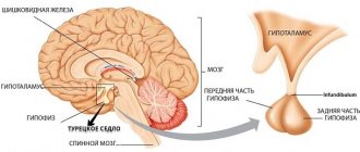
The structural features of the brain and cranium are anatomically interconnected and complement each other, since the skull is, in fact, a kind of protective box for the “central processor” of the human body. One of these anatomical features of the skull is the sella turcica - a bone formation that looks like a kind of depression in the sphenoid bone of the skull. This area received such a specific name for its visual similarity to the saddle.
The height of the saddle, measured from the deepest point of the bottom to the point of intersection with its diaphragm, is about 9 millimeters. Normally, a value of 7 to 12 millimeters is allowed. The anteroposterior or sagittal size of the sella turcica (its length) is from 9 to 15 millimeters. The ratio of these values to each other is called the saddle index, and it changes throughout a person’s life - in childhood it tends to unity, in adulthood it decreases.
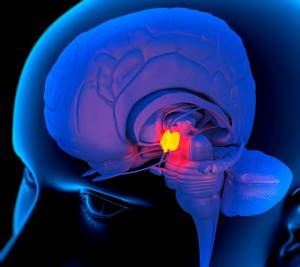
In the center of this depression is a hole where the pituitary gland is located - a gland that produces hormones involved in the processes of metabolism, growth and development of the human body. Actually, the sella turcica is a septum that separates the lower parts of the brain tissue. The size of the fossa corresponds to the size of the pituitary gland.

At the back, the saddle is limited by bone tissue - the “back”; on top of it is the dura mater - the diaphragm of the sella turcica. In the center of the diaphragm there is an opening for the pituitary stalk. The leg is a kind of canal connecting the pituitary gland with another part of the brain - the hypothalamus. In front of the tubercle sella there is a longitudinal precross stripe, behind which the optic chiasm is located.
The diaphragm performs the function of protecting the pituitary gland from subsidence of the meninges. The pituitary gland is one of the most important centers of the endocrine system of the human body. The sella turcica is its protective “shelter”, which protects this part of the brain from mechanical compression.
Features of diagnostics
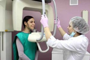
When the patient crosses the threshold of the equipped office, the specialist will lead him to the scanning machine and determine the position of the body in which the study will be carried out. Each medical institution has its own diagnostic procedure. After the person is positioned correctly near the X-ray machine, the doctor will take pictures in several projections.
Since x-rays record the current state of the brain and skull in the form of a kind of black and white photograph, a series of pictures are taken (usually 3-4) in order to comprehensively study the shape and size of the sella turcica, as well as the contours of the pituitary gland. In total, the study will last about 4–10 minutes. As soon as the necessary images from several angles are obtained, the patient will be able to leave the specialized room.
It usually takes 10 to 20 minutes to develop an x-ray. When the laboratory technician has completed his work, he sends the data to a specialist for further decoding. After a description with the appropriate diagnosis is drawn up, it is transmitted to the patient himself or his treating doctor. If the received form contains the phrase “no pathology detected,” it means that the pituitary gland and sella turcica are functioning in “normal mode.”
Before coming to the radiologist, you should make sure to turn off your phone - this will protect it from negative radiation exposure.
Why is an x-ray of the sella turcica prescribed, what pathologies can it reveal?
Any pathological processes in the area of the sella turcica have an extremely negative impact on the general condition of a person, since they become the direct cause of disturbances in the functioning of the pituitary gland.
X-ray of the sella turcica is an informative and painless diagnostic method, which doctors consider one of the most informative. X-ray images visualize bone tissue especially well, so they can be used to determine the shape, size and contours of the sella turcica as an anatomical structure of the skull, compare them with normal indicators, and identify deviations from the norm. X-rays are also prescribed to determine whether there are indications for surgery.
A referral for the procedure is issued by a therapist, endocrinologist, neurologist, oncologist, ophthalmologist, neurosurgeon or gynecologist.
Treatment
As such, there is no treatment for empty sella syndrome. Drug therapy is aimed at eliminating the symptoms of the syndrome. Therefore, if the pathology is discovered completely by chance and does not bother the patient in any way, then it does not require treatment. He registers with a specialist, undergoes periodic examinations, and, if possible, should lead a healthy lifestyle.
For those who are worried about pain and poor health, treatment is prescribed that reduces the symptomatic consequences:
- blood pressure surges;
- migraine;
- decreased immunity, etc.
Surgical treatment is rarely required. And then you can’t do without a neurosurgeon. Indications for surgical intervention:
- the need to remove the tumor;
- leakage of cerebrospinal fluid;
- sagging of the optic nerves (can cause complete loss of vision).
Treatment with folk remedies is extremely ineffective. Often it is simply necessary to eliminate some risk factors: obesity, taking hormonal contraceptives. The best prevention of the disease is a healthy lifestyle.
Forecast
It is impossible to predict the course of the disease. It all depends on the course of SPTS, concomitant diseases and the condition of the gland itself. Constant monitoring of hormone levels in order to eliminate complications in time. Engage in health promotion to help the body fight disease.
Empty sella syndrome is a pathology with an unpredictable course. It may not manifest itself in any way throughout your life, or it can cause many serious endocrine disorders. The choice of therapy can also be completely different: either the principle of non-intervention with routine observation, or surgical intervention with unforeseen consequences.
Indications for examination of the sella turcica
X-rays of the skull, including the sella turcica, can be carried out only by the decision of the attending physician, since this method is associated with a certain level of radiation exposure. The doctor refers the patient to such an examination if there are objective indications for this. All of them are divided into three general groups, which are:
- visual disturbances;
- endocrine dysfunction;
- various neurological pathologies.

The most common reasons for prescribing an x-ray of the sella turcica:
- suspicion of “empty sella syndrome”;
- cephalalgia of unknown origin;
- acromegaly (an endocrine disease manifested in abnormal production of growth hormone by the pituitary gland);
- inflammation of the brain or its membranes;
- diabetes insipidus (a disease associated with impaired carbohydrate metabolism in the body, which appears as a result of pathologies of the pituitary gland or hypothalamus);
- gigantism;
- increased intracranial pressure;
- traumatic brain injuries, if there is reason to suspect injury to the pituitary gland, as well as injuries to the cranial vault;
- visual disturbances: pain in the eye sockets, double vision in the eyes, increased lacrimation, decreased visual acuity;
- problems in the functioning of the adrenal glands and thyroid gland.
Advantages and disadvantages of the research method
In addition to being informative, x-rays of the sella turcica have the following advantages:
- low cost;
- painlessness;
- availability;
- short duration;
- lack of special training.
The most common disadvantages of radiography include:
- irradiation (albeit in small quantities);
- presence of contraindications.
Also, diagnostics do not always visualize tissues located next to the pituitary gland. Despite the many advantages, radiation examination should not be done too often. X-rays should only be taken with the direct consent of a doctor. Medical control and mutual respect between doctor and patient are the key to a speedy recovery.
What is “empty saddle” syndrome and why is it dangerous for humans?
This disease was first discovered by scientists in 1951. Since then, despite ongoing research, they have not been able to discover the causes of this anomaly. However, experimentally they were able to prove that the development of this pathology is facilitated by factors such as:
- radiation exposure or surgical interventions on the skull;
- cardiovascular diseases;
- hormonal disorders that occur during pregnancy, puberty and menopause;
- neurological disorders.
In essence, “empty sella” syndrome refers to the malposition of the pituitary gland in the fossa sella. Normally, the pituitary gland fills the fossa completely, but in the presence of the syndrome, the soft membranes of the brain, sinking, seem to press the pituitary gland to the bottom of the sella turcica, which causes its thinning.

The consequences of the appearance of the pathology are reproductive dysfunction, decreased immunity, sexual dysfunction, decreased visual acuity (in some cases, even blindness), the appearance of chronic headaches, as well as an increased risk of developing a micro-stroke.
Preparing for an X-ray
X-ray does not require extensive preparation before diagnosis. The patient is allowed to eat his usual food, but abstinence from alcoholic beverages is recommended. If radiography is required for a small child, you need to convince him a few days before the procedure that this research method is painless and necessary, using words and simple phrases familiar to him. Particular attention should be paid to the psychological state of children who will go to the diagnostic room for the first time.
We can say that an unusual device, reminiscent of one of the devices of the inventor Pin from Smeshariki, can produce a transparent picture depicting the cause of a child’s headaches. The child must understand that x-rays will help him get rid of unpleasant symptoms and return to his usual exciting life. A few minutes before the radiation examination session, you need to remove hairpins, bobby pins and elastic bands from your hair, as well as earrings, clips, necklaces, crosses and pendants. These items tend to distort x-ray images, affecting the final diagnosis.
In what cases does a gynecologist refer for the procedure?
The entire human body is a system with a complex structure. In it, all parts are interconnected, even if at first glance the connection is not obvious. Therefore, when a gynecologist sends you for an X-ray examination of the sella turcica - a bony depression in the structure of the skull - you should not be surprised.
The condition of the sella turcica directly affects the health of the pituitary gland. The pituitary gland, in turn, is part of the human endocrine system, and in the female body it produces various hormones that are closely related to the sexual and reproductive health of women, including:
- thyroid-stimulating;
- follicle-stimulating;
- luteotropic (prolactin);
- luteinizing;
- gonadotropic;
- adrenocorticotropic;
- oxytocin;
- melanostimulating hormone.
Each of these hormones plays its role in the functioning of the female genital organs. Some are involved in the ovulation process; without others, follicles cannot form in a woman’s body; others are formed during pregnancy and contribute to its normal course.
A gynecologist may send a patient for an X-ray examination of the sella turcica if:
- menstrual irregularities;
- infertility;
- increasing the level of prolactin in the blood.

Disturbances in the functioning of the pituitary gland in women are also manifested by general malaise, increased formation of wrinkles, and deterioration of skin elasticity, which is not associated with age-related changes. In such patients, hair begins to fall out rapidly, it becomes dull and brittle, appetite worsens and frequent constipation appears.
In addition to x-rays of the sella turcica, the gynecologist may recommend visiting an endocrinologist, as well as testing for hormones of the pituitary gland and other endocrine glands.
Are there any contraindications for the procedure, and how to prepare for it?
The main prohibition that doctors adhere to when choosing this method of examination for a patient is the incompatibility of radiography and pregnancy. Indeed, it is not recommended to irradiate pregnant women, especially in the early stages, when the fetus is developing all future organs and systems. If possible, alternative diagnostic tests should be performed, in particular magnetic resonance imaging.
Regarding the implementation of radiography for children, in each specific case the doctor weighs the danger of radiation with the objective need for diagnosis. If it is impossible to conduct an examination in any other way, in exceptional cases, x-rays can be taken even for children under one year old.
The procedure does not involve the introduction of a contrast agent, so allergic reactions cannot occur due to it. The only potential danger from x-rays is radiation exposure to the body. Therefore, it is not recommended to take x-rays more often than once every 6 months.
No special preparation is required for an x-ray. Women must be informed about their pregnancy before undergoing the procedure.
Contraindications
It is generally accepted that x-rays of the sella turcica have no contraindications, but this hypothesis is not true. Radiation diagnostics is not recommended:
- people in extremely difficult general condition;
- patients with leukemia;
- small children;
- to expectant mothers in early gestation.
The last two categories of people have the opportunity to enter the X-ray room only if there are strict indications, including the impossibility of conducting research using more advanced and safe types of diagnostics.
At the time of the x-ray, a pregnant woman is asked to wear a lead apron, which will protect the developing baby from radiation.
How is radiography of the sella turcica performed?
The procedure is carried out in radiographic rooms equipped with appropriate equipment.
Immediately before the examination, the patient must remove all jewelry and accessories from the neck and head: glasses, headbands, hairpins, earrings, chains, piercings.
The subject is placed between the recording plate and the X-ray tube. Pictures can be taken while sitting or lying down. First, the patient is positioned sideways to the device and the plate - this image has a lateral projection. Next, the image is captured in the frontal projection. Usually, an x-ray of the sella turcica in two projections is enough for doctors, but in some cases it is necessary to take several more photographs from other projections.

The procedure itself usually lasts no more than five minutes and is completely painless.
Which doctors can prescribe a referral for an x-ray?
Since the pituitary gland regulates the activity of almost every part of the body, disruption of its functioning can cause significant complications in any system of organs, tissues and bones. Therefore, at the moment of identifying the corresponding symptoms, specialists from several subspecialties at once can issue a referral to the X-ray room, namely:
- neurosurgeon;
- neurologist;
- gynecologist;
- endocrinologist;
- neurologist;
- ophthalmologist.
If, during a routine examination, one of the above doctors asks to undergo an x-ray of the sella turcica, it is necessary to follow this recommendation in order to avoid the consequences caused by the neglect of an imperceptible disease. To get a high-quality result, you need to find out in advance where you can get an x-ray. It is necessary to choose trusted medical centers with good characteristics and reputation, which can be confirmed by real people, and not website commentators, since positive reviews are often bought by unscrupulous institutions from copywriters.

The health care professional should be informed in advance if the patient has a hearing aid or cochlear implant.
Features of interpretation of survey results
The resulting images - lateral and frontal craniograms - are analyzed by a radiologist.
The main categories used in the process of decoding images:
- sagittal size;
- vertical size;
- sella turcica shape;
- location, shape and dimensions of the saddle back.
For an objective analysis of the results, the images must be recorded taking into account the requirements of correct placement and centering. Correctness is determined by the complete coincidence of the projections of the anterior sphenoid processes, internal and external auditory openings.
Normally, the plane of the sphenoid eminence and the pituitary fossa in the sella turcica are visualized on photographs as single lines with a clear, intense contour.
D. G. Rokhlin proposed his own method for assessing the size of the pituitary fossa of the sella turcica from X-ray images. For this purpose, an image of the skull in a frontal projection is used. The Rokhlin technique is considered one of the most objective and accurate schemes.
The sagittal dimension is defined as the maximum distance between the posterior and anterior walls of the fossa. In this case, the line that connects the outermost points of the fossa has a direction parallel to the plane of the wedge-shaped eminence. The normal sagittal size in an adult is no more than 14 millimeters.
The process of clarifying the vertical size of the fossa is impossible without drawing a projection line of the diaphragm of the sella turcica on the image. Normally, it connects the middle and posterior sphenoid processes. If the media are not visualized on the image, the projection is determined by connecting the tubercle sella and the posterior processes. In some cases, for example, in children, the posterior sphenoid processes may not be visible on the radiograph, so the projection is carried out by connecting the tubercle sella and the top of the dorsum sella with a line. In addition to this line, an auxiliary tangent line is established to the lowest point of the edge of the pituitary fossa. It is located parallel to the surface of the wedge-shaped eminence. A perpendicular is laid from the indicated tangent to the midpoint of the bottom of the pituitary fossa. The vertical size of the fossa is measured from the tangent to the intersection of the perpendicular with the projection of the diaphragm. Its normal length is up to 12 millimeters.
The shape of the sella turcica is a very individual category. In order to be able to generalize and evaluate it, scientists were proposed to calculate the sella index - the ratio of the vertical and sagittal dimensions. A flat fossa is one in which the sagittal dimension is larger than the vertical. If both dimensions are equal, the hole is considered round. If the larger size is vertical, then the hole is deep.
It is noteworthy that both sizes of the fossa tend to change, starting from childhood until the end of the organism’s maturation, and the sagittal size can increase even after the entire organism stops growth processes.
In children under 2-3 years of age, a flat shape of the pituitary fossa is usually found. By the age of 4-5 years, the vertical size becomes equal to the sagittal one, that is, the fossa acquires a round shape, and then acquires a larger size than the sagittal one. The round or deep shape of the fossa, starting from this age, persists up to 15 years in boys and up to 8 years in girls, after which the sagittal size begins to rapidly increase, which is why in adults the flat shape of the pituitary fossa sellae can most often be found on the picture.
The back of the saddle in the pictures appears as a flat or concave clear line. Basically, it has a vertical location, but a slight tilt forward or backward is allowed. Its thickness ranges from 1 to 10 millimeters.
To study the features of the shape and location of the back of the sella turcica, the doctor needs to have targeted radiographs in the nasal-frontal and posterior occipital projections. In such photographs, the back is defined as a rectangle that expands slightly upward. The transverse size of the back in an adult can reach 15 millimeters.
In addition to these values, the radiologist also pays attention to the structure of the wedge-shaped processes. The front ones are usually equal in size, while the rear ones can be located at different angles or vertically, and have different sizes. Technically correct images also show the middle sphenoid processes.