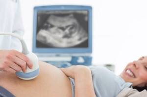
Future parents want to be sure that their child is developing fully. And in obstetric practice there are methods that allow assessing the perinatal development of the fetus even at the initial stages of embryogenesis. This opportunity became possible thanks to early ultrasound screening, carried out from the 10th to the 14th week of conception. During the study, such important indicators as the thickness of the nuchal translucency and the fetal calf size are determined. The norm for weeks of pregnancy is compared with the results obtained, which makes it possible to assess the development of the child.

Fetal CTE allows assessment of fetal development.
What is KTR
Literally, the abbreviation KTP stands for coccygeal-parietal size of the fetus. It is a straight line drawn from the highest point of the crown to the lower sacrococcygeal section of the future spine (and at this stage, the notochord) of the embryo at the moment of its maximum extension. Measured in millimeters.
This is an important indicator that allows you to assess the initial condition of the fetus. It is informative only at 7–12 weeks of pregnancy. Before this period, the embryo is too small to take measurements, and later it acquires individual structural characteristics, and CTE ceases to be a diagnostic factor.
The coccygeal-parietal size of the fetus in the first trimester depends on the gestational age, and not on hereditary characteristics - race, nationality, living conditions of the mother.
Its parameters allow you to reliably establish:
- Gestational age with minimal error. This is especially important for women with irregular menstrual cycles, when it is difficult to determine the expected date of ovulation. The results of some studies used in prenatal medicine depend on the precisely established gestational age.
- Date of expected birth. Correct determination of the period allows you to avoid the use of medications that provoke the onset of labor.
- Deviations in the dynamics of fetal growth: intrauterine growth retardation, chromosomal mutations.
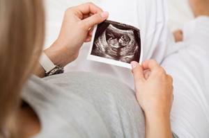
When performing several ultrasounds, the coccygeal-parietal size shows the dynamics of fetal development.
Determination of CTE occurs through ultrasound examination and does not pose any threat to the woman and the fetus.
CTE is an important indicator of the well-being of the embryo and the course of pregnancy.
KTP calculator by week
CTE is measured from 6 to 13 weeks of pregnancy. In this online CTE calculator, you can calculate the length of the fetus from the crown to the tailbone by the number of weeks.
In general, the CTE is about 0.4 cm at 6 weeks, 1 cm at the 7th week and up to 8 cm at 14 weeks. Fetal length is measured during a special medical examination using ultrasound.
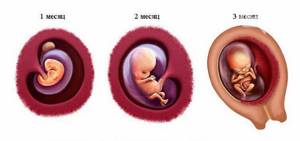
Embryo 10 weeks
At the end of the 10th week, the size will be about 4 cm. The eyelids will close before the eyes; the baby will be able to open them himself at 7 months. The respiratory system is almost ready for use. The rudimentary tail disappears, and buttocks form in its place. The baby moves freely inside the uterine area.
The skeleton and its structure fully correspond to a person. The arms and legs become longer to the appropriate proportions. But the head will occupy almost half the length of the body. This is explained by the active development of the cerebral hemispheres, the cerebellum is growing. The external genitalia develop according to gender. You will soon find out if he or she is growing in your belly. The baby's blood acquires its own group and Rh factor.
How is it measured?
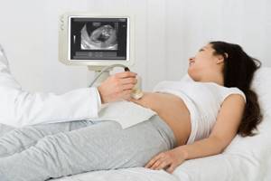
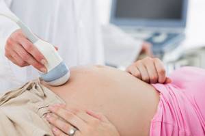
The coccygeal-parietal size is determined during the first screening ultrasound examination performed at 8–12 weeks of pregnancy. No special preparation is required for the manipulation on the part of the expectant mother. At the time of the examination itself, it is important that the pregnant woman relax completely, because... excitement can be transmitted to the embryo, and to take measurements it is important that the fetus does not move.
The scan is performed through the mother's anterior abdominal wall in the supine position. The doctor waits for maximum extension of the fetal head, takes a freeze frame, and draws a segment from the crown to the coccyx in the sagittal plane. If the fetus is actively moving, then take several pictures and select the largest indicator.
The line is measured by a special program embedded in the scanner. The obtained value is correlated with existing tables that indicate normal CTE values depending on the stage of pregnancy.
The values of the coccygeal-parietal size directly depend on how much time has passed since fertilization. The longer the period, the higher the CTE of the fetus.
Carrying out several ultrasounds in the early stages allows specialists to assess the dynamics of changes in the coccygeal-parietal size.
The calculation is carried out according to the obstetric gestational age - the time from the day the last menstruation began until the ultrasound was performed. It differs from the actual one by 2 weeks or more.
During the ultrasound examination, the woman should relax.
The embryonic period is counted from ovulation, since this is the moment of immediate conception. Ultrasound indicators CTE is the obstetric period.
CTE of the fetus - norm by week of pregnancy
At the early stage of embryogenesis, the fetus grows rapidly. In the first two months, the daily increase in height is 1 mm, and in the third month – 2–2.5 mm. And the values of the coccygeal-parietal size also become larger.
Modern ultrasound machines with highly sensitive sensors can indicate CTE with an accuracy of tenths. Older equipment defines the value in integers. If deviations from standard indicators are minimal, then development is complete.
Table of normal CTE values and correspondence to gestational age:
| Gestation period (week + day) | Lowest CTE value, mm | Average CTE value, mm | Maximum CTE value, mm |
| 5+0 | Not determined | 1,2 | 4,3 |
| 5+1 | 1,4 | 4,6 | |
| 5+2 | 1,7 | 4,9 | |
| 5+3 | 2,0 | 5,3 | |
| 5+4 | 2,3 | 5,7 | |
| 5+5 | 2,7 | 6,1 | |
| 5+6 | 3,1 | 6,6 | |
| 6+0 | 3,5 | 7,1 | |
| 6+1 | 0,4 | 4,0 | 7,7 |
| 6+2 | 0,8 | 4,6 | 8,3 |
| 6+3 | 1,3 | 5,1 | 8,9 |
| 6+4 | 1,9 | 5,8 | 9,6 |
| 6+5 | 2,5 | 6,4 | 10,4 |
| 6+6 | 3,1 | 7,1 | 11,1 |
| 7+0 | 3,8 | 7,9 | 11,9 |
| 7+1 | 4,5 | 8,7 | 12,8 |
| 7+2 | 5,3 | 9,5 | 13,7 |
| 7+3 | 6,1 | 10,4 | 14,7 |
| 7+4 | 6,9 | 11,3 | 15,6 |
| 7+5 | 7,8 | 12,2 | 16,6 |
| 7+6 | 8,7 | 13,2 | 17,7 |
| 8+0 | 9,7 | 14,2 | 18,8 |
| 8+1 | 10,7 | 15,3 | 20,0 |
| 8+2 | 11,7 | 16,4 | 21,1 |
| 8+3 | 12,8 | 17,5 | 22,3 |
| 8+4 | 13,9 | 18,6 | 23,5 |
| 8+5 | 15,0 | 19,9 | 24,7 |
| 8+6 | 16,2 | 21,1 | 26,1 |
| 9+0 | 16,3 | 22,0 | 27,0 |
| 9+1 | 17,0 | 23,0 | 29,1 |
| 9+2 | 18,1 | 24,2 | 30,0 |
| 9+3 | 19,0 | 25,0 | 31,0 |
| 9+4 | 20,2 | 26,1 | 32,0 |
| 9+5 | 21,0 | 27,0 | 34,1 |
| 9+6 | 22,1 | 29,0 | 36,0 |
| 10+0 | 24,2 | 31,1 | 38,0 |
| 10+1 | 25,3 | 33,1 | 41,0 |
| 10+2 | 26,0 | 34,0 | 42,0 |
| 10+3 | 27,1 | 35,0 | 43,2 |
| 10+4 | 29,0 | 37,1 | 45,0 |
| 10+5 | 31,0 | 39,3 | 47,2 |
| 10+6 | 33,0 | 41,1 | 49,0 |
| 11+0 | 34,0 | 42,2 | 50,1 |
| 11+1 | 35,1 | 43,0 | 51,0 |
| 11+2 | 36,0 | 44,2 | 52,1 |
| 11+3 | 37,0 | 45,3 | 54,1 |
| 11+4 | 38,1 | 47,0 | 56,0 |
| 11+5 | 39,0 | 48,2 | 57,1 |
| 11+6 | 40,1 | 49,0 | 58,3 |
| 12+0 | 42,0 | 51,3 | 59,0 |
| 12+1 | 44,0 | 53,0 | 62,0 |
| 12+2 | 45,1 | 55,0 | 65,1 |
| 12+3 | 47,0 | 57,1 | 67,1 |
| 12+4 | 49,1 | 59,3 | 69,2 |
| 12+5 | 50,0 | 61,2 | 72,0 |
| 12+6 | 51,0 | 62,0 | 73,4 |
| 13+0 | 52,1 | 63,0 | 75,0 |
| 13+1 | 53,1 | 65,2 | 77,0 |
| 13+2 | 54,0 | 66,0 | 78,2 |
| 13+3 | 56,0 | 68,1 | 80,0 |
| 13+4 | 58,0 | 70,0 | 82,1 |
| 13+5 | 59,2 | 72,0 | 85,2 |
| 13+6 | 61,0 | 74,0 | 87,2 |
| 14+0 | 63,0 | 76,0 | 89,0 |
Experts evaluate the coccygeal-parietal size until the 14th week. In the second trimester, this indicator is no longer important. Other parameters of the fetus come to the fore.
Average size of the fertilized egg in the first trimester of pregnancy
| Date of last menstruation (weeks) | Time at conception (weeks) | Inner diameter (mm) | Area (mm2) | Volume (mm3) |
| 5 | 3 | 18 | 245 | 2187 |
| 6 | 4 | 22 | 363 | 3993 |
| 7 | 5 | 24 | 432 | 6912 |
| 8 | 6 | 30 | 675 | 13490 |
| 9 | 7 | 33 | 972 | 16380 |
| 10 | 8 | 39 | 1210 | 31870 |
| 11 | 9 | 47 | 1728 | 55290 |
| 12 | 10 | 56 | 2350 | 87808 |
| 13 | 11 | 65 | 3072 | 131070 |
Examination of the mother for abnormalities
If the doctor determines a deviation of the coccygeal-parietal size from the norm, the woman is prescribed additional examinations:
- tests for the presence of urogenital infections;
- tests for chronic and hereditary diseases.
To do this, do the following:
- carry out smears;
- take blood, urine and feces;
- study the pregnant woman’s medical record.

Blood is taken to test the mother for abnormalities.
In some cases, X-ray contrast methods may be required, such as Dopplerography of the vessels of the uterus and placenta. A differential diagnosis must be prescribed. They attract narrow specialists in the required profile: cardiologists, geneticists, nephrologists, neurologists, infectious disease specialists, etc.
If no pathologies are detected in the expectant mother, they begin to examine the fetus.
Several methods can be used:
- Amniocentesis - sampling of amniotic fluid to study its parameters.
- Cord blood analysis.
- Chorionic villus biopsy.
After identifying the reasons for stopping or slowing down the child’s development, appropriate treatment is prescribed. In most cases, it is aimed at maintaining pregnancy.
If a frozen pregnancy is suspected, specialists perform an ultrasound to detect heartbeats in the fetus.
If they are not detected, then the woman is provided with emergency medical care.
If chromosomal pathologies are confirmed, experts suggest that patients undergo an abortion at an early stage. Women themselves decide whether to follow doctors’ recommendations or continue the pregnancy.
Deviations from the norm
CTE is considered the main criterion for the full development of the fetus in the early stages. Deviations from normal parameters can be either smaller or larger. If the obtained indicator differs slightly from the table, do not get nervous ahead of time. It is better to consult a doctor who will analyze the results and assess the risks. At the same time, it is important to correctly decipher the reason for deviations in KTE indicators.

The doctor can assess the risks of CTE deviation from the norm.
Average length of long bones at different stages of pregnancy (according to last menstruation)
| Duration (weeks) | Length (mm) | |||
| femoral | humeral | ulnar and radial | tibia and tibia | |
| 14 | 16 | 14 | 14 | 14 |
| 15 | 19 | 18 | 16 | 16 |
| 16 | 22 | 21 | 19 | 18 |
| 17 | 24 | 24 | 22 | 21 |
| 18 | 28 | 27 | 24 | 23 |
| 19 | 31 | 30 | 27 | 26 |
| 20 | 34 | 33 | 30 | 29 |
| 21 | 37 | 36 | 32 | 32 |
| 22 | 40 | 39 | 33 | 34 |
| 23 | 43 | 42 | 35 | 36 |
| 24 | 46 | 46 | 37 | 39 |
| 25 | 48 | 46 | 39 | 41 |
| 26 | 51 | 48 | 41 | 43 |
| 27 | 53 | 48 | 43 | 45 |
| 28 | 55 | 50 | 45 | 47 |
| 29 | 57 | 53 | 47 | 50 |
| 30 | 59 | 54 | 49 | 52 |
| 31 | 62 | 55 | 52 | 54 |
| 32 | 63 | 56 | 54 | 55 |
| 33 | 65 | 58 | 56 | 57 |
| 34 | 66 | 60 | 58 | 59 |
| 35 | 67 | 62 | 61 | 60 |
| 36 | 69 | 64 | 62 | 62 |
| 37 | 70 | 65 | 64 | 63 |
| 38 | 72 | 67 | 66 | 65 |
| 39 | 74 | 69 | 67 | 67 |
| 40 | 77 | 71 | 69 | 70 |
Tags: ultrasound
Indications for unscheduled measurement of CTE
The first ultrasound screening is performed on the expectant mother between the 12th and 14th weeks of gestation. But there are situations when a woman needs additional research to determine the coccygeal-parietal size of the fetus.
This happens in the case:
- suspicion of multiple births;
- in vitro fertilization;
- complicated previous pregnancies and childbirths;
- maternal infectious diseases;
- hereditary anomalies in the family;
- congenital disorders in children from previous pregnancies;
- chronic maternal diseases.

An unscheduled CTE measurement may be prescribed if multiple births are suspected.
Average dimensions of the abdominal cavity of the fetus (gestational age - at the last menstruation)
| Duration (weeks) | Dimensions (mm) | ||||
| transverse | anterior-posterior | average | perimeter | area (mm2) | |
| 14 | 26 | 20 | 23 | 72 | 396 |
| 15 | 30 | 24 | 27 | 78 | 548 |
| 16 | 34 | 25 | 32 | 96 | 760 |
| 17 | 40 | 32 | 36 | 108 | 972 |
| 18 | 44 | 36 | 40 | 120 | 1210 |
| 19 | 50 | 40 | 45 | 138 | 1452 |
| 20 | 52 | 41 | 47 | 140 | 1600 |
| 21 | 55 | 43 | 49 | 144 | 1728 |
| 22 | 59 | 49 | 54 | 162 | 2180 |
| 23 | 63 | 52 | 57 | 168 | 2350 |
| 24 | 68 | 55 | 62 | 186 | 2880 |
| 25 | 73 | 60 | 66 | 198 | 3260 |
| 26 | 77 | 62 | 69 | 204 | 3675 |
| 27 | 81 | 65 | 73 | 216 | 3888 |
| 28 | 84 | 68 | 75 | 228 | 4107 |
| 29 | 86 | 71 | 79 | 240 | 4330 |
| 30 | 89 | 75 | 82 | 248 | 5042 |
| 31 | 93 | 79 | 86 | 259 | 5547 |
| 32 | 97 | 82 | 89 | 270 | 6074 |
| 33 | 100 | 84 | 92 | 278 | 6348 |
| 34 | 102 | 86 | 94 | 288 | 6625 |
| 35 | 104 | 88 | 96 | 290 | 6912 |
| 36 | 107 | 92 | 99 | 300 | 7495 |
| 37 | 110 | 94 | 102 | 306 | 7803 |
| 38 | 113 | 97 | 105 | 310 | 8425 |
| 39 | 116 | 98 | 107 | 324 | 8748 |
| 40 | 118 | 100 | 109 | 325 | 9074 |
| 41 | 119 | 101 | 110 | 327 | 9192 |
| 42 | 119 | 102 | 111 | 330 | 9408 |
How the research is carried out
A pregnant woman does not require any special preparation. The only thing the doctor may ask is to come for the examination with a full bladder and on an empty stomach. To do this, the expectant mother is recommended to drink 0.5 liters of still water before going to the ultrasound. In this state, when the bladder is full, it is easier for the diagnostician to conduct a visual examination of the pelvic organs and embryo.
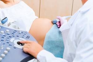
At the time of screening, the woman lies on the couch, and a specialist uses an ultrasound sensor to scan the internal genital organs in the sagittal plane through the anterior abdominal wall.
When taking measurements, it is important to ensure complete immobility of the embryo and maximum extension of the head end: this will ensure the accuracy of the study. Otherwise, the doctor takes several pictures and takes measurements on where the size of the fetus is largest.
On the scan, a segment is drawn from the crown to the tailbone, measured in millimeters. This is KTR. The obtained result is compared with tabular standards and a conclusion is made about the absence or presence of deviations.
During the first ultrasound screening, the fetal heartbeat, the quantity and quality of amniotic fluid, the condition of the umbilical cord and the degree of maturity of the placenta are also assessed.

The study is carried out using an ultrasonic sensor.
Decoding the results
In the first trimester of gestation, embryos grow approximately the same, regardless of race, heredity and environmental conditions. But this happens only until the 14th week, when the genetic characteristics of the future person appear. Therefore, the growth rate is changing.
To correctly assess CTE, it is important to conduct screening before the 14th week of pregnancy, then the calculations will be quite accurate. The specialist compares the obtained dimensions with tabular indicators and makes a conclusion about the absence or presence of deviations.
The conclusion of a functional diagnostics doctor is not a diagnosis.
And the detected deviations should become an indication for additional research to confirm or refute the pathology.
Is it necessary to carry out
Throughout pregnancy, the expectant mother undergoes 3 scheduled functional and biochemical screenings. The first is carried out between the 11th and 13th weeks of gestation, the second occurs at 18–20 weeks, and the third at 3–34 weeks. During this time, the woman undergoes an ultrasound, gynecological smears, blood and urine tests. All studies are aimed at early detection of pathologies of fetal development and the course of pregnancy.
CTE is measured during the first screening. Later, this indicator loses its information content. Ultrasound examination does not harm the unborn child, but helps to detect deviations from the norm in its development in time. Therefore, it needs to be carried out. Based on the results of the diagnostician’s conclusion, additional laboratory and instrumental studies, observation and treatment may be prescribed.
Screening should be carried out throughout pregnancy.
Average sizes of the fetal chest at different stages of pregnancy (according to the last menstruation)
| Duration (weeks) | Dimensions (mm) | ||||
| transverse | anterior-posterior | average | perimeter | area (mm2) | |
| 15 | 28 | 30 | 29 | 90 | 588 |
| 16 | 34 | 32 | 33 | 99 | 768 |
| 17 | 38 | 36 | 37 | 111 | 972 |
| 18 | 41 | 38 | 39 | 117 | 1200 |
| 19 | 44 | 44 | 44 | 132 | 1452 |
| 20 | 48 | 44 | 46 | 138 | 1587 |
| 21 | 50 | 48 | 49 | 147 | 1875 |
| 22 | 53 | 50 | 52 | 156 | 2028 |
| 23 | 58 | 53 | 56 | 168 | 2352 |
| 24 | 59 | 56 | 57 | 181 | 2523 |
| 25 | 62 | 60 | 61 | 183 | 2724 |
| 26 | 65 | 63 | 64 | 192 | 3072 |
| 27 | 69 | 66 | 67 | 201 | 3468 |
| 28 | 73 | 70 | 72 | 219 | 3888 |
| 29 | 76 | 72 | 74 | 228 | 4107 |
| 30 | 78 | 74 | 76 | 234 | 4332 |
| 31 | 81 | 77 | 79 | 243 | 4563 |
| 32 | 83 | 79 | 81 | 249 | 4810 |
| 33 | 85 | 82 | 83 | 255 | 5292 |
| 34 | 88 | 85 | 86 | 264 | 5547 |
| 35 | 91 | 89 | 90 | 273 | 6075 |
| 36 | 94 | 90 | 92 | 282 | 6348 |
| 37 | 97 | 92 | 94 | 291 | 6627 |
| 38 | 98 | 94 | 96 | 294 | 6912 |
| 39 | 99 | 96 | 98 | 297 | 7203 |
| 40 | 101 | 97 | 99 | 303 | 7498 |
Possible deviations and their causes
A problem in the analysis of CTE is considered to be a deviation from the norm in the direction of increasing or decreasing, exceeding 2 weeks.
The reasons for this may be:
- Fetoplacental insufficiency syndrome. This complication is diagnosed in many pregnant women. It disrupts the blood supply to the placenta. The exchange between the fetus and the mother is not maintained at the proper level.
- Frozen pregnancy. In this condition, fetal development stops. The death of the embryo occurs due to some disturbance in the normal course of pregnancy. To confirm the diagnosis, fetal motor activity and heartbeat are assessed. The condition requires emergency medical curettage followed by antimicrobial therapy.
- Intrauterine infection of the fetus. Pathogenic microorganisms negatively affect the development of the embryo. To identify infections, pregnant women are prescribed the necessary tests and examinations. If the presumptive diagnosis is confirmed, appropriate treatment is carried out.
- Hormonal deficiency. Due to a lack of the pregnancy hormone, progesterone, fetal development stops. There is a possibility of miscarriage. To prevent the condition, hormonal status is diagnosed and replacement therapy is prescribed.
- Chromosomal pathologies (Edwards, Patau, Down syndromes). In such cases, future parents will have additional consultations with geneticists and donation of DNA markers.
- Mother's bad habits. If the expectant mother constantly smokes and drinks alcoholic beverages, then the fetus is in conditions of intoxication. Its growth slows down, the CTE lags behind the norm.
Deviation from the norm is a problem in CTE analysis.
The discrepancy between the CTE and the norm is not always pathological; there are also harmless reasons that influence it:
- Small stature of both parents. This will cause a natural decrease in the coccygeal-parietal size in the child. If this fact becomes clear when collecting a family history, additional examination and treatment will not be required.
- Incorrect gestational age. Obstetricians calculate this indicator based on the first day of menstruation preceding pregnancy. Women with long or irregular cycles ovulate later than in a normal 28-day cycle. This fact is not taken into account when setting the obstetric due date, which postpones the actual due date by several days. In this case, dynamic observation is carried out.
CTE is an important indicator of fetal development in the first trimester.
The future growth of the unborn child is assessed by other parameters: girth of the abdomen and skull, length of the thigh and lower leg, etc.
CTE is less than normal
A lag in the indicator from the norm may be of a physiological nature (the gestational age is set incorrectly, there is a genetic predisposition).
But the reasons may be less harmless:
- Frozen pregnancy. It is detected by the absence of heartbeat and fetal movement. Repeated scanning is carried out after 5-6 days. Lack of growth is a sign of embryonic death. The condition is an indicator for obstetric curettage and further treatment. During the procedure, a biopsy of embryo tissue is taken for genetic analysis and to determine the causes of its death, which often lie in severe chromosomal abnormalities that are incompatible with life.
- Hormonal deficiency in a pregnant woman. This is a threat of miscarriage. The fruit is growing and developing, but too slowly. A timely examination and properly prescribed hormone therapy will allow the embryo to catch up.
- Infectious diseases of the mother. They are identified by additional tests. If ureaplasma, cytomegalovirus, etc. are detected, treatment is necessary.
- Chronic non-infectious diseases of a pregnant woman. Autoimmune diseases and diabetes require monitoring throughout the entire gestational period.
- Disorders and genetic changes of the fetus. Various congenital pathologies are possible: Down syndrome, Patau syndrome, Turner syndrome, etc.
- Problems with the endometrium. The condition occurs with frequent early abortions and with some chronic diseases of the woman’s reproductive system.
CTE is less than normal - this is a sign of an incorrectly set period.
CTE is growing slowly
The slowdown in CTE growth occurs for reasons such as:
- Congenital chromosomal pathologies (Down, Patau, Edwards, Turner syndromes). A slowdown is diagnosed if, with signs of vitality (heartbeat, movement), the baby’s growth does not keep up with the existing standards and the lag progresses every week. The diagnosis is confirmed or refuted using a prenatal DNA test using maternal venous blood. The child's red blood cells are separated from the biological fluid and the karyotype is assessed for the presence of chromosomal abnormalities.
- Insufficiency of the inner layer of the uterus. The problem arises if a woman had a curettage shortly before conception. Therefore, after an abortion, you need to wait several months before planning your next pregnancy, otherwise the risk of miscarriage increases. The condition requires hospital treatment.
- "Conflict" pregnancy. If a woman who has a negative Rh factor gives birth to one or more Rh-positive children, then she already has antibodies in her blood against her own “positive” children. The next conception will cause a sharp jump in them, which can cause miscarriage.
CTE is more than normal
Reasons for high rates:
- Incorrect gestational age based on the date of expected ovulation. Early ovulation is possible due to hormonal therapy in the treatment of infertility, recent viral infections, etc. In this case, the actual period is longer than previously calculated.
- The expected development of a large fetus due to diabetes mellitus in the mother or hereditary causes. In the first third of pregnancy it is too early to confidently make a conclusion about a large fetus, but a trend is already emerging. In this case, after one and a half to two weeks, a repeat ultrasound is prescribed to assess the dynamics. If the CTE increases in proportion to the term, we can talk about a large fetus and prepare the woman for a caesarean section.
- Activation of child growth through the use of medications. Certain vitamins in large quantities can cause a similar reaction. Therefore, before using them, it is better to seek advice from a gynecologist.
- Rhesus conflict, when a “negative” mother carries a “positive” child.

CTE may be higher than normal due to diabetes.
CTE is growing too fast
Normally, the growth of the embryo until the 12th week of pregnancy is about 1 mm per day, later the fetus adds 2.5 mm per day. If the daily increase exceeds these figures, we are talking about rapid growth.
The reasons for this may be:
- Metabolic disorders in the mother: Graves' disease, diabetes mellitus, obesity. Hormones secreted in excess in a woman’s body accelerate the growth of the fetus.
- Congenital pathologies of the fetus, for example hydrocephalus, gigantism.
- Rhesus conflict.
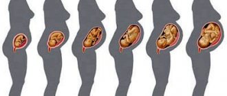
How does the human embryo develop?
Embryo at 1-2 weeks of pregnancy
In the first days, the follicle matures, from which the egg appears. It moves along the fallopian tube, where it meets the sperm. Fertilization occurs in which the 23 chromosomes of the egg combine with a similar number in the sperm. The sex of the unborn child depends on the presence of an X or Y chromosome in a male cell. Further along the course, the genome of the embryo is formed. On day 5, the embryo is ready to move into the uterus. There are cases when, after fertilization, the egg does not have time to reach the uterus, then an ectopic pregnancy occurs. Having reached the uterine region, the gastrula, which has transformed from the blastula, attaches to the walls. On day 9, the cells form a three-layer structure. As a result, the nervous system and skin will be formed from the outer layer. The middle layer will form the musculoskeletal system, muscles, blood vessels, internal organs, etc. The inner layer will create the cavity of the gastrointestinal tract. In the middle of the second week, a pregnancy test can already give a positive effect.
Embryo at 3 weeks of pregnancy
At week 3, cell division and growth into the uterine wall continues. At the same time, the formation of the umbilical cord and placenta begins, and amniotic fluid appears in the amniotic area. The size of the embryo reaches 4 mm.
Embryo at 4 weeks of pregnancy (1 month)
During this period, the heart begins to pump blood throughout the fetal body. The creation of the brain, spinal and head, from the neural tube begins. In addition, the initial stage of internal organs, eyes and limbs is formed. Nutrients were initially consumed from the yolk sac, and future reproductive cells also float in the yolk. From the end of the month, the functions of the pouch gradually weaken, and eventually it disappears.
Embryo. 5 weeks from fertilization
The fruit size is up to 2.5 mm and weighs 0.4 g. The development of the nervous system continues, sections for the stomach, brain, lungs, and trachea appear. The circulatory systems are expanding. At the same time, the mother develops drowsiness, nausea, and odor intolerance - the main signs of toxicosis.
The body's systems are rapidly developing: the neural tube is improving, future parts of the brain, lungs, stomach, trachea are being distinguished, blood vessels are growing. At this moment, the woman experiences signs of toxicosis: nausea, drowsiness, intolerance to certain odors.
Embryo. 6 weeks from fertilization
The embryo here looks like a “fry”. Its size is up to 6 mm. The brain is divided into hemispheres. The heart already has two chambers. It is already pumping blood, which is enriched with nutrients and oxygen. Limbs are forming. The excretory, respiratory, and digestive systems are formed. The placenta is formed, and amniotic fluid is around the embryo. The baby can already move in the shell. Pigment is acquired in the retina.
Embryo at 7 weeks of pregnancy
At this stage, the size of the embryo can reach 13-15 mm. Gaps appear between the fingers, although they are practically not developed. The head increases in size. Its body still has an arched shape, with a “tail” remaining on the pelvic part. The baby's breathing and nutrition come from the mother's blood. The pregnant woman’s belly is not yet visible, but urination becomes frequent due to an excess of fluid in the body.
Embryo 8 weeks
The dimensions are already about two centimeters. The face already looks like a human one - eyes, ears, lips and nose are distinguishable. The rudiments of the lungs, the digestive system, the heart, and the brain with two hemispheres have already been formed. Almost all the most important things have been formed. The bones are still in the form of cartilage.