According to WHO statistics, cardiovascular diseases are the number one cause of death worldwide. Experts believe that this particular group of diseases will remain in the fatal category for a long time, but the risk of developing pathology can be minimized by leading a healthy lifestyle, giving up bad habits and regularly visiting a doctor. One of the ways to diagnose the heart is computed tomography. What do you need to know about tomography, in what cases is it prescribed and how does X-ray radiation affect the human body?
General characteristics of tomography
Computed tomography is a method of non-invasive layer-by-layer examination of internal organs and systems of the human body. It is based on X-rays and complex computer algorithms that process the effects of the rays on the body. During radiodiagnosis, the anatomical features of the heart, the norms and pathologies of its functionality are assessed. Additionally, the CT machine covers the condition of the arteries, veins and the entire cardiovascular blood supply system down to the smallest capillaries (the entire chest is scanned).
Content:
- General characteristics of tomography
- Contrast tomography
- Indications/contraindications for the study
- Methodology and preparation for diagnosis
- Alternative diagnostic methods
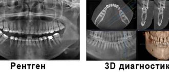
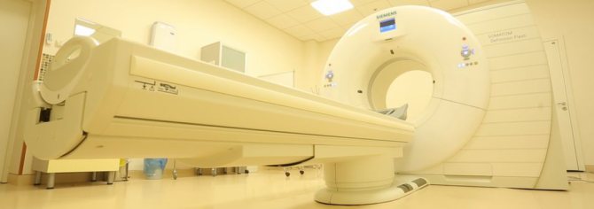
How is information collected? Pictures are taken using modern CT equipment, which consists of a table and X-ray emitters that scan the body layer by layer at intervals of 1 to 5 mm. The emitters penetrate tissues of different densities, and special sensors record this data and transmit it to a computer, where it is systematized by a program and collected into one image. Each image obtained in this way can be viewed individually, or integral three-dimensional models can be formed that are displayed on the screen.
Today, two types of CT scans are widely used: conventional (spiral) and multislice. The first is more affordable and widespread. This can be done in any city and in most private clinics. Although multispiral is more expensive, it is distinguished by a reduced radiation dosage, higher delivery speed and increased accuracy. If you have a choice between these two methods, then it is better to choose MSCT - less risk, more informative. But conventional CT is still relevant, as it allows you to see various diseases, neoplasms, injuries and pathologies. There are no closed areas for such an examination, and organ tissues do not overlap each other.
A heart scan is different from a CT scan of other organs. This is due to the fact that the heart is mobile. Therefore, a camera is used that is synchronized with the contraction of the heart, keeping in time with it. As a result, the pictures are static. The image on the screen is motionless and quite clear, which allows you to see all the structural features of the organ. The situation becomes more complicated if the patient has arrhythmia. In order for a person’s heart to work stably with a contraction frequency of 65 beats per minute, he is given beta-blockers.
In some cases, vasodilators are also required, which are administered immediately before the scan.
The next step in the development of CT was the invention of devices with two radiation sources. They were first introduced in 2005. Since at a high heart rate it is difficult to synchronize the camera with heart contractions, the use of 2 X-ray tubes solved this problem, making it possible to process absolutely any images of the organ, regardless of the heart rate: the device penetrates the patient’s body with rays in layers of 1-2 mm. There are a total of 64 of them in different planes. In this way, you can see the structure of the organ not only from the outside, but also from the inside. This is a good way to identify serious diseases at their inception.
CT scan of the heart with contrast – what does it show?
Computed tomography of the cardiovascular system is performed according to the following indications:
- Determination of areas of narrowing of aortic and coronary artery stenosis in coronary heart disease. Contrast CT scan of the heart clearly shows atherosclerotic plaques inside the vessel, areas of calcium deposits inside the coronary artery;
- Suspicion of inflammatory heart diseases (pericarditis, myocarditis, rheumatism), fluid accumulation in the pericardial cavity, inflammation of the heart valves;
- Study of the condition of the aorta, determination/exclusion of dissection, aneurysm (wall dissection);
- Diagnosis of thromboembolism (blockage of the lumen by a blood clot, air embolus);
- Malformations (pathological anastomosis between arteries and veins), developmental anomalies, malformations, vascular tumors.
The list of indications for CT of the cardiovascular system can be expanded.
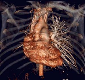
Scheme of MSCT of the heart and blood vessels
Contrast tomography
Contrast agents help enhance the overall picture, visually separate organs from each other, and highlight the necessary areas of the body for further study. Contrast is indispensable when checking the cardiovascular system. It helps to examine in detail the anatomical features of the organ and makes the scanning results more accurate and reliable. In most cases, iodine-based preparations are used today - they easily penetrate soft tissues without causing side effects, and just as easily come out of them. Although these drugs are safe, in rare cases they cause allergic reactions in patients with individual intolerance and serious kidney pathologies. In the first case, a person may develop an allergic reaction, in the second, the body will not be able to quickly and effectively eliminate the substance. If their use is still necessary, the patient is given antihistamines. The contrast agent is administered intravenously or orally, depending on the area being examined. When diagnosing the heart, the first option is used.
The procedure with contrast lasts about 2-3 minutes, without it even less - 1-2 minutes. Usually the patient does not experience discomfort, but due to the contrast, unpleasant sensations are possible: itching, nausea, urticaria. All this passes without a trace within a day. If health deteriorates significantly, the patient must inform the doctor, who is observing the procedure in the next room through the viewing window, and the scanning will be stopped. Communication is carried out via a special built-in speaker.
How is the examination performed?
A cardiac CT scan usually takes about an hour. This time is necessary to communicate with the medical staff, change clothes, prepare for the examination procedure and undergo the examination itself.
To undergo a CT scan, it is advisable to come to the clinic in clothes without metal fasteners. Otherwise, you will have to change into a robe or shirt provided by the clinic. The fact is that any metal objects leave a mark on the images, which makes diagnosis difficult. You will also have to remove chains, pendants, beads and brooches.
The patient is placed on the moving table of the CT scanner in the supine position. You will have to lie still during the entire examination. It's almost 20 minutes.
To administer a contrast agent, a special catheter is inserted into a vein in the patient's arm, which is connected to a dispenser using a thin tube.
The heart is constantly in motion, so for imaging it is necessary to select certain moments of the cardiac cycle. Also, the mediastinal organs can shift during breathing. To avoid the appearance of distortions and artifacts in the images, ECG synchronization and holding the patient's breath are used to obtain images.
When the pictures are ready, the doctor checks their clarity and detail. If the images do not contain artifacts, unclear or blurred areas, then the examination procedure is considered complete.
Indications/contraindications for the study
| Indications | Contraindications |
| Clinical manifestations of angina with ambiguous results of ultrasound, ECG, various tests and samples | Renal failure, individual intolerance to iodine-containing substances (with contrast study) |
| Acute chest pain without an identified etiology (to assess the degree of threat to life and begin a therapeutic course) | Excess weight that exceeds the norm that the CT equipment can support |
| Confirmation/refutation of such diagnoses as: myocardial infarction, dissecting aortic aneurysm, pulmonary embolism | Claustrophobia and mental disorders (the patient will not be able to follow the instructions of the laboratory assistant) |
| Non-invasive assessment of the condition of coronary vessels | Pregnancy (due to the harmful effects of X-ray radiation on fetal development) |
| Preoperative examination of the cardiovascular system | Thyroid pathologies |
| Clarification of the results obtained during selective interventional coronary angiography (used if the artery cannot be catheterized or contrasted) | The general serious condition of the patient (inform the attending physician even about a slight ailment. Perhaps he will decide to postpone the diagnosis and a new therapeutic course) |
| Diagnosis of congenital malformations of the coronary arteries and aneurysms | Diabetes |
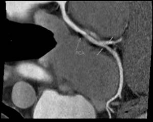
Angiography of the coronary arteries
This method is an invasive research method that allows one to determine the patency of blood vessels.
Contrast angiography is performed using a contrast agent containing iodine. The contrast fills the lumen of the vessel and becomes visible on an x-ray. The presence of a defect during filling of the vessel may indicate stenosis of the walls, their narrowing. The method determines the patency of blood vessels, the presence of plaques, and blood clots. Angiography is indicated for:
- angina manifestations;
- congenital heart defects;
- mitral valve prolapse;
- heart failure.
Due to the fact that the method is invasive, it may be accompanied by a number of complications. Hematomas and prolonged bleeding are likely.
Methodology and preparation for diagnosis
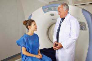
Conducting the study is within the competence of the x-ray laboratory technician. He provides preliminary instruction, reviews the patient's medical records, helps him position himself on the extendable table, and monitors the scanning process.
Approximately 30 minutes before the procedure, the patient takes an oral medication. It reduces heart rate and is consumed only once. Typically, drugs from the group of B-blockers are used. During a contrast study, a catheter is inserted intravenously into a peripheral vein (mainly the elbow bend). A contrast agent is injected through it, based on scanning cycles. Once the diagnosis is complete, the catheter is removed.
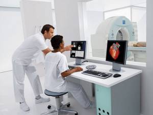
As soon as the patient has assumed the required position on the retractable table, the laboratory assistant adjusts the location of the equipment, gives specific recommendations (don’t move, hold your breath, etc.) and leaves for the adjacent room. In it, a medical worker monitors the functionality of the machine and the patient’s condition. Once the CT machine is started, a gantry begins to rotate over the area being examined. This is a special device with sensors that emit X-rays and transmit the received information to the main computer.
Important: When scanning your heart, you will have to hold your breath several times. The laboratory assistant warns the patient about this in advance and advises to listen carefully to the prompts through the loudspeaker.
You must lie still during the scan so as not to affect the results of the study. Even the slightest careless movement will not allow you to form a high-quality three-dimensional image. Also, before the procedure, it is necessary to remove all metal elements from clothing and body. X-rays simply won't be able to pass through metal components, leaving blank white traces in the final image. Metal must be removed only from the area being diagnosed. When scanning your heart, a metal belt buckle or trim on your pants will not affect the overall result.
The duration of the procedure depends on a number of factors and is communicated to the patient before the diagnosis begins. Typically the time frame is 2-5 minutes. CT scan results can be obtained on the same day, depending on the workload of the medical staff. The results are assessed by a radiologist. The doctor must evaluate the images, compare them with the medical history, and take them into account when making a diagnosis.
Decoding the received data
To decipher the data obtained during a cardiac CT scan, the radiology doctor needs to evaluate the structure and condition of a large number of anatomical structures: individual structures of the heart, such as valves, chords, septum, cardiac cavities, cardiac muscle, pericardium, coronary arteries, aorta, pulmonary veins and arteries. A large number of indicators need to be assessed, since only a cumulative accounting of a number of pathological changes often allows a correct diagnosis to be made.
Data from previous examinations, expert opinions, prescribed treatment and its effectiveness can help the doctor decipher the results, so it is advisable to take the medical documentation you have on hand to the clinic.
It can take a lot of time to decipher, so you can usually get the results of the examination only the next day after the CT scan.
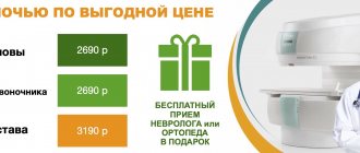
Price for cardiac CT scan with contrast in Moscow and St. Petersburg
Computer scanning of the heart with contrast differs in price in different diagnostic centers due to the equipment, qualifications of specialists, characteristics of the examination, type of contrast, and rental of premises. Factors also determine the cost of scanning in Moscow and St. Petersburg.
Approximate prices for CT types:
- With calcium calculation in the coronary arteries – 1500;
- CT angiography – 2350;
- Diagnosis of oncopathology of the mediastinum – 3200;
- Comprehensive scan of the chest, brain, intestines, pelvis – 6500.
Readers will be able to choose the optimal diagnostic center in the “medical centers” section, where an algorithm has been created for the optimal search for a medical center in St. Petersburg and MSK using dozens of criteria.
CT coronary angiography
CT coronary angiography of the vessels of the heart (general, selective) - a method for examining the coronary artery after intravenous administration of a contrast agent
Read the article