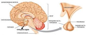
The sella turcica in the brain takes part in the vital functions of organs.
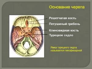
It is a slight notch located in the sphenoid bone strictly under the hypothalamus. Scientists compare it to a backrest.
The site provides reference information. Adequate diagnosis and treatment of the disease is possible under the supervision of a conscientious doctor. Any medications have contraindications. Consultation with a specialist is required, as well as detailed study of the instructions! Here you can make an appointment with a doctor.
Various shapes of this anatomical region
This is the area in which the venous sinus lies. There are optic nerves on both the right and left sides of it. The carotid arteries emerge from this place. Thanks to them, blood supply occurs to both hemispheres.
If for some reason pathological disorders of neuro-ophthalmological, neuroendocrine or neurological function occur, prolapse of the meninges begins to develop in the sellar area.
The pituitary gland spreads along its walls. The sella turcica also has another name – “pituitary fossa”. This part of the brain got its name because of the shape of the saddle.
Find the answer
Are you having any problem? Enter “Symptom” or “Name of the disease” into the form, press Enter and you will find out all the treatment for this problem or disease.
Can take different forms:
- A flat shape is defined by a significantly smaller vertical diameter than the same distance between the front and rear walls.
- Deep - develops in strictly opposite ratios than with a flat form.
- The round shape occurs when the mentioned diameters are equivalent to each other.
The slightest disturbances concerning the structure or functioning of the pituitary fossa provoke changes in the pituitary gland. In old age, thinning of the sections occurs. In newborn children, the structure of this section is cartilaginous.
Dimensions of norm and pathology at different periods of life
The structure, size and shape of the pituitary fossa depend on the structure of the skull. Confirmation of this theory can be found in pathological changes when the bones of the skull develop differently than in most people.
In a healthy newborn baby, it has a cup-shaped shape, with a characteristic wide opening and cartilaginous structure. During the first 3 years of life, the form becomes larger, and by 5 years it reaches 8-10 mm.
During adolescence, the formation of the sella turcica occurs, as well as other parts of the body and organs. Its normal size by the age of 13-15 is 9-12 mm. Its individual structure is fully formed by the age of 19.
For men, the value should not exceed 15 mm, for women – 12 mm. On average, according to statistics, normal sizes for men correspond to 12 mm, for women – 9 mm.
The saddle can increase in size due to microadenoma of the pituitary gland, less often due to hyperplasia of the adenohypophysis. A progressive tumor causes severe headaches, obesity and other disorders.
The process of premature ossification of the sphenoid bone causes a decrease in the volume of the pituitary fossa. This is often preceded by accelerated puberty. All pathologies require treatment.
What it is?
Inside the human skull there is a hollow formation called the sella turcica, which has a rounded shape measuring 8-12 mm. Inside it is the pituitary gland, a gland that regulates the body’s performance by producing hormones. The process of hormone production is also under the control of the hypothalamus. These two organs are connected by the so-called pedicle, lowered into the saddle.
The pituitary gland is protected by the diaphragm of the sella turcica - a rigid plate that separates the formation cavity from the subarachnoid space. This space is filled with liquor. The purpose of the sella turcica is to protect the delicate pituitary gland from various types of mechanical influences (concussions, blows).
In a healthy person, the pituitary gland completely fills the space of the saddle allocated for it by nature. If there are pathologies, the membrane of the brain descends and begins to put pressure on the pituitary gland. This can happen if the diaphragm is thinned or the stem hole is too large in diameter. In this case, the meninges simply presses the pituitary gland and spreads it along the bottom of the saddle. This creates an empty saddle. This is exactly how the pathology was discovered by a pathologist who was studying completely different diseases.
In some cases, the reverse process is observed: the diaphragm of the sella turcica seems to be underdeveloped, but sella turcica syndrome has not been identified. From this it was concluded that intracranial hypertension plays an important role in the occurrence of the syndrome. That is, the cerebrospinal fluid fills the entire space of the saddle and puts pressure not only on the pituitary gland, but also on the stalk. Because of all this, the regulation of the hypothalamus is disrupted, which causes disruption of the endocrine system.
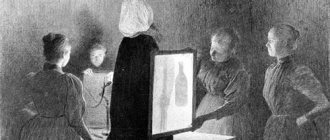
The need for an x-ray examination
To identify changes in shape and contours due to the formation of tumors or after injuries, to determine the size of this area, radiography is prescribed. This study is relevant if a pituitary tumor is suspected.
These examinations are prescribed to patients with increased intracranial pressure, pathologies of the thyroid gland and adrenal glands.
X-ray is necessary if the blood test shows prolactin levels higher than normal.
X-rays are indicated in cases of headaches of unknown origin.
Gigantism, diabetes insipidus, acromegaly require careful diagnosis and correct diagnosis, which is impossible without examining changes in the pituitary gland.
X-rays are necessary after injuries to the cranial vault or inflammation of any nature. Using X-rays, an “empty sella turcica” can be established.
The examination can be done at any medical institution that has an X-ray machine. Digital installations with enhanced radiation protection systems are the most accurate and safest for health.
Ionizing effects from such devices are contraindicated for women during pregnancy. As a last resort, X-rays are prescribed for children. This is possible if other methods do not allow a correct diagnosis to be made.
The images are interpreted by a radiologist. Features of changes in bone structures are revealed. The shape, size, and deformation of the walls of the pituitary pit are important.
If it has increased in size, or the walls have become thinner, a neoplasm has occurred. Benign tumors do not provoke changes in the structure of the walls. You must take the results and photographs with you for further consultation with specialists.
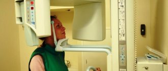
How is radiography of the sella turcica performed?
Digital X-ray diagnostics does not require any preparatory measures. Before taking pictures, the patient should remove earrings, hairpins, glasses and other items that may reduce the information content of the study.
The patient may be in a standing or lying position at the time of diagnosis. Pictures are taken in several projections: direct frontonasal; right and left side; straight back.
In some cases, a lateral radiography of the skull may be indicated. This allows you to evaluate the size and shape of the sella turcica relative to the size and shape of the skull as a whole.
After digital X-ray diagnostics, the laboratory assistant passes it on to a radiologist for a description. The patient is given a printout of the images and the results of the study are recorded on a CD.
Computed tomography capabilities
Computed tomography of this anatomical area is often performed in conjunction with the study of general pathological changes in the brain.
If the patient has indications, a separate examination is possible.
Computed tomography (CT) is indispensable for vision problems, the provoking factors of which doctors consider compression of the chiasm.
The reason for this is the deformation of the saddle as a result of formations of a benign or malignant nature.
Prerequisite factors for prescribing CT:
- Possible neoplasms in the pituitary gland (adenomas and adenocarcinomas);
- Hormonal disorders associated with the pituitary gland;
- Various developmental anomalies.
Types of CT:
- No contrast;
- With contrast to indicate detailed changes in the pituitary gland, carefully identify the subtleties of the blood supply and features of the tumor process.
CT is recognized as the most alternative to radiography. The organ under study is presented in a three-dimensional image. The results are recorded on electronic media.
In addition to this, the radiologist provides a written report. Another feature of CT is that it allows you to identify the most minor lesions.
X-ray results of the sella turcica
An X-ray of the sella turcica in one projection or another can be used to evaluate its shape, wall structure and size.
The presence of a pituitary tumor may be indicated by:
- Local or total osteoporosis of the walls of the sella turcica with preservation of the normal structure of other bone formations.
- Local or total atrophy of the walls of the sella turcica.
- Local or total thinning of the wedge-shaped processes of the sella turcica.
- Uneven internal contour of the wall of the sella turcica.
In the diagnosis of pituitary tumors, the symptom of double contours of the walls and/or bottom of the sella turcica is of great importance. Clear, even both contours, as a rule, indicate the absence of a pathological process in the pituitary gland. In cases where the internal contour is unclear or blurry, it is likely that there is a tumor in the area of this anatomical formation or the patient suffers from empty sella syndrome.
After a trauma to the base of the skull or as a result of a complicated birth, hematomas may be found in the area being examined. These are heterogeneous formations of different shapes and sizes that do not have any specific structure. Their shadow is intense, their outlines are uneven, but quite clear. These types of formations are usually localized at the bottom or walls of the sella turcica.
Craniopharyngioma (benign tumor of the pituitary tract) is visualized as a large calcification with unclear contours, often localized above, in some cases under, the diaphragm of the sella turcica.
Longitudinal streaks of high intensity represent calcified walls of blood vessels.
With tuberculous meningitis above the sellar diaphragm, the doctor sees small-sized tuberculomas with large- and small-spotted medium-intensity calcifications in them.
With tumors of the pituitary gland, especially with prolactinomas, small punctate, medium- or low-intensity calcifications are localized under the diaphragm sellae.
Calcifications of the dura mater in the area near the sella turcica and calcifications of its diaphragm may also be found.
There is a fairly rare pathology - osteomas of the fundus or wall of the sella. They are localized in the same place as hemorrhages (radiologically - calcifications), but their structure is different and they are located, as a rule, associated with bone tissue.
Indications for MRI
Violation of the diaphragm provokes a decrease in the pituitary gland. He finds himself crushed against the walls of his hole. This cannot be detected using an x-ray.
An MRI of the brain is indicated. Severe headaches, rapid decrease in vision, decreased tone are symptoms that require this procedure.
This clinical picture may indicate dangerous pathologies or changes in the functioning of the pituitary gland that can be effectively treated. Violations almost always have a hidden course.
It is easy to confuse such pathologies with other diseases. Careful diagnosis is of paramount importance in the treatment of brain abnormalities of this origin.
Indications for MRI of the pituitary gland:
- Deviations of a neurological nature, with a difficult to determine etiology;
- Endocrine disorders;
- Significant visual impairment.
Among the most characteristic neurological manifestations, doctors:
- Constant headaches;
- Failures associated with blood pressure, in which unreasonable feelings of fear and anxiety increase;
- “heart pain” of a stabbing nature;
- Sudden increase in body temperature, chills for no specific reason;
- Fainting and semi-fainting states, recurring frequently.
Vision problems can develop with lesions of the pituitary gland and a number of other brain pathologies.
The group consists of symptoms accompanying disorders of the sella turcica:
- Pain in the orbital area;
- When an object before your eyes doubles or triples;
- Unreasonable and excessive tearing;
- Distortion of the field of view;
- Loss of visual acuity.
Disturbances in this part of the brain are accompanied by unrelated symptoms. You shouldn’t delay getting an MRI, especially if it’s advised by specialists.
Indications for x-ray of the sella turcica
An X-ray may be prescribed to clarify the cause of a persistent headache.
Radiography of the sella turcica may be recommended for patients in the following cases:
- When diabetes insipidus is diagnosed in order to identify its causes.
- For traumatic brain injuries, especially localized in the cranial vault.
- For persistent headaches, especially of unclear localization and accompanied by visual impairment.
- For abnormal development of the skull.
- For symptoms of acromegaly, gigantism, dwarfism.
- For menstrual irregularities and infertility in women.
- For sexual dysfunction in men.
- With an increase in the blood level of the hormone prolactin.
- With constant weakness, loss of strength, exhaustion of the body, which is not associated with other diseases.
- If you suspect empty sella syndrome.
X-ray of the sella turcica has virtually no contraindications.
Research in gynecology
The reproductive functions of the female body are controlled by the pituitary gland and hypothalamus. Any disturbances in the activity of these 2 glands instantly provoke hormonal changes.
A set of disorders of this nature is called neuroendocrine syndromes in medicine.
Of these, the following list is presented:
- Premenstrual syndrome;
- Menopausal syndrome;
- Polycystic ovary syndrome;
- Post-castration syndrome;
- Adrenogenital syndrome.
Research is necessary when there is a combination of signs indicating a particular syndrome. X-ray or MRI may be prescribed if the patient has problems conceiving or there is an increase in prolactin levels in the blood.
There are hormonal imbalances in the body that require correction. Pathological disorders of the pituitary gland in the female half are manifested by many ailments.
The most common health problems are associated with a heavy menstrual cycle and the period preceding it.
Wrinkles begin to appear early, the elasticity of the skin deteriorates, and pigment spots form. The hair structure changes and hair loss begins.
Dyspeptic syndrome develops, which is characterized by:
- Constipation;
- Loss of appetite;
- Weakness;
- Fast fatiguability;
- Migraine;
- Headache;
- Drowsiness.
To make a correct diagnosis, it is important to conduct other examinations. These include hormone tests, ultrasound diagnostics, and pelvic tomography.
Gynecological examinations are necessary. Disturbances in the functioning of the pituitary gland require an integrated approach to treatment.
Prolapse of the suprasellar cistern into the sellar cavity is defined in medicine by the term “empty sella syndrome,” abbreviated as PTS.
The symptomatic picture is accompanied by:
- A series of neuroendocrine disorders;
- Violations in the organs of vision;
- Severe headaches.
Almost 50% of people suffer from underdevelopment of the sellar diaphragm. A diagnosis of empty sella syndrome is made in a maximum of 23% of patients.
There are many factors that provoke the development of this anomaly, the most important of which are:
- Hereditary predisposition to connective tissue pathology.
- Autoimmune diseases.
- Infectious lesions of the body of complex etiology.
- Physiological (hormonal) changes in the body.
- Formation of arachnoid cysts.
- Significant increase in intracranial pressure associated with pulmonary-cardiac problems, hypertension or traumatic brain injury.
- Pituitary infarction or sudden necrosis of a pituitary adenoma.
- Long-term use of oral contraceptives or hypofunction of the endocrine glands.
Diagnostics consists of the use of instrumental and laboratory methods. Clinical manifestations are characterized by a relapsing course. Early diagnosis allows the use of adequate therapy in treatment without prescribing drastic surgical methods.
Empty sella syndrome
The sella turcica is an anatomical area of the skull where the pituitary gland is located (the part of the brain responsible, among other things, for the functioning of the endocrine glands). If, when analyzing MRI or CT images, the doctor does not find the pituitary gland in the right place, you may hear a diagnosis of “empty sella syndrome.” It sounds impressive and serious, but you don’t seem to have any unpleasant symptoms. What's the matter?
Causes
There are two main groups of reasons for the formation of the “empty sella” syndrome:
- This condition can be physiological (for example, in women after repeated childbirth, during menopause, during long-term use of oral contraceptives). Also, sometimes the pituitary gland is displaced from the area of the sella turcica due to the congenital structural features of the skull bones. In these situations, they speak of the primary “empty sella” syndrome. In the primary syndrome, the functioning of the pituitary gland is not significantly impaired. The prognosis is favorable.
- The pituitary gland sometimes becomes displaced due to certain diseases (for example, tumors, infections, after radiation therapy to this area), then they speak of secondary “empty sella” syndrome. In this case, the functioning of the pituitary gland is disrupted. Depending on the manifestations, treatment by an endocrinologist, neurologist, or neurosurgeon may be required.
Who has it more often?
Empty sella syndrome is not such a rare occurrence. When examining the brain of healthy people using MRI, this syndrome can be observed in 10–40% of cases.
It is known that in representatives of the fairer sex aged 35–55 years, this syndrome is detected 5 times more often than in men. Another risk factor is obesity.
Manifestations of the syndrome
In most cases, the “empty sella” syndrome, accidentally detected by MRI or CT, does not manifest itself in any way. You may not even suspect that anything is wrong with you.
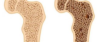
Osteoporosis
Osteoporosis is a life-threatening disease. With osteoporosis of this anatomical part of the skull, irreversible destruction of the bone component of the skull occurs. The size of the pituitary gland, according to scientists, remains normal or slightly reduced.
Diffuse osteoporosis of the sellar wall occurs due to natural causes of aging of the body. The process of bone tissue breakdown can also occur due to calcium and vitamin D deficiency.
Local osteoporosis is diagnosed as a secondary disease caused by pituitary tumors. The risk group consists of patients weighing less than 50 kg. Age category is people over 35 years old.
Thinning bone structure can be diagnosed in the earliest stages. An MRI is most often prescribed. Doctors emphasize that the symptoms of this disease should not be ignored and if headaches of unknown etiology develop, immediately consult a doctor.