Most often, a doctor sends patients for an MRI when it is necessary to find out detailed information about their health condition for diagnosis. This examination costs more than a simple x-ray and provides the doctor with significantly more information. Therefore, it is prescribed only if other examination methods cannot reflect the full picture of the condition. In milder cases, when the diagnosis is made by x-ray or ultrasound, tomography is not necessary.
You can find out how much an MRI costs in our center in the “Price” section. Such an examination can be carried out on any area of the body to determine the condition of the internal organs. One of the most common areas is MRI of the spine, in which each section can be examined in detail on the image. Usually, at the first consultation, the doctor determines the localization of the problem and then prescribes a diagnostic procedure in this department. Sometimes it is necessary to examine the entire spine for pathologies or injuries.
For example, an MRI of the lower back, or lumbar spine, is prescribed for pain in this area, which can be caused by neuralgia or diseases of the internal organs. An MRI of the cervical spine will help diagnose the cause of headaches and back pain, the presence of curvatures and incorrect position of the vertebrae.
MRI of the back is a popular procedure, since this is where many problems associated with imbalances in the body originate. A slight change in the position of the vertebrae can lead to a deterioration in a person’s well-being and the functioning of the entire body, including internal organs.
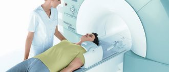
The procedure is comfortable for the patient even with severe health conditions. All that is required of the patient during an MRI of the thoracic spine or other area is to lie quietly for a few minutes so that the tomograph takes pictures in the desired projections.
X-ray or MRI of the spine – which is better?
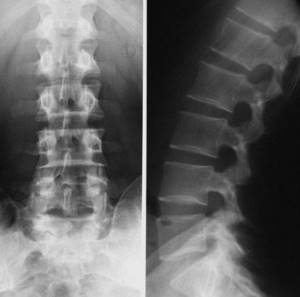
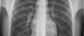
Compared to MRI, x-rays are a more economical and accessible type of examination. Thanks to the effects of radiation, it better visualizes bone tissue, which is why it is successfully used in the diagnosis of vertebral fractures and other consequences of injuries, anomalies in the development of vertebrae, tumors of bone structures, hematomas in the area of the vertebral bodies and the accumulation of fluid in them. If it is not possible to perform an MRI, x-rays can diagnose osteochondrosis, especially if it occurs without complications in the soft tissues. X-ray reveals infectious lesions of the spine in syphilis and tuberculosis, and also helps to detect unstable variants of osteochondrosis (radiography with functional tests is performed).
The main and significant disadvantages of x-rays are the low information content in the study of intervertebral discs, soft tissues, vessels, spinal cord and the presence of radiation exposure.
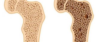
What is osteochondrosis?
The human spine performs three main functions: support, protection and shock absorption. Due to the intervertebral discs, it is able to absorb any load. But over the course of a person’s life, these cartilaginous structures gradually wear out. They, like all tissues of the body, lose moisture, become brittle, the process of fiber disintegration begins, and microtears appear. As a result, the intervertebral discs begin to come into contact with each other and put pressure on the nerve endings, which causes pain. This is how osteochondrosis of the spine is formed.
Ultrasound of the spine or MRI - which is better?
Ultrasound, like MRI, does not involve any radiation and can be performed on patients of any age. The advantage of ultrasound is greater availability and low cost compared to MRI.
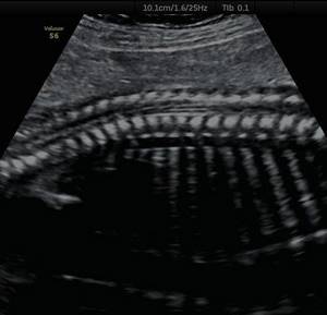
Ultrasound shows the size and location of intervertebral hernias, the condition of the discs, joints and adjacent vessels. This diagnosis is carried out if there is a suspicion of protrusion, osteochondrosis, narrowing of the spinal canal, defects in the development of the spine and vascular disorders.
Due to the inability of ultrasound to penetrate dense tissue, ultrasound of the thoracic region is not performed, which is a significant drawback of the method. Another disadvantage of ultrasound is the requirement for special preparation for diagnosing the lumbar region, since the study is performed through the abdominal cavity, and increased intestinal gas formation will lead to a decrease in the information content of the screening.
Also, ultrasound is based on the diagnostician’s subjective assessment of the picture seen, so the overall result of the study will depend on his qualifications and professionalism.
MRI does not have all these disadvantages. Along with the fact that MR scanning is characterized by the greatest information content and accuracy, its results can be recorded on a medium and transferred to any specialist for independent review of the study in controversial cases.
Why MRI is more effective in detecting spinal problems
The phenomenon of nuclear magnetic resonance (NMR) has been used in MRI techniques since 1938. After Chernobyl, nuclear magnetic resonance imaging was changed to MRI, so that there would be no fear of the nuclear origin of examining internal organs without surgery. In 1960, in the USSR, inventor A.A. Ivanov submitted an application for his device with detailed descriptions, but he received a certificate only in 1984. 1971, Armenian-American R. Damadian received a patent for the idea of “Tumor Detection Using Nuclear Magnetic Resonance.” A scientist used MRI to study spinal diseases
, created an MRI scanner.
Comparison of MRI and X-ray
For fractures, an X-ray examination is used, where a person is exposed to an ionizing flow in small doses. If the procedure is performed frequently, tissue damage may occur.
The patient experiences severe pain in the head due to osteochondrosis of the cervical spine. With an x-ray, the doctor may not see the abnormality. MRI, as it were, checks and reflects the functioning of organs in the images; it can measure the speed of blood flow and the condition of the cerebrospinal fluid. X-rays are prohibited for pregnant women because they involve radiation. MRI has a higher degree of protection, but doctors do not recommend examination in the first trimester.
Thanks to the examination using the device, you can see the whole picture of the disease; spinal pathologies are clearly visible to the specialist conducting the examination. This means that the attending physician will be able to select a more effective treatment and relieve the patient’s pain syndrome.
Possibilities of MRI for spinal pathologies
A person encounters manifestations of the disease in elementary school, and sometimes even earlier. treat osteochondrosis
Modern examination helps qualitatively and productively, identifying signs of a degenerative disease. It usually progresses over a lifetime, so symptoms need to be monitored.
After a certain time, the patient undergoes diagnostics, where the doctor visually determines the behavior of the nerve roots, what changes occur during protrusion of the intervertebral discs, and the proliferation of the vertebrae. On MRI, intervertebral hernias are immediately visible, as well as their size and structure. It is impossible to see the migration of the hernia on an X-ray, but an MRI shows the spinal canal and the displacement of the vertebrae, down to a millimeter. The degree of kyphosis can be determined. After the examination, the doctor can immediately see any deformation of the vertebrae due to this disease. Treatment after MRI is more effective and of higher quality.
Do you need to prepare for magnetic resonance imaging, what requirements should you follow before examining the spine?
- No special medical training.
- There is no need to fast before the procedure.
- There is no need to stop taking medications.
- Be sure to empty your bladder.
- Be patient; you need to lie still for 15-45 minutes.
There is no need to be afraid of the tomograph, it is not scary, and the procedure is completely painless. For those with claustrophobia, operators try to support such patients with conversations.
How is the research process going?
The operator, a medical worker, gives instructions where he tells how the study will take place. It is necessary to remove all metal objects and some clothing. The patient will be alone in the room. He is placed on a special table, headphones are put on, and the process begins. If discomfort or back pain occurs, you can signal to the staff to stop the study. After fixing the problem, the device is turned on and the process continues.
You will not feel the effects of magnetic waves. The study consists of the behavior of hydrogen in diseased and healthy tissues of the body. Signals from organs produce images that are seen by a diagnostic specialist. MRI provides fairly clear diagnostic data for neurosurgeons, neurologists, surgeons, oncologists and other specialists.
Indications for MRI examination
Doctors recommend examinations in the following cases:
- Benign and oncological diseases.
- Problems of the body's nervous system.
- Disruption of the brain, cardiovascular system, breast disease, headache, inflammation of the kidneys and liver.
- Disease of the joints and spine.
This is not a complete list of all diseases that can be diagnosed using magnetic resonance imaging.
Contraindications
What harm does MRI cause to the body? This is a question many patients ask. Now that doctors can help a diseased spine with medications in the early stages of the disease, people have begun to trust the diagnosis using MRI.
The study is sometimes compared to the effects of a cell phone. Of course, there are certain prohibitions. In the first trimester of pregnancy, when the fetus is forming, doctors do not risk conducting research. From the second trimester there is no such risk.
When a person has diseased kidneys or liver and does not tolerate the contrast agent well, detoxification may occur. Here you need the help of an allergist. There are patients with enormous weight, more than 150 kg; unfortunately, it is impossible for them to have an MRI. Pacemakers and all iron objects that are in the human body are completely prohibited. Under the influence of magnetic fluxes, they will cause harm to the patient.
There is no evidence in medical science that the procedure is harmful to health or causes complications in patients.
Among the millions of people who underwent magnetic resonance imaging, there were no cases of poor health or exacerbation of diseases. Author: K.M.N., Academician of the Russian Academy of Medical Sciences M.A. Bobyr
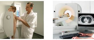
Which is better - MRI or CT scan of the spine?
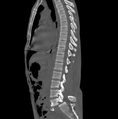
As a result of CT, the doctor also receives layer-by-layer images of the area of interest. However, the main difference between CT and MRI diagnostics lies in the different operating principles of the equipment. MRI does not use the influence of X-rays, while the work of a computed tomograph is based on recording the attenuation of X-ray radiation as it passes through tissues of different densities. In this regard, CT imposes limitations in the diagnosis of pregnant women, children and patients with cancer.
Unlike MRI, CT better visualizes bone tissue and large vessels, which is why it is used to diagnose compression fractures, bruises, cracks and other consequences of spinal injuries. MRI, in turn, is excellent for diagnosing disc herniations, multiple sclerosis, tumors and degenerative processes.
How else does CT differ from MRI of the spine? Both methods have different contraindications, as well as cost - doing a study on a CT machine is somewhat cheaper than an MRI.
Symptoms of osteochondrosis
If you feel the symptoms listed below, think about the condition of your intervertebral discs and the possibility of developing osteochondrosis:
- constant discomfort in the spine, stiffness of movement;
- pain in the back, in the neck, chest, usually aching in nature;
- tinnitus, persistent headache;
- weak back muscle tone;
- numbness of hands and feet;
- pain when changing position or during movement.
Unfortunately, osteochondrosis develops to one degree or another in any person with age. This degenerative change in the spine does not pose a threat in itself. However, complications from it can have irreversible consequences for the body. The consequence of advanced osteochondrosis can be a hernia or the occurrence of diseases associated with impaired blood supply to the brain. In addition, it is impossible to live with persistent back pain all your life. If back pain and discomfort have appeared in your life, do not put off visiting a neurologist and get an MRI of the spine to understand the condition of your intervertebral discs.
| Service | Price according to Price | Discount Price at Night | Discount Price During the Day |
| from 23.00 to 8.00 | from 8.00 to 23.00 | ||
| MRI of the cervical spine | 3300 rub. | 2690 rub. | 2990 rub. |
| MRI of the thoracic spine | 3300 rub. | 2690 rub. | 2990 rub. |
| MRI of the lumbosacral spine | 3300 rub. | 2690 rub. | 2990 rub. |
| MRI of the craniovertebral junction | 3300 rub. | 2690 rub. | 2990 rub. |
| MRI of the sacroiliac joints | 4000 rub. | 3190 rub. | 3690 rub. |
| MRI of the coccyx (MRI of the sacrococcygeal region) | 3300 rub. | 2690 rub. | 2990 rub. |
| MRI of the cervicothoracic spine | 6600 rub. | 5380 rub. | 5980 rub. |
| MRI of three parts of the spine (MRI of the cervical, thoracic and lumbosacral regions) | 9900 rub. | 6900 rub. | 7900 rub. |
| MRI of the entire spine / MRI of the back | 9900 rub. | 6900 rub. | 7900 rub. |
| MRI of the central nervous system (MRI of the brain, MRI of the cervical, thoracic and lumbosacral regions) | 13200 rub. | 9590 rub. | 10890 rub. |
| Appointment with a neurologist | 1800 rub. | free after MRI | free after MRI |
| Comprehensive body diagnostics (MRI of the thoracic spine, MRI of the lumbar spine, ultrasound of the abdominal organs, ultrasound of the kidneys, ultrasound of the bladder, consultation with a neurologist, consultation with a therapist) | 11700 rub. | 7000 rub. |