If a gynecologist has diagnosed you with an ovarian cyst, do not be alarmed: as a rule, these formations are benign and do not pose a danger; surgery is not required. Often they do not need to be treated at all and disappear on their own within a few months. It is enough to periodically visit a gynecologist and undergo an ultrasound examination.
- What types of ovarian cysts are there?
- When is laparoscopy performed?
- When is laparoscopy not performed?
- Benefits of laparoscopy
- Preparation
- How is laparoscopy performed?
- Postoperative period
- Complications
- Is it possible to get pregnant after removing an ovarian cyst?
Sometimes ovarian cysts still have to be removed. Operations can be performed without an incision, through several punctures in the abdominal wall - laparoscopically. Euroonco employs expert-level gynecological surgeons who have extensive experience in performing such interventions. Come for a consultation with our specialist: he will tell you whether surgery is necessary in your case, and prescribe an examination that will help distinguish a benign neoplasm from a malignant one.
A cyst is not a specific disease. This term refers to any pathological cavity with fluid. The reasons for its formation are different. According to statistics, ovarian cysts are diagnosed in every tenth woman. They most often occur during puberty and menopause. Some girls have them from birth.
What types of ovarian cysts are there?
All ovarian cysts are divided into two large groups: functional , which occur in most cases, and pathological , which doctors have to deal with much less frequently.
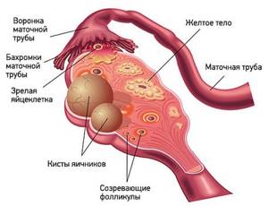
Functional cysts are the result of disturbances during the menstrual cycle. Usually they do not cause complications and disappear on their own. They come in three types:
- Follicular . follicle matures in a woman’s ovaries . Normally, it should open and release the oocyte. If this does not happen and the follicle continues to grow, it turns into a cyst.
- Corpus luteum cysts . After the follicle releases the egg, it turns into a special gland - the corpus luteum . It produces the hormones estrogen and progesterone. If pregnancy does not occur, the corpus luteum atrophies. And if fluid accumulates in it, it turns into a cyst.
- Theca-luteal cysts often develop as a side effect of infertility treatment with hormonal drugs.
Pathological ovarian cysts are not associated with the menstrual cycle. They are always characterized by the appearance of “wrong” cells that should not be present normally. The most common types of pathological cysts:
- Endometrioid . This is a form of endometriosis, a condition in which endometrial tissue (the lining of the uterus) gets into unusual places and grows there. In the ovaries, it can form cavities with fluid. They often appear as “chocolate” cysts filled with dark blood.
- Dermoid cysts , or teratomas , are a special type of benign tumors made from embryonic cells. There may be different tissues inside them, for example, skin, hair, nails. Dermoid cysts become malignant very rarely.
- Cystadenomas are benign neoplasms of epithelial cells. Usually they are filled with contents of a mucous or watery nature.
For more information about malignant cysts, please visit the ovarian cancer page on our website.
When is laparoscopy performed?
If the ovarian cyst is small, does not cause symptoms and does not look cancerous, it can be left alone. No surgery needed. The gynecologist will prescribe periodic examinations and control ultrasounds. Moreover, postmenopausal women will have to do this more often, because they have a higher risk of malignant tumors.
Are there effective medications?
In some cases, hormonal contraceptives may be helpful. They help prevent the formation of new cysts in the ovaries, but do not affect the growth of existing ones. If a woman is bothered by pain, the doctor may prescribe drugs from the group of nonsteroidal anti-inflammatory drugs (NSAIDs). But this is only symptomatic treatment. The only way to get rid of the formation is surgery.
If the cyst is “problematic,” then the doctor will definitely say that it needs to be removed. Indications for surgery:
- Large size of formation. In most cases, ovarian cysts have a diameter of 1–3 cm. Very rarely they reach 15–30 cm.
- Presence of symptoms: abdominal pain, pelvic pain, bloating, feeling of heaviness in the abdomen, heavy periods, vaginal bleeding not associated with the menstrual cycle. A large cyst can compress the intestines and bladder. This leads to problems with stool and urination.
- Suspicion of a malignant nature of the cyst - the risks are increased in postmenopausal women.
- Continued growth over 2–3 menstrual cycles.
- Pathological ovarian cyst.
What happens if the cyst is not removed?
If the doctor said that the cyst needs to be removed, then delaying the operation is primarily dangerous for postmenopausal women. They, as we have already mentioned, have a higher risk that the formation may turn out to be malignant. And with cancer, time is critical. The later treatment is started, the lower the chances that it will be successful and that remission will be achieved. The prognosis worsens and survival rate decreases.
At the Euroonko clinic you can receive treatment for ovarian cancer according to modern standards. Our doctors perform operations of any complexity; we have access to all the latest generation antitumor drugs registered in Russia. We work according to modern European, American, Israeli treatment protocols. For ovarian cancer complicated by peritoneal carcinomatosis, we use an innovative technique - hyperthermic intraperitoneal chemotherapy (HIPEC).
With benign ovarian cysts, serious complications can also occur, although they are rare. Large cysts may rupture. In this case, severe bleeding develops into the abdominal cavity, and severe abdominal pain occurs.
As the cyst grows, the risk of ovarian torsion increases. Due to compression of the blood vessels, it stops receiving blood supply, severe abdominal pain, nausea, and vomiting occur. The result may be necrosis (death) of the ovary, and it will have to be removed urgently.
If you experience symptoms such as severe abdominal pain, nausea, vomiting, or a fever of more than 38 degrees, you should immediately seek medical help.
Indications and contraindications for laparoscopic tubal surgery
Indications for laparoscopic operations on the fallopian tubes are:
- occlusion of the lumen of the fallopian tubes;
- the presence of peritubar adhesions;
- tubal pregnancy;
- salpingitis, sacto(pio)salpinx;
- sterilization.
Laparoscopic operations on the fallopian tubes (tuboplasty) are contraindicated if:
- inflammatory process in the lower genital tract;
- active inflammatory process of the pelvic organs (in less than 4-6 months);
- fallopian tube length less than 4 cm;
- the presence of a huge sactosalpinx, after removal of which the remainder of the tube will be less than 4 cm;
- obstruction in several parts of the fallopian tube, including in the isthmic section.
To determine the type of infertility and indications for surgery on the fallopian tubes, as well as choose the correct tactics of surgical treatment, you must send me a , blood results for hormones, and indicate age and chief complaints. Then I will be able to give a more accurate answer to your situation.
When performing laparoscopic operations in gynecological practice, the following instruments and equipment are used:
- endovideo system, including an optical system with a video camera, a light guide and a medical monitor;
- laparoscope;
- gas insufflator;
- aspirator-irrigator;
- electrosurgical unit;
- Veress needle;
- 5 mm trocars – 2;
- 10 mm trocar – 1;
- soft and hard 5 mm clamps, 5 mm dissector, 5 mm endhook;
- monopolar scissors;
- bipolar forceps;
- needle holder;
- suture material: “Polysorb” or “Vicryl” 4-0, 6-0;
- uterine retractor "Clermontferan" (Germany) or hysterograph, which ensures a change in the position of the uterus during the operation and the introduction of contrast.
Surgical correction of tubo-peritoneal infertility is carried out in the 1st phase of the menstrual cycle in order to ensure optimal conditions for tissue regeneration and the possibility of carrying out rehabilitation measures.
In this case, you must be guided by the following provisions:
- all operations should be performed using 2-3 manipulators;
- all manipulations and displacement of organs should be performed only with the help of atraumatic instruments;
- tissue dissection in all cases is carried out after preliminary coagulation;
- all types of plastic surgery on the fallopian tubes should be performed after tightly filling them with methylene blue;
- At the end of the intervention, the pelvic cavity should be washed with sterile saline and anti-adhesion gel should be injected.
The tubes are inspected before the contrast is introduced (methylene blue solution), and then the movement of the latter through the tube and its outflow from the fimbrial region are traced.
Before attempting any surgical manipulation related to the fallopian tubes, it is advisable (if technical capabilities are available) to perform a tuboscopy to assess the condition of the endosalpinx and identify the degree of disruption of its folding, which has a very great prognostic value. Attempts to restore the patency of the fallopian tube when its mucosa is in poor condition do not give a positive effect; the method of choice for treating these patients is in vitro fertilization.
Through the umbilical access, a Veress needle is used to apply carboxyperitoneum in a volume of 3-4 liters to an intra-abdominal pressure of 12-15 mmHg. Interventions are performed from three approaches, using two 5-mm trocars and one 10-mm trocar for optics (Fig. 1). The fourth, 5-mm trocar, can be inserted as needed (in the case of severe adhesions in the pelvis). A uterine manipulator or hysterograph is inserted into the uterus, fixed on bullet forceps, which ensure a change in the position of the uterus during the operation, which makes it easier to manipulate the abdominal cavity and perform chromosalpingoscopy.
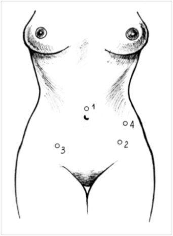
Fig. 1. Points of insertion of trocars during operations on the fallopian tubes (1 - 10 mm trocar, 2,3,4 - 5 mm trocars), the 4th trocar is placed extremely rarely
Preparation
Ovarian cysts are usually detected during an ultrasound examination. In order to better assess the size, location and internal structure of the neoplasm, the condition of the ovary, they usually resort to not only transabdominal (through the abdominal wall), but also transvaginal (using a special sensor inserted into the vagina) ultrasound.
In rare cases, usually when a malignant nature of the formation is suspected, computed tomography and diagnostic laparoscopy are prescribed. of cancer antigen 125 (CA 125) in the blood testifies in favor of cancer . But this analysis is unreliable, since a positive result can be obtained for uterine fibroids, endometriosis and inflammatory processes in the pelvic organs.
Based on the examination results, the doctor advises the woman and explains which treatment tactics will be optimal in her case. If laparoscopic surgery is indicated, a date for hospitalization is set. You need to undergo a preoperative examination. It usually includes the following diagnostic methods:
- General and biochemical blood tests.
- General urine analysis.
- Blood test for hormone levels.
- Tests for infections.
- Cervical smears - cytological, flora.
- Blood clotting study.
- Determination of blood group AB0, Rh factor.
- Electrocardiography.
- X-ray of the lungs.
Laparoscopic removal of ovarian cysts is performed under general anesthesia - endotracheal anesthesia. The woman is hospitalized in the hospital the day before surgery. You can’t eat anything for 8 hours before surgery, and you can’t drink anything in the morning. Some time before anesthesia, premedication : the woman is given drugs that help her relax and calm down.
Laparoscopy in gynecology
Laparoscopy
is one of the minimally invasive types of surgical intervention in gynecology, with the help of which it is possible to diagnose and treat the organs of the reproductive system without an abdominal incision. This method is suitable for most gynecological operations.
To access the operated organs, the doctor makes punctures measuring 5-7 millimeters. After the operation, the patient does not require long-term rehabilitation, since the sutures heal quickly enough and cause less discomfort than during a full abdominal operation.
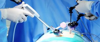
During laparoscopy, a special device is inserted into the operated area - a laparoscope, which is a flexible tube equipped with a system of optical lenses, a video camera, special sensors and lighting.
The image from the video camera, an image enlarged 40 times, is transmitted to the monitor, which allows the operating doctor to examine the reproductive organs that cannot be seen during a regular gynecological examination, as well as to identify disorders and carry out surgical treatment.
Types of laparoscopy
Depending on the scope of surgery, there are several types of laparoscopy:
- diagnostic - used to clarify or establish an accurate diagnosis and prescribe further treatment tactics, since during a detailed examination of internal organs the doctor can identify abnormalities that are not visible during ultrasound diagnostics
- minor laparoscopic operations, these include laparoscopic sterilization, ovarian biopsy, puncture and aspiration of ovarian cysts, coagulation of endometriotic lesions, adhesiolysis with a minimally expressed adhesive process (removal of adhesions from the pelvic organs)
- Major laparoscopic surgeries are used in the treatment of stage 2 and 3 endometriosis, plastic surgery on the fallopian tubes with a diagnosis of infertility, tubectomy and salpingotomy for ectopic pregnancy, cystectomy
- Laparoscopic operations of increased complexity, this type of laparoscopy is used in the treatment of severe and chronic diseases, such as treatment of endometriosis grades 3 and 4, treatment of genital prolapse, myomectomy, hysterectomy, lymphadenectomy, radical hysterectomy, removal of ovarian cysts, ovarian resection.
Indications for laparoscopy
Laparoscopy is prescribed exclusively by a doctor, after conducting research, as a result of which the indications for its implementation are divided into several types:
Planned indications:
- tubo-peritoneal infertility
- formations in the uterus, ovaries and uterine appendages (tumors, tumor-like formations, fibroids, adhesions, polyps)
- endometriosis of the ovaries or pelvic peritoneum;
- developmental anomalies of the internal genital organs;
- pain of unknown etiology;
Indications for emergency laparoscopy:
- ectopic pregnancy;
- ovarian apoplexy;
- acute inflammatory diseases of the pelvic organs
- suspicion of torsion of the pedicle or rupture of formations in the ovaries, as well as torsion of subserous fibroids;
- differential diagnosis between acute surgical and gynecological pathologies.
Contraindications for laparoscopy
Contraindications before laparoscopy can be absolute and relative: Absolute contraindications include:
- hemorrhagic shock;
- diseases of the cardiovascular and respiratory systems in the stage of decompensation;
- uncorrectable coagulopathy;
- diseases for which it is unacceptable to place the patient in the Trendelenburg position, for example, previous brain injuries or cerebral vascular lesions;
- acute and chronic hepatic-renal failure;
- ovarian cancer and RMT (with the exception of laparoscopic monitoring during chemotherapy or radiation therapy).
Relative contraindications are:
- polyvalent allergy;
- diffuse peritonitis;
- pronounced adhesive process after previous operations on the organs of the reproductive system;
- gestational age (starting from 16-18 weeks);
- suspicion of the presence of malignant tumors in the area of the uterine appendages.
The following are also contraindications to elective laparoscopy:
- acute infectious and colds suffered less than 4 weeks ago;
- degree 3-4 purity of vaginal contents;
- Poorly conducted examination in the treatment of infertility.
How to prepare for laparoscopy in gynecology
Laparoscopy requires mandatory patient preparation. As prescribed by the attending physician, the patient must undergo a number of laboratory tests, including a mandatory ECG, ultrasound of the pelvic organs, standard urine and blood tests, as well as a vaginal smear.
During the seven days of the scheduled operation, doctors at the OXY-center clinic recommend that the patient exclude smoked, fatty and high-carbohydrate foods from the diet. During the last 3-4 days before surgery, the amount of food consumed should also be strictly controlled and gradually reduced.
List of non-recommended products:
• milk and black bread; • fatty meat and potatoes; • apples and plums; • all legume products; • fresh and salted cabbage; • eggs and black bread.
During this period, it is better to eat only low-fat foods, for example, dairy products, fish, broths and cereals. The day before surgery, a cleansing enema is prescribed. Medication preparation depends on the disease, the planned volume of surgical intervention and can only be prescribed by the attending physician.
The minimum scope of examination for surgical treatment (hysteroscopy) according to the order of the Ministry of Health of the Russian Federation 572N:
- Microscopic examination of the discharge of the female genital organs for aerobic and facultative anaerobic microorganisms (smear for flora)
- Ultrasound examination of the pelvic organs (genitals)
- Ultrasound of the mammary glands (once a year) or mammography (first mammography at 35-36 years old, once every 2 years at 35-50 years old, once every 2 years over 50 years old)
- Colposcopy, smear cytology (PAP test)
- UAC
- OAM
- Biochemical blood test: study of the level of total blood protein, creatinine, level of alanine transaminase (ALT), aspartate transaminase (AST), urea, total bilirubin, direct bilirubin, blood glucose, cholesterol, sodium, blood potassium
- Coagulogram
- Determination of blood group and Rh factor
- Determination of antibodies to Treponema pallidum in the blood
- HIV
- HBsAg, HCV
- X-ray examination of the chest organs (fluorography) - once a year
- Electrocardiography (ECG)
- Consultations: general practitioner, related medical specialists (as indicated)
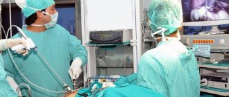
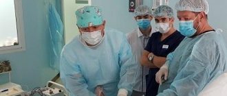
How is laparoscopy performed?
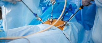
Laparoscopic intervention is performed through several punctures in the abdominal wall. Through one of them, in the navel area, a laparoscope - an instrument with a miniature video camera. It broadcasts an enlarged image onto the screen. For better visualization during surgery, the abdominal cavity is filled with gas.
Special laparoscopic instruments are inserted through additional punctures, and the cyst is removed with the help of them.
Depending on the specific situation, the scope of the operation varies:
- Often it is possible to remove only the cyst, preserving the ovary.
- In some cases, it is necessary to remove the entire ovary - to perform an oophorectomy (oophorectomy) - while the second ovary can be saved. This has to be done if a malignant tumor is suspected, or if the cyst is in an “inconvenient” location, when it is difficult to remove it separately.
- In rare cases, both ovaries may have to be removed. This operation is performed only in extreme cases, especially in women of reproductive age, since menopause occurs after removal of both sex glands. The levels of female sex hormones decrease, and this can lead to symptoms such as headaches, dizziness, nausea, hot flashes, etc.
Don't hesitate to ask your doctor questions before surgery. Ask what is the likelihood that during the operation you will have to remove both ovaries, what negative consequences this threatens, and how to cope with them.
Laparoscopic surgery - salpingo-ovariolysis
The purpose of the operation is to restore normal topographic relationships by cutting the adhesions around the fallopian tube and ovary, isolating them from each other. Salpingo-ovariolysis is performed either as an independent intervention or as a preparatory step for surgery on the fallopian tubes. The fallopian tube (ovary) is picked up with atraumatic forceps and moved upward if possible. The adhesions are cut with endoscissors after their preliminary coagulation. Rough adhesions are excised and removed from the abdominal cavity. Special precautions are necessary when working near the intestines, ureters, and large vessels. The risk of their damage can be reduced by observing the following conditions: the operation is performed under general anesthesia with sufficient relaxation, the absence of defects in the insulating braid of the instruments, short-term switching on of the electrosurgical unit. After complete release of the fallopian tube from adhesions along its entire length, ovariolysis is performed. In this case, it is imperative to lift the ovary and inspect its surface facing the broad uterine ligament, since adhesions can often be localized there.
Operation of salpingostomy (neostomy) and methods of laparoscopic fimbrioplasty
Fimbryolysis is performed for phimosis of the fimbrial part of the fallopian tube. While maintaining tight filling of the fallopian tube, endoscissors are used to gradually dissect along the radial scars and the center of the stellate scar, if any. After that, atraumatic forceps are inserted into the lumen of the tube in a closed state, the jaws are opened to a width of 2.5-3 cm and removed in this position. The procedure is performed 2-3 times.
Salpingostomy is performed when the tube is obstructed in the ampullary section. The ampullary section is fixed between two clamps against the background of tight filling with a methylene blue solution, which will allow you to correctly determine the location for the incisions. Endo-scissors are used to crosswise dissect the sealed ampullary section of the fallopian tube with two incisions 2-3 cm long along the scar changes so that they intersect at the pole of the hydrosalpinx (Fig. 2). Thus, 4 flaps (petals) are formed.
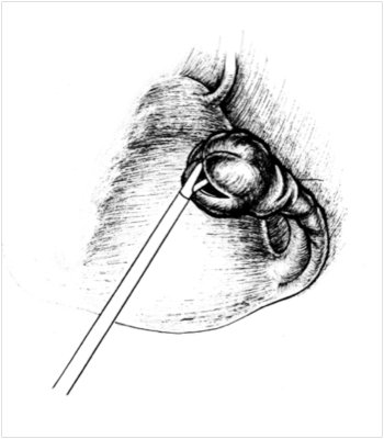
Figure 2. Cross-shaped dissection of the ampullary part of the fallopian tube during salpingostomy (Puchkov et al, 2005)
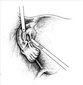
Figure 3. Inversion of the petals and their fixation with sutures for salpingostomy (Puchkov et al, 2005)
A soft clamp is inserted into the lumen of the fallopian tube and fixes its wall. Using a dissector, grasping the edge of the fimbrial section, the tube is turned out for 2-3 cm. To prevent it from returning to its previous position, the petals are fixed with 2-3 sutures with threads no thicker than 4-0 gauge, using the technique of intracorporeal knot tying (Fig. 3). If it is not possible to apply sutures, to maintain the tube in a passable state, you can use bipolar coagulation, which will “glue together” the serous cover of the peritoneum of the inverted section of the tube in the form of a duplicate. Hemostasis is achieved by point coagulation with a dissector. If possible, bleeding vessels of the mucous membrane are not coagulated to avoid damage.
There is a simpler salpingostomy technique - point endocoagulation (both mono- and bipolar coagulator) of the peritoneum of the fimbrial section dissected crosswise at a distance of 0.5-1.0 cm from the edge of the tube mouth along its perimeter. The endocoagulation areas should be separated from each other by a distance of 0.7-1.0 cm. As a result of endocoagulation, the outer layer of the pipe wall contracts and the edges of the stoma turn outward, which prevents them from gluing in the postoperative period. Salpingoneostomy involves the creation of a new artificial opening in the ampullary section of the fallopian tube. The operation is performed when it is impossible to open the tubal lumen in the fimbrial region. After filling the tube with methylene blue at the site of the intended dissection of the wall on the side opposite the mesosalpinx, linear coagulation is performed with a point coagulator for 2-3 cm along the ampullary section, followed by opening the lumen with an endo-hook. Inversion and fixation of the edges of the neostomy is carried out using one of the above methods.
Mechanical restoration of fallopian tube patency , assessed by laparoscopy, does not guarantee its normal functional activity. Tube function can be assessed by examining pregnancy rates. The final result depends on many factors: the duration of occlusion, the severity of the adhesive process in the pelvis, the degree of damage to the organ - the state of the mucous and muscular layers that ensure the transport of the egg to the uterus, the severity of the cilia and the smoothness of the endosalpinx folds, the presence of polyps of the mucous membrane, the degree of thickening of the tube wall, the presence of functionally complete fimbriae, the length of the tube, the size of the hydrosalpinx (more than 3 cm in diameter), as well as the surgical technique used. The simultaneous use of means to prevent postoperative adhesions (introduction of anti-adhesive gel, antibacterial and physiotherapeutic treatment) helps to improve the results of the intervention.
Fallopian tube resection surgery - salpingectomy and salpingotomy
Salpingectomy is an operation to remove the fallopian tube from one or both sides, which is performed in the following cases:
- with a disturbed tubal pregnancy, accompanied by pronounced changes in the tubal wall;
- for chronic salpingitis that is not amenable to conservative treatment, especially in the presence of hydrosalpinx.
Removal of the fallopian tube, which has changed due to inflammatory processes and has no prospects for restoring its function, can be a preparatory step for subsequent in vitro fertilization.
A number of salpingectomy methods have been described in the literature.
Using an instrument inserted through the left lateral port, the fallopian tube is grasped and pulled up, creating exposure. Using an electrosurgical dissector in the mode of alternating monopolar coagulation and cutting, the fallopian tube is crossed at a distance of no more than 1 cm from the angle of the uterus and its mesentery in close proximity to the latter. Knowledge of the anatomical location of r.tubarius will help avoid damage to the vessels of the mesosalpinx and the occurrence of intraligamentary hematoma (Fig. 4).
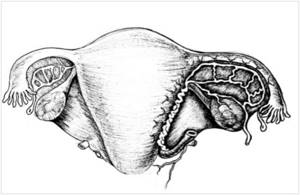
Figure 4. Location diagram of r.tubarius indicating areas of coagulation and intersection during salpingostomy (Puchkov et al, 2005)
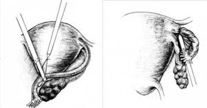
Figure 5. Performing salpingectomy using a loop ligature: a) applying a ligature to the fallopian tube; b) tightening the ligature (Puchkov et al., 2005)
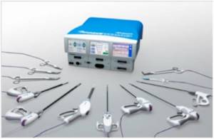
Fig.6. Force Triad LigaSure device with a set of tools
Mobilization of the above-mentioned formations can also be performed by step-by-step coagulation with bipolar forceps followed by dissection with endoscissors. The third option for tubectomy is to apply a loop ligature with a 2-0 absorbable thread to the mesosalpinx and the uterine end of the tube, followed by cutting off the tube with endoscissors (Fig. 5 a, b). The proximal section of the tube is pre-coagulated as close to the uterus as possible in order to exclude the possibility of subsequent pregnancy developing in the stump of the removed tube. At the end of the operation, careful sanitation of the entire abdominal cavity is necessary, including the subdiaphragmatic space, lateral canals, and surgical field.
Recently, in operative gynecology, electrosurgical platforms of a completely new generation Force Triad “LigaSure” (Switzerland) have begun to be used for practical use. This device includes both monopolar and bipolar electrosurgical units, as well as an improved system of dosed ligation technology for vascularized tissue, which provides dosed energy supply depending on the properties of the coagulated tissue (tissue impedance). According to a number of foreign and Russian authors, dosed bipolar electrocoagulation makes it possible to stop bleeding and reliably close vessels with a diameter of up to 7 mm. When the device acts on the tissue, the liquid component is evaporated, collagen and elastin are dried and denatured until a dense homogeneous mass is formed that reliably closes the lumen of the blood vessels. Thus, the use of the LigaSure device for hemostasis during endosurgical mobilization of organs and closure of large vessels can serve as a universal way to stop bleeding. The use of this device when performing tubectomy reduces the operation time to 5 minutes and reduces the amount of blood loss to zero.
Performing salpingotomy with preservation of the fallopian tube during ectopic pregnancy is advisable:
- when the diameter of the fertilized egg is no more than 4 cm;
- in the presence of a developing pregnancy in the ampullary region;
- in the absence of tissue imbibition and arosion of the vessels of the pipe wall.
After dissecting the peritubar adhesions and fixing the tube with a soft clamp, the antimesenteric edge is opened longitudinally with a needle electrode or scissors above the proximal area of dilation, where the fertilized egg is usually located (the distal part of the dilation of the fallopian tube is filled with blood clots). The fertilized egg is easily removed by pressing from the side of the serosa or peeled off using hydraulic preparation. With the correct choice of incision site and precision surgical technique, it is possible to completely remove the fertilized egg and detach the chorion without bleeding. The lumen of the fallopian tube is thoroughly washed and hemostasis is controlled. If there are bleeding areas in the placental area, excessive coagulation should be avoided and carried out using microsurgical forceps. If it is not possible to identify individual vessels, then, according to V.I. Kulakov, L.V. Adamyan, a continuous suture or coagulation can be applied to the mesosalpinx in this area. The hole in the fallopian tube is usually not sutured. Both edges of the wound are usually well matched. Studies conducted by Tulandi T., Guralnick M., Nelson L. have proven that suturing during these operations worsens the effectiveness of surgical treatment, compared with sutureless surgery, due to deformation of the fallopian tube. In this regard, they believe that suturing the tubotomy incision is inappropriate. Complete healing of the wound with restoration of integrity and patency occurs in 3-6 months. At the final stage of the operation, chromosalpingoscopy is performed to determine the patency of the fallopian tube.
A specific postoperative complication of salpingotomy performed for tubal pregnancy is persistent ectopic pregnancy - the growth of trophoblast tissue after incomplete removal of the fertilized egg. This dictates the need to determine the beta-hCG titer over time after surgery. If it decreases, then you can stick to wait-and-see tactics. If the level of beta-hCG remains the same or increases, surgical intervention is indicated to remove the remaining part of the ectopia or the prescription of a short course or a single dose of methotrexate.
Complications
Laparoscopic operations are associated with a low risk of complications. As after any surgical intervention, in rare cases, infection and suppuration in the area of surgical sutures and bleeding are possible. If you are concerned about severe pain, vaginal bleeding, or increased body temperature, you should immediately consult a doctor.
Adhesions may occur after surgery. Very rarely, during surgery, damage to neighboring organs (intestines, bladder) is possible.
At the Euroonko clinic, operations are performed by experienced gynecological surgeons. Our operating room is equipped with modern equipment, multifunctional laparoscopic stands from leading manufacturers. This helps to minimize risks and perform the operation as safely as possible.
After removal, the cyst may recur, in the same ovary or on the other side. Only removal of both ovaries helps to completely eliminate relapse. But such an operation leads to menopause and undesirable consequences, so it is resorted to only in extreme cases.
Is it possible to get pregnant after removing an ovarian cyst?
You can get pregnant even with an ovarian cyst. Most often, they do not interfere with pregnancy, but they can make it difficult to conceive a child.
If during surgery only the cyst is removed or at least one ovary is left, the woman’s fertility is preserved. She may become pregnant in the future. Of course, you need to understand that reproductive function is affected not only by surgery. The ovarian reserve (the number of eggs in the ovaries - it constantly decreases with age) and concomitant diseases play a role.
Get a consultation with a doctor at the Euroonko clinic. Our doctor will tell you whether treatment is needed in your case, what type of surgery is indicated for you, and what is the likelihood that you will be able to conceive and carry a pregnancy in the future.
Book a consultation 24 hours a day
+7+7+78