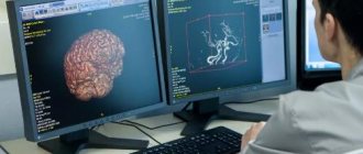
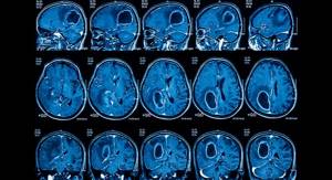
Magnetic resonance imaging of the brain is a highly informative study that allows you to study in detail brain tissue, blood vessels and other intracranial structures, and diagnose many diseases.
Our expert in this field:
Sergeev Pyotr Sergeevich
Oncologist, surgeon, chemotherapist, Ph.D.
Call the doctor Reviews about the doctor
During this procedure, a strong magnetic field is applied, which produces slice-by-slice images of the head and three-dimensional images. In some cases, contrast is used for better visualization. MRI of the brain and cervical spine is widely used in neurology, neurosurgery, when tumors and vascular pathologies are suspected.
We invite you to undergo an MRI of the brain at the Medica24 International Clinic using a modern machine. Based on the results of the study, an experienced medical specialist will consult you, establish an accurate diagnosis and prescribe effective treatment.
In what cases is MRI of the brain performed? Types of research.
Typically, patients are referred for brain MRI by neurologists and neurosurgeons. You can, of course, sign up for the study yourself, but if you are bothered by certain symptoms, it is better to first visit a doctor, he will prescribe the necessary types of diagnostics.
Magnetic resonance imaging helps diagnose many diseases of the central nervous system:
- hydrocephalus (“dropsy of the brain”);
- various developmental anomalies;
- infectious processes;
- causes of seizures in patients with epilepsy;
- tumors of the brain and spinal cord;
- intracranial hemorrhages;
- causes of visual and hearing impairment;
- multiple sclerosis, Alzheimer's disease, Parkinson's disease;
- arterial aneurysms, arterial occlusions (blockages), venous thrombosis.
We will call you back, leave your phone number
Message sent!
expect a call, we will contact you shortly
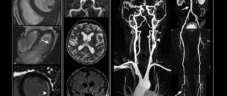
MRI of the cerebral arteries is performed with contrast. A special drug is injected intravenously, which will “paint” the blood vessels and allow them to be clearly outlined in the photographs.
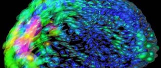
Using functional magnetic resonance imaging, you can evaluate the activity of different areas of the brain. This helps to obtain valuable information in a number of pathologies, such as neurodegenerative diseases.
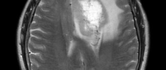
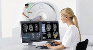
What does MRI diagnose?
The innovative diagnostic MRI device General Electric Optima MR360 has a large number of programs that allow you to examine any part of the body. MRI is divided according to the area being examined.
MRI of the head includes examination:
- brain matter
- pituitary gland
- soft tissue
- large vessels
- contents of the orbit
- ENT organs (nasal cavity, nasopharynx, larynx, middle and inner ear, paranasal sinuses)
This type of diagnosis is one of the most advanced and effective ways to diagnose head diseases, including diseases of neurological origin.
MRI of the spine includes examination:
- intervertebral discs
- lumen of the spinal canal
- spinal cord and surrounding tissues
- vertebral bone tissue
- ligamentous apparatus
- nerve plexuses
MRI of soft tissues includes examination:
- lymph nodes
- vessels of intermuscular and synovial tissue
- skin-fat layer
- connective tissue layers
- striated muscles
MRI of joints includes examination:
- temporomandibular joints
- shoulder joint
- elbow joint
- wrist joint
- brushes
- hip joints
- knee joint
- ankle and foot
MRI of the abdominal cavity includes examination of the following organs:
- liver
- kidneys and adrenal glands
- pancreas
- stomach, large and small intestines
- gallbladder and bile ducts of the liver
- spleen
- The lymph nodes
- blood vessels
- soft tissues of the peritoneum, retroperitoneum
MRI of the pelvic organs
Area to be examined in women:
- rectum
- bladder
- ovaries
- vagina
- uterus
- blood vessels, lower spine, soft tissue, lymph nodes
Area to be examined in men:
- rectum
- bladder
- prostate
- vas deferens
- scrotum
CT or MRI?
Both studies work approximately the same way and create highly informative images with layer-by-layer sections of a certain area of the body, three-dimensional images. But during a CT scan, X-rays are used, and during an MRI, a strong magnetic field is used.
Computed tomography better visualizes the bones of the skull and vertebrae, and intracranial hemorrhages. In addition, it is cheaper and simpler than MRI, so it is more preferable for quickly examining people who have suffered a traumatic brain injury.
MRI images clearly show ligaments and tendons, nerve fibers, and tissue of the brain and spinal cord. This study is better suited than CT for diagnosing benign and malignant neoplasms of the nervous system.
Come to an appointment with medical specialists at the Medica24 International Clinic, they will assess your situation and tell you whether you need to do an MRI of the head and cerebral vessels, or undergo other diagnostic procedures.
We will call you back, leave your phone number
Message sent!
expect a call, we will contact you shortly
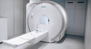
Equipment in the clinic
MRI at the Moscow clinic is performed using the latest American tomograph General Electric Optima MR360 .
Advantages of the device:
- Expert-level research thanks to 1.5 Tesla power
- scanning area 48 cm
- allows you to take pictures with a slice step of 1 mm
- Thanks to OpTix RF technology and gradient coils, the tomograph allows you to obtain high-precision, clear images and track the function of the internal organ over time
- allows you to visualize any area of the body and scan tissue even in hard-to-reach places, for example, mammary glands, small vessels
- allows detection of small tumors
- supports patient weights up to 150 kg
General Electric Optima MR 360 provides environmental safety for patients and allows you to obtain accurate detailed information about the organ/group of organs being examined.
Specialists conducting diagnostics using this device can receive and process data in both two-dimensional (2D) and three-dimensional (3D) planes.
How to prepare for the procedure?
Magnetic resonance imaging does not require special preparation. Some clinics give patients advice on what to eat the night before and on the day of the test. If your doctor does not advise you to do so, you can eat and take your medications as usual.
Before the procedure at the Medica24 International Clinic, you will be consulted by a doctor. If you suffer from any diseases, allergies, or have recently undergone surgery, you must inform your doctor about this. If a patient has kidney problems, blood tests may be needed to evaluate kidney function. In some cases, radiography may be required to check whether there are metal implants or foreign bodies in the patient's body.
When going for the procedure, you do not need to wear any metal jewelry - they will still have to be removed before the study, because a strong magnetic field is used during MRI. Do not wear clothes with metal buttons or fasteners.
Preparing for an MRI
Before performing the procedure, the specialist will ask the patient about the presence of contraindications. To perform an MRI of the spine, the specialist may ask the patient to change his clothing to a disposable robe. Before the examination, the patient is asked to remove clothing that contains metal parts.
When diagnosing the gastrointestinal tract, you must completely abstain from eating 7-8 hours before the procedure. The large intestine is checked only for “clean” intestines, that is, it will be necessary to first perform a cleansing enema.
To conduct an MRI of joints, muscle fibers, chest, and brain, no preliminary steps are required.
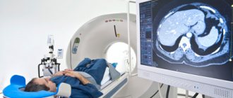
How is an MRI of the brain performed?
If an MRI of the brain is being performed without contrast, you will immediately be asked to go into the office and lie down inside the machine. If contrast is required, you will first be given gadolinium intravenously.
You must lie still during the examination to ensure clear images. The diagnostic table moves, while the device takes a series of images. The doctor is in the next room and communicates with the patient through a microphone.
How long does a brain MRI take? On average, the study lasts 45 minutes, the total time depends on the need to use contrast and perform a functional MRI.
Where can I get a referral?
A referral for an MRI can be prescribed by a doctor of any specialization. Most often, such examinations are prescribed by neurologists. Thus, they look for the cause of frequent headaches in patients. MRI examinations are performed before or after neurosurgery. An endocrinologist can also prescribe a tomography of the head. The results will help him determine if there are problems in the pituitary gland.
In order to receive a free referral, you need to contact the public medical institution to which you are assigned. Every year quotas are allocated for these purposes. After a preliminary examination, the doctor will determine how urgently you need the procedure. After that you will be able to get into the queue.
Today, the following procedure has been developed for referring patients for free MRI:
- After the examination and necessary tests, the specialist makes a conclusion about the need for research.
- The results are sent to the medical commission. Based on its results, the need for a free MRI examination is determined.
- If the application is approved, it is transferred to the specialists of the MRI room. They set a specific date for the procedure.
- The completed referral is certified by the head of the medical institution.
- The patient must appear for examination at the agreed time. At the same time, in addition to the referral, he must have test results and recommendations from the attending physician.
The specialist who referred for the examination will subsequently receive its results and develop a competent treatment strategy.
To receive a free referral, you will need to provide the following documents to the medical institution:
- Conclusion from your attending physician.
- Passport of a citizen of the Russian Federation.
- SNILS.
- Original compulsory health insurance policy.
If your medical institution has quotas for free procedures, you will definitely be put on a waiting list.
How safe is a head MRI? Are there any contraindications?
Magnetic resonance imaging does not use X-rays, so it is safer than x-rays and computed tomography. However, there are some contraindications.
First of all, this is the presence of any metal objects in the patient’s body, such as pacemakers, defibrillators-cardioverters, metal clips on blood vessels, embolic coils, some types of cochlear implants, and foreign bodies.
There have been many studies on the safety of MRI during pregnancy. No risks were found, however, in pregnant women this study is carried out with caution and only in cases where it cannot be avoided. Magnetic resonance imaging is especially not recommended in the first trimester, when the organs of the unborn child are forming.
During an MRI of the brain, only the head is inside the machine, however, difficulties may arise for people suffering from claustrophobia.
The procedure is problematic if the patient suffers from a mental disorder and is in a state of agitation and cannot lie still. Most devices are not designed for people weighing more than 150 kg.
The main contraindications for MRI with contrast are the patient's allergy to the drug and severe renal impairment.
We will call you back, leave your phone number
Message sent!
expect a call, we will contact you shortly
Risks of Magnetic Resonance Imaging
Because MRI uses powerful magnets, the presence of metal in your body may pose a safety risk. Metal objects can distort the MRI image. Before your MRI, you will likely fill out a questionnaire that will tell you whether you have metal or electronic devices in your body.
If your device is not certified as MRI safe, you will not be able to have an MRI. List of devices that need to be reported:
- Metal joint prostheses
- Artificial heart valves
- Implantable cardiac defibrillator
- Implanted infusion pumps
- Implanted nerve stimulators
- Pacemaker
- Metal clamps
- Metal pins, screws, plates, stents, or surgical staples
- Cochlear implants
- Bullet, shrapnel, or any other type of metal fragment
- Intrauterine device
If you have tattoos or permanent makeup, ask your doctor if they may affect your MRI. Some types of ink contain metal.
Pregnancy is a relative contraindication to MRI . The effect of magnetic fields on the fetus has not been sufficiently studied. Your doctor may recommend alternative testing or postpone the MRI. Also tell your doctor if you are breastfeeding, especially if you want to have a procedure with contrast.
It is also important to discuss kidney or liver problems with your doctor and MRI technologist because problems with these organs may limit the use of injectable contrast agents during the scan.
How much does a brain MRI cost?
Prices for MRI of cerebral vessels in Moscow vary, depending on the pricing policy of a particular clinic, the model of the device (more modern and high-precision ones are usually more expensive), and related medical services (consultation with a doctor, blood tests, radiography). In our International Clinic Medica24, you can conduct a routine examination and MRI of the brain with contrast in Moscow using one of the latest generations of equipment and get a consultation from an experienced doctor cheaper than in many other medical centers.
For more information, to schedule an initial consultation and to undergo a head MRI at our clinic, please contact us by phone
Is it possible to get an MRI without a referral?
Do I need a referral for an MRI? The answer is clear: yes. Today, many paid clinics offer such examination. But even for money, without a doctor’s referral, this cannot be done. Specialists need at least minimal information about your health. Before being examined, you will need to provide the following information:
- Are there any contraindications to MRI?
- Will it be necessary to administer a contrast agent?
- The results of some tests and a preliminary examination by the attending physician.
Without such information, no specialist will undertake an examination. Therefore, if you are sure that an MRI is necessary, first consult your doctor.
MRI and pregnancy
Despite the fact that MRI is partially contraindicated during pregnancy, there are cases when it saves the lives of both mother and child. In one of the regions of Udmurtia, a woman who was 32-33 weeks pregnant came to the maternity hospital with complaints of abdominal pain. She was hospitalized due to risks of premature birth.
The results of the examination and ultrasound of the pelvic organs did not allow us to determine treatment tactics and make a decision - to allow childbirth or to prolong treatment. The lady was sent for an MRI, where it was revealed that the pregnancy was ectopic - ovarian. The lady was sent for surgery. Only a miracle and an MRI saved both mother and child.