The NEARMEDIC clinic offers diagnostics and high-quality treatment of sinus pathologies. Advantages of our clinic:
- the latest Italian CTE General Electric tomograph with expert-level capabilities, ensuring both patient comfort and high quality results;
- qualified personnel with extensive experience in radiology and CT diagnostics;
- the presence of doctors, candidates and doctors of medical sciences who can interpret the examination results and prescribe treatment;
- our own laboratory and a wide range of equipment, thanks to which you can undergo all the necessary research in one place;
- 8 clinics within Moscow, appointments by appointment and no queues.
CT scanning of the paranasal sinuses is one of the best studies for assessing the extent of the pathological process and searching for tumors. The basis for conducting the study is the presence of a referral from an ENT specialist.
general information
Computed tomography (CT) is a cross-sectional imaging technique based on the X-ray principle.
A tomograph's X-ray tube is not static, unlike a conventional X-ray machine. Together with a digital detector, it rotates around the patient at high speed (up to 4 revolutions per second) inside the tomograph body, which is called a gantry. During scanning, the patient moves on a special table along the longitudinal axis of the body into the gantry aperture, so the X-ray beam “draws” a spiral trajectory on his body, hence the name spiral and multispiral computed tomography. From the data obtained by the detector, a three-dimensional model of the area under study is formed, and then sections in various planes are formed.
MSCT of the sinuses in detail
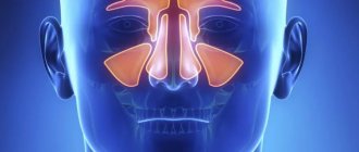
This is a non-invasive and highly accurate method for identifying a number of pathologies. The procedure relates to radiation diagnostics. As a result, you get a detailed image of the structures of the maxillary, frontal, ethmoid, and sphenoid sinuses.
Good to know: MSCT differs from CT in higher accuracy. After all, the equipment is equipped with several X-ray tubes and specific sensors. This allows you to achieve maximum detail in your images.
Typically, a CT scan of the paranasal sinuses is prescribed to detect the following problems:
- inflammatory processes in the sinuses, including sinusitis, frontal sinusitis, ethmoiditis, sphenoiditis;
- neoplasms with differential definition of etymology;
- consequences of injuries;
- identification of foreign bodies.
This procedure is often performed without contrast. But to assess the condition of blood vessels, the use of a dye is necessary. Contrast is also shown when clarifying the degree of germination of neoplasms into adjacent structures,
Why is a CT scan performed?
Medscan centers perform CT scanning, which allows you to visualize internal organs, bone structures, blood vessels, and analyze their condition in different projections and modes. All this is necessary for:
- identifying tumors, assessing their prevalence and the effectiveness of antitumor treatment
- diagnosis of vascular diseases such as atherosclerosis or vasculitis
- visualization of inflammatory changes, such as sinusitis
- clarification of the nature and differential diagnosis of various lung diseases, including viral pneumonia
- diagnosis of acute conditions such as strokes, thrombosis and thromboembolism, intestinal obstruction
- monitoring the results of surgical treatment (diagnosis of complications, for example, inflammatory changes in the abdominal cavity or foreign bodies after abdominal surgery)
- carrying out invasive manipulations (biopsy followed by histological examination) for diagnostic purposes under CT control
Preparing for a CT scan
A CT scan of the sinuses does not require any preparation unless the use of contrast is planned. Before the procedure with contrast, the level of creatinine in the blood is determined. If the indicators are too high, this indicates problems with the kidneys, for which the procedure is contraindicated.
Our “Ear, Nose and Throat Clinic” conducts computed tomography using a new generation device, NewTomVGievo. It provides radiation exposure tens of times less than when performing procedures on computer tomographs from other manufacturers.
Sign up with us for a computed tomography scan of the paranasal sinuses!
Types of CT
Globally, computed tomography options can be divided into non-contrast and contrast. The first does not require the introduction of a contrast agent into the body and is intended for visualization of radiopaque structures. The most common CT scans performed without contrast include:
- CT lungs
- CT scan of paranasal sinuses and teeth
- CT scan of bones
- CT scan of the spine
- CT scan of the brain for injuries (to exclude brain contusions, intracranial hematomas and skull fractures)
If the primary target of the study is soft tissue structures or vessels, it is necessary to intravenously administer a special contrast agent to the patient, which, as its name suggests, increases the contrast of soft tissue structures and vessels on CT images. Among the most common contrast CT studies are:
- Abdominal CT
- CT scan of the kidneys and urinary tract
- CT angiography of the brachiocephalic arteries
- CT angiography of the aorta
- CT coronary angiography
- CT scan of the chest for cancer
The price of a CT scan depends on the part of the body being examined and the need to use a contrast agent.
What can you see on a CT scan?
What does a CT scan of the sinuses show:
- pathology, its size and volume;
- characteristics of damage, if injury occurred;
- involvement of neighboring structures in the pathological process, their condition;
- condition of the bones that make up the paranasal sinuses, density, structure.
Thanks to these capabilities, the study makes it possible to demonstrate fractures of bones, septa, cartilage, various types of bleeding, anatomical defects resulting from long-standing trauma or received at birth, inflammation and tumors. Tomography of the paranasal sinuses records ethmoiditis, sinusitis, frontal sinusitis and various types of formations - cysts, polyps, malignant and benign tumors, the spread of metastases. The specialist finds the reasons for the violation of ventilation of the sinuses and nasal cavity, and in preparation for the operation determines the tactics and scope of the intervention.
Why does the doctor recommend a CT scan:
- one of the most reliable methods for visualizing the sinuses, painless and non-invasive;
- is faster than MRI and costs less;
- allows you to simultaneously obtain images of bones, blood vessels and soft tissues;
- helps identify internal bleeding in emergency cases;
- the device is less sensitive to patient movements than MRI;
- the presence of implants, pins, insulin pumps and other devices are not contraindications to the study;
- The radiation is not as intense as with regular x-rays.
Contrast helps to examine the paranasal sinuses in more detail. The drug is administered before diagnosis using a dropper. There are several contraindications for contrast studies - kidney and liver pathologies, pregnancy, diabetes mellitus, thyroid disease, individual intolerance to contrast components. The decision on the possibility of tomography is made by the attending physician on an individual basis.
How is the CT scan procedure performed at Medscan in Moscow?
Before the examination, the x-ray technician carefully interviews the patient, collects an anamnesis of life and illness (information about previous diseases). Sometimes, before a contrast study, results of a test of renal excretory function (creatinine test) may be needed. If the patient has data from previous studies (CT, MRI, PET-CT, etc.), then their images are imported into the information system, and scans of extracts and analyzes are attached to the medical history.
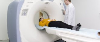
The patient lies down on the tomograph table and the first “marking” low-dose scan (topogram) is taken, on which the rest of the study is marked; the table moves back and forth, and the patient may be asked to hold their breath through a speaker.
If a contrast study is required, after the topogram an injector (a special automatic syringe with a contrast agent and saline solution) is connected to the intravenous catheter.
Next, one or more CT scans are performed, and between them, a contrast agent is injected intravenously into the patient using an injector at high speed; The table moves forward or backward with each scan and instructions to hold your breath may be heard again. The patient can hear the tube and detector rotating inside the gantry, but the noise level is low and does not cause discomfort. The patient does not experience any sensations from the operation of the X-ray tube.
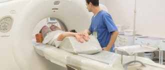
The x-ray technician controls the examination from the next room, which has a special window through which he can see the patient; The speaker and microphone built into the tomograph allow the laboratory assistant and the patient to be constantly in touch.
The entire CT procedure lasts from 3 minutes if the study is non-contrast, to 20 minutes if contrast is required.
The contrast agent is safe for the body and is excreted by the kidneys within 12 hours.
After the examination with contrast, we monitor the patient’s well-being for 20 minutes, during which time we prepare a digital medium on which all obtained images are recorded. After a 20-minute wait, the intravenous catheter is removed from the patient and the procedure is completed.
Then a team of radiologists comes into action, analyzing hundreds and thousands of images obtained during each study. The doctor carefully examines all the sections obtained, compares them with previous studies and analyzes what he saw, taking into account the anamnesis, medical history and treatment performed. The result of image analysis is a protocol (conclusion), which describes the identified changes and draws conclusions about their nature. It is ready within a few hours, maximum within a day.
This is not an easy job, but our centers employ real professionals who love their job; Our radiologists constantly improve their qualifications by participating in consultations, seminars, webinars and conferences, reading modern foreign literature in their specialty.
Indications and contraindications
To cleanse itself from pathogens, dust and foreign bodies, mucus is constantly produced in the paranasal sinuses. During inflammatory processes, the secretion of mucous masses increases, which can cause blocking of narrow passages. Pressure on surrounding tissues increases, and this causes pain in the eye sockets, bridge of the nose and head. To find out the causes of pain, patients are prescribed MSCT of the sinuses.
Other indications:
- prolonged runny nose (rhinitis);
- difficulty breathing through the nose;
- impaired sense of smell;
- atypical discharge and frequent nosebleeds;
- suspicion of sinusitis, congenital anomalies, trauma, neoplasms, presence of foreign bodies;
- postoperative observation.
There are few contraindications to the procedure. Multispiral diagnostics using X-ray radiation is not prescribed to pregnant women and children under 7 years of age. If MSCT of the paranasal sinuses with contrast is planned, it is necessary to exclude allergies to iodine-containing substances. It is not always possible to use contrast in case of hyperthyroidism, renal and liver failure.
The essence of the study
No preparation for the study is required. If a decision is made to administer a contrast agent, a break from eating and drinking for at least 4 hours is necessary.
The study is carried out in a specially equipped room. Before starting the diagnosis, medical personnel explain all subsequent actions.
The patient lies with his back on the diagnostic table and is fixed with soft fastenings (to maintain complete immobility). Particularly sensitive patients can use headphones to not hear the noise of the operating device.
The nurse places an intravenous cannula (Venflon) to automatically administer contrast. The contrast agent causes a feeling of heat at the injection site, and a metallic taste may be felt in the mouth.
The table with the patient is gradually moved into the tunnel of the device, and the scanner rotates around it, executing a given program. Each tomograph has a feedback device through which the patient can inform the staff about any discomfort that has arisen (in practice, this is almost never required). The doctor periodically asks you to hold your breath so that the picture is clearer.
The duration of the study does not exceed 1 hour. Upon completion, the patient can immediately go home or wait for the conclusion to be issued.