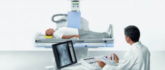
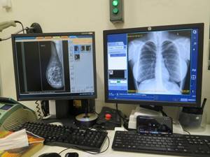
We are pleased to announce that the Kutuzov Center has installed a new ultra-modern digital X-ray machine (digital X-ray machine Brivo XR575 Premium from GE Healthcare)
One of the popular types of diagnostics is x-ray. It is used to study bone tissue and internal organs. The popularity of x-rays is easy to explain - it is a quick, painless and highly informative study. And in new digital X-ray machines, the main disadvantage of X-rays is practically absent - radiation exposure, which is potentially harmful to the body and limited the use of X-rays to only a few cases per year. There are other benefits of digital x-rays. You can take an X-ray without a referral using a modern digital device Brivo XR575 Premium from GE Healthcare any day in the diagnostic and treatment department. We will carry out this and other examinations (including comprehensive Check Up of the body) with comfort for patients, high accuracy of results and a quality guarantee!
How X-rays work
Radiography is a method of visualizing internal organs and tissues using x-rays. They penetrate the tissues of the body and, depending on their density, physical and chemical properties, are absorbed by them in different ways. This creates a contrast image of the internal organs and (x-ray), from which a specialist can evaluate their structure, location, condition, structure and other features.
Modern x-ray equipment has made x-rays less dangerous than the old analogue method of examination, and more informative. On a digital device, the image is immediately displayed on the computer screen and saved as a file. This is convenient - the X-ray image can be examined, enlarged, sent to the doctor and stored on electronic media. At the same time, the sensitivity of the digital device is higher, which means the level of radiation exposure is lower. The picture is obtained with better quality and resolution.
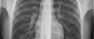
How is the chest x-ray procedure performed?
This procedure is carried out in a specialized x-ray room. There is a portable option for X-ray devices when a patient needs to be examined in a hospital bed. Pictures can be taken in frontal and lateral projections. At the signal from the laboratory worker, you will need to inhale and remain motionless for several seconds. In order not to expose areas of the body other than the thoracic region to radiation, the patient is covered with an apron made of lead. It creates a kind of reflective screen.
Types of X-rays at the Kutuzovsky Children's Center
X-ray is an indispensable method for diagnosing lungs and other hollow internal organs, as well as skeletal bones and joints.
In multidisciplinary medical X-rays are actively used, both as an independent diagnostic method and as part of comprehensive examination programs. We perform the following types of x-rays:
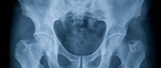
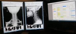
- Bones of the spine, head, limbs and joints.
- Lungs.
- Chest – assessment of the condition of the chest organs.
- Kidneys and urinary tract – urography.
- Mammary glands - mammography.
- Paranasal sinuses.
- Dental (orthopantomography) and other types of x-rays.
X-ray of the kidneys: X-ray of the kidneys with contrast - MEDSI
Table of contents
- What does it represent?
- X-ray of the kidneys: indications for examination
- X-ray (urography) of the kidneys: types of examination
- Contrast urography
- What will a kidney x-ray show?
- How is the procedure done?
- How to prepare for a kidney x-ray
- Contraindications for radiography of the kidneys
- Possible side effects of kidney x-ray with contrast
- Advantages of carrying out the procedure at MEDSI
X-ray of the kidneys is a radiation research method designed to evaluate the anatomical structure and functional state of the urinary system. One of the most common methods of examining patients with suspected urolithiasis.
What does it represent?
The method is based on the ability of X-rays to pass through body tissues, the degree of density of which determines the degree of darkening (the so-called shadow) in the finished image. Considering the harmful effects of ionizing radiation on the body, the intensity of the rays is strictly dosed, and the procedure is carried out only for medical reasons.
X-ray of the kidneys: indications for examination
- Urolithiasis or suspicion of it
- Chronic kidney diseases (for monitoring and treatment control purposes)
- Diagnosis of tumors, metastases
- Injuries of the pelvic and lumbar region
- Postoperative control
- Significant abnormalities in urine analysis: hematuria (blood in the urine), proteinuria (protein in the urine), pathological changes in odor, color, density and clarity of urine
- Pain of characteristic localization (lumbar region and lower abdomen), renal colic
- Painful urination
- Suspicion of obstruction (obstruction) of the urinary tract, urinary retention, swelling of the eyelids and face
- Hypertension
- Clarification of ultrasound examination
X-ray (urography) of the kidneys: types of examination
- Survey x-ray of the kidneys - a direct projection image without contrast agent. Allows you to evaluate the location, shape and number of kidneys, the state of the skeletal system of nearby areas, large foci of inflammation, necrosis, tumors, large calcium stones
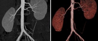
- Excretory urography - x-ray of the kidneys with a contrast agent. Allows you to evaluate the excretory (excretory) functions of the kidneys: the state of the pyelocaliceal system, the integrity and patency of the urinary tract; performed in two or more projections
- Direct pyelography - injection of contrast through a catheter from the lower urinary tract or through a nephropyelostomy directly into the kidney. Performed in a hospital
Contrast urography
The most informative, simple and inexpensive research method. It can be performed on an outpatient basis: immediately after the procedure the patient goes home. The main advantage is the introduction of an iodine-based contrast agent, which, as it is filtered by the glomerular system and excreted through the urinary tract, highlights different areas in the image.
What will a kidney x-ray show?
The method allows you to determine:
- Anatomical features of the structure of the organ: quantity, location, shape, size, homogeneity, integrity
- Functional features: rate of filtration and excretion of the substance, filling of the pyelocaliceal system inside the kidney, diameter and patency of the urinary tract
- Foreign inclusions: the presence of concrements (stones), foreign damaging particles (bone fragments after injury, splinters)
- Tumor, its size, prevalence, presence of metastases
How is the procedure done?
The patient is injected with a contrast agent, after which 3 pictures are taken at strictly defined time intervals: the first - 5-7 minutes after injection, the second - after 15-17 minutes, the third - after 20-23. Thus, the image captures the contrast in the collecting system, then in the ureter and, finally, in the bladder.
How to prepare for a kidney x-ray
- Before referral for testing, the doctor prescribes tests to rule out renal failure. Some medications should not be taken; Allergic history is important; if present, consultation with an allergist is necessary
- 3 days before the procedure, gas-forming foods are excluded from the diet.
- On the eve of the procedure (in the morning, on an empty stomach), if there is constipation, a cleansing enema is recommended. The study is carried out on an empty stomach
- Before the procedure, the patient removes metal jewelry, watches, dentures, and a belt and sits on the couch in light clothing without fasteners. Areas not involved in the study are covered with a lead apron.
Contraindications for radiography of the kidneys
The only contraindication to X-rays is pregnancy; other restrictions relate to the administration of contrast:
- Allergy to iodine-containing drugs
- History of hypersensitivity reactions
- Asthma
- Decompensated renal failure
- Heart failure without compensation
- Collapse, shock, coma
- Use of certain hypoglycemic drugs
- Pregnancy and breastfeeding
- Less than a week old
Possible side effects of kidney x-ray with contrast
The most common are allergic reactions. Also, during the administration of contrast, the following may occur: a short-term feeling of heat, nausea and an unusual taste in the mouth.
Advantages of carrying out the procedure at MEDSI
- Availability of latest generation X-ray machines that allow individual exposure
- The equipment and technical capabilities of the equipment provide more than 50 types of radiographic examinations
- The images are described by radiologists with extensive practical experience.
- Emergency radiography is possible for injuries and acute conditions
- Carrying out radiography for children of different ages
Some MEDSI clinics do not require an appointment for x-rays. For more details, call 24/7 + 7 (495) 7-800-500
Advantages
It is profitable to take an X-ray for a fee using a digital machine. Here are just the main advantages:
- Safety of the examination for health due to the low dose of radiation exposure.
- Speed of obtaining results - pictures appear on the computer almost immediately, they can be viewed, and, if necessary, taken again or enlarged.
- High information content due to good image detail.
With all the advantages, the cost of x-ray services is quite affordable for everyone (cost details are in the price list on the website).
Contraindications
Why are x-rays harmful? The potential harm lies in the exposure of the body to ionizing radiation. This is especially harmful for a growing, developing organism, so x-rays are undesirable for pregnant women and children under 15 years of age. There are also restrictions on the number of photos per year. The number of times an X-ray can be taken depends on the equipment - a larger number of studies are allowed on a digital device.
Where to get an x-ray for a fee in Moscow
When choosing where to get a paid x-ray, pay attention not only to its cost and proximity to a medical facility, but also to the type of device - the capabilities of digital equipment are much higher. The pictures are clearer, with greater enlargement, they can be enlarged on a computer, conveniently transferred to another doctor, and stored. With such a device, X-rays can be taken several times over a short period of time, which may be needed to monitor chronic diseases or evaluate the effectiveness of treatment.
We invite you to the Kutuzovsky Children's Center for this and other examinations, if you need a quick and accurate diagnosis at a convenient time. Based on the results obtained, you can get advice from a specialized specialist immediately after completing the diagnostic procedures.
X-rays in the clinic are performed by specialists with extensive experience and qualifications. This guarantees an individual approach, care for your health, and high quality services.
For the convenience of patients, we work daily. The clinic is located at: Moscow, st. Davydkovskaya, 5. To consult on questions of interest, to sign up for an x-ray for an adult in Moscow without a referral, to see doctors to interpret the results - call: +7 (495) 478-10-03.
X-ray of the chest organs X-ray of bones and joints X-ray of the spine X-ray of the hip joint X-ray of the sella turcica X-ray of the nasal bones General X-ray of the abdominal organs X-ray of the hip X-ray of the lower leg X-ray of the bones of the facial skeleton X-ray of the lumbosacral spine
Contraindications for chest x-ray
Any x-ray examination carries a radiation dose, the intensity of which largely depends on the characteristics of the equipment used and the time of exposure of the x-rays to the body (this indicator also directly depends on the capabilities of the equipment).
X-rays are strictly contraindicated in cases of severe bleeding, open pneumothorax, as well as in patients in severe or critical condition. The procedure is also unfavorable for pregnant women - despite the fact that the abdomen is protected by a “lead apron” at the time of the procedure, subsequently it is theoretically possible that abnormalities will develop in the fetus. Regarding the lactation period of mothers, experts have no arguments against it, although many doctors advise avoiding radiological diagnostics of nursing mothers.
Children under 14 years of age are not recommended to have a chest x-ray for preventive purposes. However, there are a number of situations when this is inevitable:
- entry of a foreign body into the respiratory system and the need to determine the location and shape of the object for the purpose of its further removal;
- the clinical picture indicates the likelihood of pneumonia;
- suspicions of tuberculosis arising on the basis of controversial results of mantoux samples;
- the need to exclude the appearance of malignant neoplasms in the lungs;
- signs of cardiopulmonary failure;
- chest injuries.
The decision to send a child for an x-ray should be the result of a careful and comprehensive analysis of the clinical picture by an experienced doctor. Parents can go to a clinic that uses digital equipment that provides up to 50% less radiation exposure compared to traditional analog equipment used in public medical institutions.