X-ray of the sinuses at the German Family Clinic
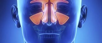
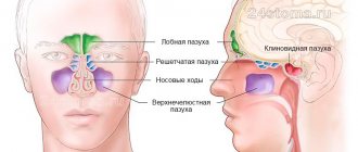
: conducting research and decoding images. Make an appointment for an x-ray by phone, (812) 337-12-12. The nose is a complex organ that is part of the upper respiratory tract. Performs respiratory, olfactory, protective and resonator functions. Each half includes four walls. The turbinates, which are cartilaginous projections of the nasal septum, divide both nasal cavities into the upper, lower and middle passages. The nasal cavity communicates with the paranasal sinuses (cavities in the bones of the skull filled with air) through the openings of the upper and middle nasal passages. According to localization, the maxillary, frontal, sphenoid sinuses and ethmoidal labyrinth are distinguished (the names correspond to the names of the bones). At birth, only the maxillary (maxillary) sinus and the ethmoid labyrinth are formed in the child. But the rest begin to form at the age of 3-4, finishing at about 25 years.
The complexity of the structure and functions performed, the location of the nose makes it susceptible to various injuries, as well as the development of various diseases. An effective method for diagnosing the paranasal sinuses is radiography.
In what cases is sinus examination prescribed?
X-ray of the paranasal sinuses (PSN) can be prescribed by an otolaryngologist if the following clinical symptoms are present:
- feeling of pressure, tension in the nose;
- severe headaches in the forehead, possibly in the temple;
- copious discharge, nasal congestion, difficulty breathing;
- elevated body temperature (38ᵒC and above);
- general malaise: fatigue, loss of appetite
In addition, a traumatologist and surgeon may prescribe an X-ray of the PPN in order to:
- determination of structural features, congenital and acquired pathologies and integrity of the nasal bones;
- to identify neoplasms (tumors, cysts, polyps), metastases;
- for visualization of foreign objects;
- as part of preoperative preparation;
- to monitor the effectiveness of the ongoing (performed) treatment
But most often, otolaryngologists and general practitioners are faced with the need to conduct an X-ray examination of the paranasal sinuses (paranasal sinuses) to confirm the diagnosis of sinusitis. This is an inflammatory process of bacterial, fungal, viral or allergic etiology that occurs in the paranasal sinuses. This process can be localized in:
- frontal lobe - frontal sinusitis;
- sphenoid bone – sphenoiditis;
- mastoid cells – ethmoiditis;
- maxillary (maxillary) sinus – sinusitis. This disease can differ in form (purulent, serous, exudative, polypous, etc.), each of which is clearly visualized on an x-ray and is not difficult to decipher.
Why do doctors prescribe x-rays of the paranasal sinuses?
Doctors order x-rays of the paranasal sinuses to detect sinusitis. Readers understood this from the above part of the article. As radiologists say, sinusitis and sinusitis are different, so here is a classification of pathology according to form:
- Exudative.
- Catarrhal, serous, purulent.
- Parietal-hyperplastic.
- Productive.
- Polypous.
Each of these forms is clearly visualized by radiography of the PPN. Only if the X-ray symptoms of the disease are unclear can the study be supplemented with CT (computed tomography).
Exudative sinusitis (frontal sinusitis, sinusitis) is visualized as a darkening with an upper horizontal border.
The parietal-hyperplastic form can be traced as parietal darkening due to thickening of the mucous membrane near the bone walls. The contour of the sinuses (paranasal sinuses) with this sinusitis is turned inward and is slightly uneven or wavy.
Polypous sinusitis is manifested by a parietal protrusion on a pedicle, facing inward.
The X-ray picture of catarrhal, serous or purulent sinusitis resembles exudative one. The only difference is the morphological substrate of the liquid that is obtained after puncture (piercing).
Depending on the location, the following forms of sinusitis are distinguished:
- Hemisinusitis is a lesion of the PPN on both sides.
- Pansinusitis is inflammatory changes in all paranasal cavities.
What is a cyst and how does a picture of the paranasal sinuses show it?
A cyst on an X-ray of PPN is an unexpected finding for the physician. It has no special clinical symptoms other than the frequent occurrence of maxillary sinusitis. When exposing paranasal formations using X-rays, the doctor can see a rounded shadow of low or medium intensity with an even, clear contour.
Cyst formation in the paranasal sinuses requires surgical treatment.
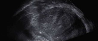
Fragment of an x-ray of the paranasal sinuses: cyst on the right
What will a sinus x-ray show?
Normally, the PPN on the X-ray image should look like this: the contours of the bone walls are clear; the filling of cavities with air is compared in orbits; the contours of the lattice cells should be traced. If the patient has sinusitis, the x-ray will show a certain level of fluid in the area of the medial and lateral walls. An indicator of chronic inflammatory processes for a specialist will be thickening of the mucous membrane and concavity of the external contour.
For a radiologist, assessing the results of radiation diagnostics is not difficult if the image was taken correctly and meets the basic quality criteria: the structure of the bone walls is clear, the placement is symmetrical, there are no additional shadows (only white-gray).
Contraindications and restrictions
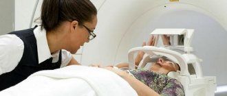
Contraindications to X-ray examination include pregnancy and children under 7 years of age. The influence of gamma rays on the fetus can provoke the development of pathologies. The procedure can be performed if it is impossible to do an ultrasound or the woman’s health justifies the possible risk to the fetus.
Are radiography prescribed for children?
It is not advisable to send children for x-rays due to the negative effects of radiation on the skeletal system. However, if there is no alternative, if the doctor considers it necessary to take an x-ray, you should not refuse. Some experienced ENT doctors and radiologists believe that the diagnostic method is suitable for children from 5 years of age.
X-ray of the sinuses: preparation and procedure
No special preparation is required for a nasal x-ray. The X-ray room laboratory technician will check to see if there are any metal objects on the body, and then help the patient to take the correct position for the procedure. It will be necessary to approach the vertigrapher (digital radiographic mobile vertical stand), take a position in which the nose and chin are in the same plane (nose-chin position), or the nose and chin are at an angle (chin position). The procedure is often performed sitting or standing, but a horizontal position of the subject is also possible (for example, nasofrontal projection). The subject will be asked to hold their breath and remain motionless.
It should be noted that there are many nuances when installing. Therefore, it happens that a radiologist, having analyzed the appointment of the attending physician, can cancel his appointment and perform x-ray diagnostics in a slightly different way.
In addition to the main two projections above (nasomental and mental), there is a frontal option, which will show only the ethmoid labyrinth, the right and left halves of the sphenoid sinuses. Therefore, this method of obtaining radiographs is rarely used to determine pneumatization of the paranasal sinuses. A targeted photograph of one of the sinuses may be taken.
For a detailed study of the structure of the paranasal sinuses, it is practiced to obtain an X-ray image using an X-ray contrast agent (usually an iodolipol solution). This method is used in particular to identify various types of intra-axillary formations, polyps, and cysts. The procedure involves puncturing the patient's sinus wall under anesthesia; rinsing the sinuses with furatsilin and administering iodlipol heated to body temperature through a needle. Nasofrontal, lateral and nasomental projections are used. However, due to the high probability of deterioration in the clarity of the images due to the mutual overlap of exposure between the nasal sinuses, images of both maxillary sinuses are not taken simultaneously.
Interpretation of the radiograph of the PPN
Approximate interpretation of the X-ray image of the PPN (for the sample):
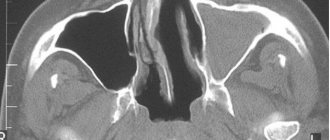
The presented radiograph of the paranasal sinuses visualizes a decrease in pneumatization of the left maxillary sinus in the lower third with the upper horizontal level. Conclusion: X-ray signs of left-sided maxillary sinusitis (sinusitis).
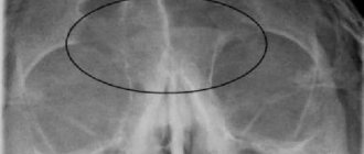
In the picture above you can see the parietal darkening of both maxillary sinuses. Its description will lead to the logical conclusion of bilateral sinusitis.
X-ray of the paranasal sinuses allows not only to diagnose pathology, but also to analyze the dynamics of treatment. When sinusitis is detected in a patient, doctors prescribe several images: the first – immediately after the inflammation is detected; the second and subsequent ones - during treatment with antibiotics.
Deciphering a radiograph of the PPN requires analysis of other anatomical structures that can be traced in the image - bones, oral cavity, eye orbits. Tumors can sometimes be found in them, which are an incidental but important finding.
Deciphering one radiograph takes about 10 minutes of a radiologist's time.
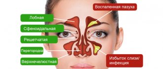
Schematic representation of PPN diseases
Contraindications to radiography of the sinuses
X-rays are contraindicated in seriously ill patients. It is also recommended that the procedure be performed on pregnant women and children under 14 years of age only for clinical indications, since such a study carries some radiation load on the body. On average, when radiography of the maxillofacial area, including the sinuses, the individual dose per procedure is 0.04 mSv when using traditional film X-ray equipment and 0.02 mSv when obtaining an X-ray using digital equipment. In addition, digital equipment makes it possible to obtain very good, clear images, eliminating the need for a repeat procedure. The doctor, after analyzing the clinical signs (based on complaints) and the patient’s medical history, will decide on the advisability of radiography of the sinuses, provided that the expected harm from the study is less than the seriousness of the consequences in the event of a false diagnosis and delay in starting treatment.
Very often, children experience nasal injuries; in babies, foreign objects can get into the nasal cavity, and emergency assistance is extremely necessary. Due to poorly developed immunity in children, the consequence of acute respiratory viral infections can be sinusitis, the purulent form of which poses a very serious danger. In such cases, radiography is an inevitable measure.
Parents should understand the value of a timely correct diagnosis. In such cases, it is possible to reduce radiation exposure in order to avoid risks by performing radiography on modern digital equipment. State medical institutions do not always have such equipment at their disposal. But many private clinics will help you undergo X-ray diagnostics safely. Thus, the German clinic’s equipment arsenal includes X-ray equipment manufactured by GE, which will make even an X-ray examination of an infant safe. Qualified clinic specialists will help you correctly determine the position required for diagnosis, experienced pediatricians will help you correctly carry out the X-ray diagnostic procedure for a child of any age, and the radiologist will give a detailed interpretation of the resulting image.
Attention: the prices presented on the website are not a public offer. Please check the cost of services by calling +7 (812) 432 32 32.
What the study shows
X-ray of the SNP helps determine:
- sinusitis in acute or chronic form;
- cyst;
- benign or malignant tumor;
- injury to the nose or facial bones;
- deviated septum;
- osteomyelitis (purulent-necrotic pathology of bone marrow and bones);
- osteoporosis.
The video talks about how and what radiography of the paranasal sinuses helps to identify. Filmed by the Moscow Doctor channel.
How does the procedure work and how long does it last?
The duration of the study is several minutes.
X-rays are taken depending on the indications as follows:
- The patient enters a special room. Children are accompanied by adults.
- In a standing or sitting position, the radiologist fixes the head in the position required to take the image. This can be a straight or lateral plane, a projection of the chin, the occipito-frontal part or the occipito-mental part. One of the parents holds the child's head.
- You need to take a deep breath and hold your breath for a few seconds.
- The doctor will inform you that the procedure is complete.
- The picture develops within 15-20 minutes.
Is X-ray dangerous? This is explained in the video from the Live Healthy channel.
Preparing for an X-ray
Preparation for the procedure comes down to two rules:
- remove jewelry and other metal objects located in the head area (chain, earrings, hairpins, hoop);
- wear a special apron (lead), which is issued immediately before the x-ray.
There are no restrictions related to food or liquid intake.
It is necessary to clarify when and how to take an x-ray when observing the disease over time. For example, if a patient is having a cuckoo sinus irrigate, x-rays are taken before the sinuses are cleared of fluid.