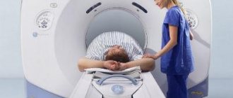
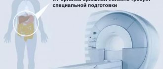
In recent months, the number of calls to tomographic methods for diagnosing the respiratory system has been growing at an incredible rate, as they are becoming the method of choice for detecting covid pneumonia. The most informative type of tomography in this direction is considered to be MSCT (CT of the chest organs). It is prescribed along with MRI, complementing magnetic resonance scanning, giving the most accurate results about current diseases.
When is it necessary to do MSCT of the chest organs?
A list of indications has been developed for tomographic diagnostics, which can be expanded when searching for non-standard pathology. If there are signs of a pulmonary disease, but suspicion of its prevalence in adjacent zones cannot be excluded, CT of the lungs becomes a targeted study within the framework of a general MSCT. Hardware diagnostics covers not only the pulmonary parenchyma, but also the bronchi, lymphatic collections, bronchi, rib and clavicle bones, the lower part of the trachea, and the thymus gland. The described areas show up well on CT and MRI scans, especially if contrast is used during the session.
The main indications for diagnosis are the following:
- symptoms of latent inflammation;
- problems with large and small blood channels of the sternum;
- tuberculosis infection;
- exacerbated or chronic asthmatic syndrome;
- growth of tumor formations;
- search for metastases from distant and adjacent organs;
- studying the degree of necrotic damage and tissue scarring;
- acute pain in the chest for no apparent reason.
What does MSCT show and who is it prescribed for?
Thanks to the procedure, it is possible to identify genetic abnormalities, malignant and benign neoplasms, foci of infections, age-related degenerative changes and the consequences of injuries.
Indications for the procedure are probable and diagnosed diseases:
- chest;
- abdominal cavity;
- urinary tract;
- gastrointestinal tract;
- kidneys and adrenal glands;
- musculoskeletal system;
- CNS and peripheral nerves;
- heart and blood vessels;
- ear, nose and throat;
- respiratory tract.
The procedure is prescribed to not only identify or confirm the presence of pathologies, but also to establish exactly where the lesion is located, which areas are affected, and whether the prescribed therapy brings good results.
What are the differences between MRI and CT of the lungs?
Standardly, for assessing the functions of the respiratory and cardiovascular systems, preference is given to MSCT of the chest organs. This is due to the peculiarities of this type of diagnosis. Since the scanning is carried out in the field of exposure to X-rays, dynamic structures that are in natural motion (cell contraction during breathing, heart pulsation, etc.) do not affect the clarity of the images taken. During MRI, artifacts can appear even when the body moves slightly from a fixed position.
The magnetic field better shows the structure of soft tissue objects; MRI of the chest organs perfectly complements CT of the lungs, and is also prescribed if the patient is prohibited from using x-ray diagnostic methods. This applies to a greater extent to women during pregnancy and young children, for whom MRI is not contraindicated from the first hours of life. Another option for ionizing scanning is PET CT of the lungs. This type of tomography is the most informative, but it is expensive, which makes it necessary to turn to it in extremely complex cases.
Preparation for MSCT
During the procedure you must wear loose clothing. It is advisable to remove all metal objects, as their presence may cause certain image distortions. At the same time, the presence of endoprostheses or cardiac pacemakers is not a contraindication for MSCT, unlike MRI. If the study is carried out on a woman, information about the presence of pregnancy is required. MSCT is usually painless and takes a short period of time (a few minutes).
When should you refuse MSCT of the chest organs?
Along with its indications, CT scan of the lungs has limitations in its use. Absolute prohibitions are specified above and apply to rapidly developing organisms during intrauterine development and active growth in childhood. At this time, it is better to completely abandon X-ray procedures so as not to provoke oncological cell growth in the future. Also, MSCT and CT have conditional contraindications that can be removed if additional study conditions are met. Most of them concern the use of a drug such as contrast. Enhanced screening with intravenous staining is prohibited for persons with:
- cross allergic reactions;
- poor functionality of the urinary system;
- thyroid diseases;
- aggravated asthma;
- severe forms of diabetes.
In these cases, MSCT of the chest organs can be performed without contrast, so as not to cause complications. There are also personal situations when the patient cannot fit inside the equipment due to excessive weight. To help obese patients, open-type equipment with increased capacity has been developed.
Radiation dose during MSCT
One of the differences between modern multislice computed tomography and conventional CT is the amount of radiation exposure the patient receives.
With MSCT, the radiation exposure is 2-3 times less, which means there is less harm to the body and it is possible to be examined more often. For example, with conventional thoracic screening, a person receives radiation exposure of 10-11 mSv, and with multispiral screening, it is only 3.9 mSv.
CT of the brain CT of the abdominal cavity CT of the teeth CT of the orbits CT of the jaw CT of the cerebral vessels CT of the abdominal aorta CT of the adrenal glands CT of the facial bones CT of the temporal bones CT of the lumbosacral spine CT of the thoracic spine CT of the elbow joint CT of the shoulder joint CT of the thoracic aorta CT of the cervical spine CT of the heart CT of the hip joint CT of the knee joint CT of the pelvis CT of the lungs CT of the kidneys CT of the sinuses CT of the larynx CT of the coronary vessels CT of the liver CT of the intestine CT of the vessels of the lower extremities CT of the hand CT of the pelvic bones CT of the foot CT of the chest CT of the vessels of the neck CT of the coccyx CT of the ankle CT bladder CT scan of the thyroid gland CT scan of the skull CT scan of the soft tissues of the neck CT scan of the wrist CT scan of the inner ear
How is a CT scan of the lungs performed?
You can apply for tomographic screening both in public clinics on a free/paid basis, and in specialized commercial MSCT CT centers. Free diagnostics are available with a direct referral from a specialist and a quota for a computer examination or MRI. Paid scanning is also considered affordable as it is comparatively cheaper than alternative scan procedures.
It will not take much time to complete the screening. A CT scan of the lungs takes a maximum of 15 minutes, even if the session is planned with contrast. The progress of the research consists of the following stages:
- Registration at a medical institution, conclusion of a service agreement.
- Preparing for the session, checking clothes and pockets for forgotten metal items.
- Brief instructions from the laboratory assistant.
- Placement in the equipment in a supine position.
- Scanning using MSCT/CT equipment with production of layer-by-layer images.
- Completion of screening, waiting for documentation.
After the diagnosis is completed, it is not necessary to stay in the medical institution waiting for a description of the images; this takes a lot of time. You can return to the center later (after 1-2 hours) or come the next day. Most modern MRI organizations offer to send completed documents in digital form to the client’s email address.
Advantages
MSCT in Krasnogorsk and other cities is in considerable demand. First of all, this is a non-invasive research method, during which tissue damage is excluded. Thanks to the presence of high resolution and the use of advanced programs, it is possible to obtain the thinnest sections in which it is possible to examine pathological changes even if they are very small in size, no more than 1 mm.
Thus, MSCT allows you to identify diseases at an early stage of their onset, which means you can prevent serious consequences and achieve a speedy recovery, provided that the therapy is selected by an experienced specialist. This diagnostic is chosen for the following positive qualities.
- Efficiency of implementation.
- The patient will not experience even the slightest discomfort.
- The images will be of the highest quality and most detailed.
- Minimum radiation exposure.
How to do a CT scan of the lungs with contrast
With a contrast study, one more stage is added to the standard session plan. After the person is placed inside the machine and the initial images are taken, he is given an intravenous injection of an iodide drug.
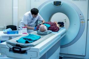
The solution quickly spreads throughout the body through the circulatory system, disperses through the tissues and accumulates in those areas where inflammation persists or tumor growth occurs. In most cases, the contrast is hypoallergenic, but it is better to do an immunological test in advance to determine the tolerance of this substance. After a CT scan of the chest with contrast, performed in enhanced mode, the patient’s well-being does not change; slight dizziness is possible immediately after the injection of the dye. It is excreted from the body imperceptibly through the kidneys. For this, 48 hours after a CT scan of the lungs is enough, but the process can be accelerated by taking an increased volume of clean water.
How much does MSCT of the chest organs cost?
Different diagnostic centers offering MSCT, CT, and MRI services place different prices in their price lists, since the cost is influenced by a large number of objective factors:
- duration of patient preparation, provision of consumables;
- the complexity of the clinical situation for decoding data;
- examination mode;
- contrast consumption per total body weight;
- written or digital presentation of the report;
- recording scans on the selected media or film printing;
- workload of center specialists;
- Availability of additional discounts and loyalty programs.
On our service you can find medical institutions that perform MSCT of the chest organs, compare their offers, and study the price lists in more detail. Average prices can be seen in the table below:
| Procedure name | Minimum price |
| CT scan of the chest | 2500 rub. |
| CT lungs | 2500 rub. |
| PET CT of the lungs | 31500 rub. |
| CT scan of the chest with contrast | 4200 rub. |
| MRI of the chest | 4115 rub. |
Where are MSCT and CT performed?

For full-fledged screening using a tomograph, you need not only specialized equipment, but also personnel trained in this type of activity and experienced diagnosticians. Specialized clinics monitor the quality of service and conduct research quickly and inexpensively. You can find a medical facility with the necessary conditions on our service. Here are complete lists of tomography medical centers in the region. Select a service, set additional criteria using filters, study the displayed options and select better according to your own criteria.
Main advantages of MSCT
In terms of speed and speed of obtaining results, multislice tomography is better than CT and MRI. Magnetic resonance imaging can last up to two hours, and is prescribed mainly for studying soft tissues. MSCT takes a maximum of half an hour, allowing you to assess the condition of bones and other dense structures.
Other advantages of the method should be noted:
- reducing radiation exposure to the patient;
- maximum reliability and accuracy;
- the most effective use of the X-ray tube by modernizing its design;
- minimal restrictions on the coverage area - you can simultaneously explore different areas without compromising image quality;
- pictures with very thin (up to 0.5 millimeters) sections;
- optimization of contrast resolution;
- higher signal to noise ratio.

