Ultrasound of the abdominal cavity (AP) with high reliability determines the presence of pathologies of the digestive organs and, if necessary, the genitourinary system in the initial stages, and also confirms the diagnosis assumed on the basis of existing symptoms. The study allows you to assess in detail the condition of the abdominal organs, their size, structure, location, and determine the presence of inflammation, fluids, cysts, stones or other neoplasms in the area under study.
COST OF ULTRASOUND OF THE ABDOMINAL CAVITY - 1000 RUB, ULTRASOUND OF THE ABDOMINAL CAVITY + KIDNEYS - 1500 RUB.
MAKE AN APPOINTMENT, TEST OR ULTRASOUND
Indications for abdominal ultrasound
The content of the article
You should contact us for an abdominal ultrasound if you have any discomfort in the abdomen. At the same time, to undergo an ultrasound examination you do not need a referral from a doctor - you can sign up for an ultrasound examination yourself. The diagnostic results are made available to you and the specialist performing the examination will definitely warn you about the pathologies found. If the condition requires additional consultation, you can always contact a gastroenterologist, taking with you the transcript of the ultrasound.
Ultrasound diagnostics of the abdominal organs is indicated for patients who have the following symptoms:
- abdominal pain of varying intensity, including suspicion of appendicitis, heaviness or pain in the right hypochondrium, lower back pain;
- a feeling of bitterness in the mouth, unpleasant belching, nagging pain or a feeling of fullness in the stomach after eating;
- increased blood pressure, provided that the exact causes of the symptom are not clear;
- aversion to fatty foods, if this is not associated with taking certain medications, renal colic, a feeling of pulsation in the abdominal cavity;
- jaundice, suspicion of it, yellowing of the tongue, uncomfortable or difficult urination;
- increased gas formation, low-grade fever;
- symptoms of the oncological process: loss of appetite, severe weight loss, lethargy, weakness.
Even in the absence of alarming symptoms, ultrasound examination of PD is recommended as part of an annual medical examination in order to promptly identify existing diseases of the digestive system.
Some pathologies, such as cancer or metastases localized in the abdominal cavity, can develop asymptomatically for a long time, leading to irreversible consequences for human health and life. Ultrasound diagnostics makes it possible to detect such anomalies even at the initial stage, when the tumor is just beginning to emerge.
Features of preparing and conducting diagnostics for children
Preparing a child, just like an adult, includes dietary restrictions. For 2-3 days, it is necessary to exclude from the menu foods that provoke increased gas formation, as well as candies and sweets. If a small patient has a tendency to flatulence, he is prescribed enzyme and absorbent drugs.
For children of any age, ultrasound is performed on an empty stomach. Mothers of infants need to calculate feedings so that the last one before the procedure is carried out no later than 3 hours before. It is not advisable to give your child fruit or vegetable purees before the test, as they take a long time to digest.

The process of performing an ultrasound examination of the abdominal organs in children is no different from a similar procedure in adults. Diagnosis is carried out with a special ultrasound probe using conductive gel.
For what diseases is BP ultrasound prescribed?
Doctors prescribe an ultrasound of the abdominal organs to determine the condition of the organs if the following diseases are present or suspected:
- cirrhosis is a chronic liver disease in which normal tissue is replaced by fibrous tissue;
- hepatitis - inflammatory damage to the liver;
- jaundice is a disease whose main symptoms appear due to increased levels of bilirubin in the blood;
- pancreatitis is an inflammatory pathology affecting the pancreas, cholecystitis is an inflammatory lesion of the gallbladder;
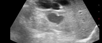
- cysts in the abdominal organs are pathological formations of a cavity nature, filled with fluid;
- cholestasis - stagnation of bile in the abdominal cavity;
- calculi – stones that form in the gall bladder or kidneys;
- benign and malignant neoplasms in the organs of PD;
- diseases in which there is an enlargement of the liver or spleen: malaria, infectious mononucleosis, sepsis, aplastic anemia.
Ultrasound diagnostics of PD organs must be carried out immediately before surgery on organs in this area.
In what cases is it prohibited to prescribe the procedure?

There are patient conditions when performing an ultrasound becomes impractical or impossible, even despite the complete safety of the method itself.
Contraindications to the study are:
- pustular rashes located in the examined area;
- acute infectious diseases;
- acute pathologies of blood circulation in the brain;
- heat;
- the presence of a significant area of the wound surface on the abdomen, as well as a violation of the integrity of the skin.
As for age restrictions, the procedure does not have them. Ultrasound examination of the abdominal organs is prescribed for children of any age, even infants.
Pregnancy is not an absolute contraindication for an abdominal ultrasound. This method is prescribed much more often during pregnancy than, for example, computed tomography or radiography. In some cases, this diagnostic method is also prescribed to study the state of fetal development.
In the first time after colonoscopy and gastroscopy, ultrasound of the abdominal organs is not prescribed, since the procedures spasm smooth muscles and distort the results. After fluoroscopy with a barium contrast agent, the results of ultrasound are also doubtful due to the fact that the contrast accumulated in the folds of mucus is displayed in the examination picture, so ultrasound is not performed immediately after fluoroscopy with contrast.
How is an abdominal ultrasound performed?
An ultrasound diagnostic doctor must study the structure and features of the liver, gall bladder, retroperitoneum, pancreas, spleen and blood vessels. But according to indications or if any pathology is suspected during ultrasound, other organs of the digestive and genitourinary system can be examined.
The protocol for ultrasound diagnostics of the abdominal organs is standard and includes the following described parameters:
- determination of localization and, if necessary, features of the location of PD organs;
- determining the size of the organs under study, studying their internal structure;
- confirmation of the presence or absence of free liquid in the space being examined;
- identification of changes characteristic of chronic pathologies;
- in the presence of stones, cysts and neoplasms, determining their location and size;
- identification of the inflammatory process if it exists.
If there is a need and the technical equipment of the ultrasound room allows for more complex manipulations, under ultrasound control they can remove fluid from the retroperitoneal space or perform a biopsy of the kidneys or liver.
Advantages of MRI in the study of abdominal organs
To examine internal organs, along with MRI, CT (computed tomography) and ultrasound (ultrasound) are also used.
Each of these methods has its own indications and contraindications:
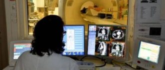
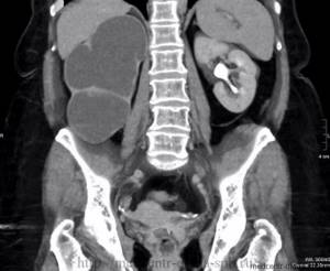
CT allows you to obtain a detailed picture of the structure and location of the internal organs of the abdominal cavity only with the use of a contrast agent, which significantly increases the cost of the study. MR diagnostics of the abdominal organs is quite informative even without the use of contrast. Contrast agents used for MRI are much easier to tolerate by the patient's body than similar drugs for CT.
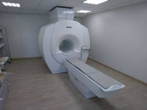
Magnetic tomograph Philips
CT is more appropriate to use for studying hollow organs: the stomach, intestines, veins and arteries of the abdominal cavity, as well as in cases where there are objective contraindications to magnetic resonance imaging.
MRI is a safer technique than CT. It does not use X-rays, and magnetic waves are absolutely harmless to humans.
Ultrasound is usually prescribed as the first choice test for pain and discomfort in the abdominal area. MRI is preferable to ultrasound when blood, urine and physical examination of the patient raises suspicion of the presence of neoplasms or when abnormalities in the structure and location of organs need to be examined in detail
Additional examinations
According to indications, if an inflammatory or tumor process is suspected, during ultrasound the BP can be examined:
- Prostate gland in men.
- Uterus in women.
- Kidneys. They are not a mandatory organ to be diagnosed during an ultrasound scan of the PD, but can be examined according to indications. Ultrasound of the kidneys and adrenal glands allows you to determine their size, relative position and exact location. The examination may reveal cysts, stones, inflammation or tumors.
- It is necessary to take a biochemical blood test.
- If cancer is suspected, an analysis is performed for tumor markers.
What diseases are detected during abdominal ultrasound?
Based on the nature of the changes present in the organs, the characteristics of their location, size and structure, as well as the presence of pathological formations, the ultrasound specialist can make an entry in the report on suspicion of the following diseases:
- cirrhosis of the liver;
- fatty liver;
- focal changes in the liver;
- congestive liver circulatory failure;
- liver cancer;
- cholelithiasis;
- chronic or acute cholecystitis;
- chronic or acute pancreatitis;
- cholestasis;
- tumor process in the pancreas.
To finally confirm the diagnosis, the patient may be prescribed additional diagnostic procedures.
Interpretation of abdominal ultrasound results
Decoding the results obtained during an ultrasound of the abdominal cavity is a set of digital values and characteristics of the ultrasound reflected from a particular organ. This information is reflected in the study protocol. With it, the patient turns to the attending physician, who will make an accurate diagnosis. However, before visiting a doctor, you can independently understand all these numbers and have a rough idea of the state of your health.
To do this, you need to compare the ultrasound results with indicators of normality and deviations. They are different for each organ.
Interpretation of abdominal ultrasound: liver
| Normal indicators | Deviations |
| Right lobe: length - 10 cm, thickness - up to 7 cm; Left lobe: length - 5 cm, thickness - 12-13 cm: Oblique vertical size: up to 15 cm. | Rounded edge, increased echo density, increased size, dilation of the splenic and portal veins, uneven contours, free fluid in the abdominal cavity. |
Explanation:
- If there is a rounding of the edges with an increase in the size of the liver, this may be a sign of stagnation of harmful substances in the liver. This happens against the background of heart disease.
- The presence of abscesses, tumors and cysts is indicated by areas of the organ with a disturbed echostructure.
- Increased echo density is observed with fatty hepatosis, which is also accompanied by compaction of the organ and the inability to visualize blood vessels.
- Liver cirrhosis is characterized by uneven contours, an increase in the size of the organ and dilation of the portal and splenic veins.
Interpretation of ultrasound: gallbladder
| Norm | Deviations |
| Pear-shaped or cylindrical shape of the organ; Width - 3-5 cm, length - up to 10 cm; Wall thickness - up to 4 mm; Volume - 30-70 cm³; Absence of formations in the lumen. | Wall thickening (“double contour”), fluid around the gallbladder, acoustic shadow, uneven contours, dilation of the bile ducts. |
Explanation:
- An acoustic shadow on the monitor indicates the presence of a tumor or stones in the gallbladder. This can be determined by the nature of the shadow.
- Signs of acute cholecystitis are: thickening of the walls, changes in size both up and down. And the presence of fluid means the development of peritonitis and the need for surgical intervention.
- With chronic cholecystitis, the contours of the bladder will be clear and dense.
- A sign such as dilatation of the bile ducts indicates that the free flow of bile is blocked by a stone.
Bile ducts
| Norm | Deviations |
| Common duct diameter: 6-8 mm; The hepatic ducts are not dilated inside. Pancreas: Head length - 35 mm, body - up to 25 mm, tail - 30 mm; Smooth contours; Homogeneous echostructure; Echogenicity is within normal limits; Lack of education. | Change in echo density; The Wirsung duct is dilated; Uneven contours; Displacement of the aorta. |
Explanation:
- Low echo density may be a sign of acute pancreatitis.
- If echo density increases, cancer or a chronic form of pancreatitis occurs.
- Signs such as uneven contours of the pancreas and increased size also indicate a malignant tumor.
Ultrasound interpretation: spleen
| Norm | Deviations |
| Length - about 11 cm, width - 4-5 cm; Longitudinal sectional area: up to 40 cm²; Homogeneous structure; Splenic index: up to 20 cm²; Splenic vein at the hilum | Densified organ tissue, increased size, |
Explanation:
- Dense splenic tissue is noted during a splenic infarction, when a certain area dies due to injury or thrombosis.
- On an ultrasound, you can see a rupture of the spleen with an increased size, which is also caused by injuries or bruises.
Interpretation of ultrasound of hollow organs - kidneys and gastrointestinal organs (stomach, intestines)
| Norm | Deviations |
| Buds: Width - 5-6 cm, length - up to 11 cm, thickness - up to 5 cm; The pelvis is not dilated; The thickness of the kidney parenchyma is up to 23 mm; Absence of foreign structures in the lumen of the ureters | Fluid accumulation, lesions |
Ultrasound of the gastrointestinal tract is performed to determine possible damage to their structure. The accumulation of fluid near the organs indicates deviations from the norm, which are identified through additional examinations. The stomach and intestines are not necessarily examined. Ultrasound of these organs may be prescribed by the attending physician for special indications.
Interpretation of ultrasound lymphatic structures in the abdominal cavity
| Norm | Deviations |
| Not rendered | Increase in size |
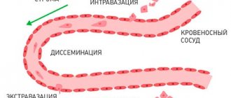
If they are enlarged, then there is a suspicion of an infection of the abdominal cavity, a malignant tumor, or the presence of tumor metastases in nearby organs.
Research technique
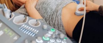
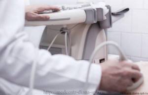
Before starting the ultrasound procedure, the examinee removes metal jewelry. He is placed on the couch in a supine position. During the process, the doctor will ask the patient to change position, turning over to the left or right side.
The examination is carried out abdominally, that is, through the anterior wall of the abdomen. A special conductive gel is applied to the abdomen, which improves the glide of the sensor over the body and helps reduce interference in the image.
The procedure is completely painless and takes no more than 20-30 minutes.
The examination algorithm involves performing an ultrasound of larger organs first. For example, a physician might start with the liver. It evaluates and records the size of the liver, its location and shape, structural features and ability to transmit ultrasonic waves, as well as the condition of the blood vessels.
Next, the doctor proceeds to examine the gallbladder and bile ducts:
- their forms;
- structure and condition of the walls;
- presence or absence of foreign inclusions;
- bile conditions.
The diagnostician checks the position of the pancreatic duct, the lumen of the biliary tract, the portal, inferior and splenic veins. Next, he evaluates the condition of the spleen, pancreas and kidneys according to a similar scheme. At the same time, the doctor must remember that examining the pancreas is often difficult due to the peculiarities of its anatomical location - it is partially overlapped by the intestines and stomach.
To visualize the spleen, the patient is placed on the right side. Access to it is also a little difficult, since it is surrounded by ribs and lungs.
In some cases, during an ultrasound of the abdominal cavity, the bladder is also examined.
How to prepare for research
In order for ultrasound diagnostics to be as informative as possible, it is imperative to follow the stages of preparation for the study, which the doctor warns the patient about in advance:
- Medication preparation. Includes taking an adsorbent for 3 days, which will reduce the process of gas formation in the intestines. For these purposes, espumisan, activated carbon, and enterosgel may be prescribed. To improve food digestion, your doctor may recommend taking enzyme preparations such as festal, mezim and their analogues.
- If the patient has a tendency to constipation , the day before the ultrasound you need to take a laxative orally: senade, senadexin or their analogues. If such drugs are ineffective, you will need to give an additional enema 12 hours before the test.
- 2 hours before an ultrasound scan of BP, you need to drink 2 capsules of a drug based on simethicone or 5-10 tablets of activated carbon, depending on the patient’s weight.
Diet. Preparing for ultrasound diagnostics necessarily involves following a diet:
- 3 days before the ultrasound, you should eat small portions every 3 hours;
- Avoid foods that increase flatulence: baked goods, bread, soda, fermented milk products, fresh vegetables, fruits;
- You should eat boiled, stewed or steamed porridge, lean meat, cheese, lean fish, 1 egg per day; drink enough water;
- On the eve of an abdominal , you can eat a light dinner, but before 8 pm, it is not recommended to choose fish and meat, even lean ones, as dinner;
- On the day of the ultrasound, you cannot have breakfast, if the study is planned after 15:00, you can have a light breakfast, but no later than 11:00.
Only strict adherence to medical recommendations will allow you to obtain a reliable result of an ultrasound examination.
Special requirements for preparation for the study
Ultrasound examination of the abdominal organs can give accurate and informative results only if the person being examined begins to fulfill in advance all the requirements prescribed by the doctor to prepare the patient. Otherwise, during diagnostics, noise may appear in the image, distorting the ultrasound picture.
The doctor may verbally list all the preparatory requirements to the examinee, and in some medical institutions these lists are given to patients in printed form for convenience.
The procedure can only be performed on an empty stomach. If it is scheduled for the first half of the day, the patient needs to refuse breakfast. If the ultrasound is performed after 15:00, you can afford a light breakfast no later than 9:00 am. It is not recommended to consume any liquid an hour to an hour and a half before the start. Also, an hour or two before the ultrasound, you should not chew chewing gum or smoke - these actions provoke spasms of smooth muscles.

People who are prone to constipation should take a laxative the evening before the procedure as part of their preparation.
All medications taken at this time must be reported to the diagnostician in advance. During the ultrasound, the patient takes with him the results of all previously performed tests and examinations, if any.
Those who have a tendency to increased formation of gases in the digestive tract are prescribed a special slag-free diet with a reduced content of coarse dietary fiber. They begin to adhere to it 2-3 days before the examination date.
In addition to dietary restrictions, taking sorbents, for example, activated carbon after each meal, helps reduce the level of gas formation.
A slag-free diet, as a component of preparation for the procedure, involves avoiding the following foods:
- milk and dairy products;
- fresh bread, especially black bread;
- fresh fruits and vegetables;
- fatty and fried foods;
- smoked, spicy and pickled products;
- baking and muffins;
- alcoholic and carbonated drinks, strong tea and coffee;
- legumes
The diet before the study consists of lean meat, poultry and fish, cereals, yeast-free stale white bread, baked or boiled vegetables, vegetable broths, non-concentrated clear juices, compotes. Be sure to drink at least one and a half liters of clean water per day.
All these rules are irrelevant in cases where urgent diagnosis is necessary, when it is not possible to wait 2-3 days, for example, in case of acute appendicitis.