Computed tomography of the abdominal organs is a method of examining internal organs, often used in modern medicine. CT examination appeared in the diagnostic field relatively recently, in the 70s of the 20th century. It is based on the principle of the penetrating power of X-rays. The discovery of X-ray radiation occurred at the end of the 19th century, after which mathematical and technical research gradually led scientists to develop the possibility of scanning the human body with X-rays and converting the received information into tangible media. The first commercial tomography machines were developed in 1971. Since then, computed tomography techniques have improved every year.
What is a CT scan of the abdominal organs and what is it used for?
Content:
- What is a CT scan of the abdominal organs and what is it used for?
- Indications and prohibitions for performing computed tomography
- The process of preparing for a CT scan of the abdominal cavity
- What does a CT scan of the abdominal cavity without contrast show?
- Why do you need contrast injection?
- How does a computed tomography scan of the abdominal organs work?
- Study of the results of CT scan of the abdominal cavity
- Advantages of computed tomography over other types of examinations
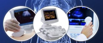
Timely diagnosis of the disease is one of the main factors in the success of combating it. To do this, doctors have a whole range of different examination tools and techniques at their disposal, for example, ultrasound, magnetic resonance and computed tomography. The latter is considered more accurate and in-depth than ultrasound, and at the same time costs less than MRI, so doctors usually give it preference when it is necessary to obtain data not only on the size and location of an organ, but also on the internal structure of deep tissues, cavities, and formations.
The essence of the examination is that X-rays, penetrating the human body, make it possible to obtain a layer-by-layer image of organ tissue in increments of 0.5 to 10 millimeters. The information displayed on the tomograph monitor makes it possible to display a three-dimensional image of a specific organ.
In some cases, the doctor prescribes a CT scan with contrast - for this, a special drug is injected into the patient after the scan, a second scan is performed, after which the structure and structure of the organs is visualized in great detail on the monitor and in the pictures.
A CT scan of the abdominal organs provides a cross-sectional image of each organ, without the image being superimposed on one another. A computed tomogram shows the presence of diseases of the gastrointestinal tract and retroperitoneal space, such as: cancerous tumors, stones, foreign bodies, injuries, cystic formations.
When not to do it?
Since computed tomography of the stomach and esophagus is an x-ray diagnostic method, it has several important contraindications:
- Pregnancy . The tomograph produces radiation that can lead to disruption of the formation and development of the fetus.
- Chronic renal failure with decreased filtration function. If the GFR (glomerular filtration rate) drops <30 ml/min, a contrast agent should not be administered.
- Obesity . Most tomographs are not designed to examine patients weighing more than 120-160 kg.
- The need for emergency resuscitation measures , stopping active bleeding, and surgical intervention. In such a situation, a CT scan is done after the patient’s general condition has stabilized.
- Mental disorders that are accompanied by a pronounced fear of conducting research. It can be done only after special medical preparation of the patient (administration of sedatives).
- Active epileptic seizure . In this condition, it is not possible to keep the patient immobilized in a safe position.
The listed contraindications require the appointment of an alternative diagnostic technique (ultrasound, FGDS or MRI), or specific treatment, after which a computed tomography may be performed.
During menstruation

X-ray irradiation does not affect the course of menstruation and does not increase the severity of pain.
However, if reproductive system is being examined , the image will show changes that may be misinterpreted. Therefore, you should always warn doctors who prescribe and conduct examinations about the onset of menstruation.
If a CT scan is performed routinely to examine the uterus, then it is better to postpone the diagnosis to another day. If an acute pathology of the reproductive system is suspected (for example, endometritis or pelvioperitonitis), it is done despite menstruation. The examination of other organs is not affected by menstruation.
Help During menstruation,
an hour before the start of diagnosis, it is advised to take non-steroidal anti-inflammatory drugs (ibuprofen, ketoprofen) and antispasmodics (drotaverine). This will reduce the severity of unpleasant symptoms and relaxes the smooth muscle fibers of the uterus.
Indications and prohibitions for performing computed tomography
Considering that tomography is directly related to the need to irradiate the body, such a procedure is carried out only as prescribed by a doctor in cases where there is an objective need for this.

Indications for CT scan of the abdominal cavity are:
- determining the need for surgical intervention or an already prescribed operation: this way the doctor receives the most accurate data on the degree of change in organs and the localization of such change;
- the appearance of acute pain, jaundice, sudden weight loss of unknown etiology;
- disruption of the urinary system;
- digestive disorder;
- suspicion of the presence of neoplasms;
- manifestation of liver diseases;
- suspicion of vascular disorders;
- control over the implementation of prescribed treatment;
- the appearance of symptoms of lymph node damage.
Some conditions and pathologies in humans completely exclude the possibility of examining the abdominal organs using a computed tomograph. For example, during pregnancy and breastfeeding, the use of X-ray radiation is strictly not recommended - it can harm the baby.
Some tomographs have a patient weight limit of 140-180 kilograms. If your body weight exceeds this mark, the procedure may be in question.
Conducting a CT scan of the abdominal organs, as well as other parts of the body and organs, is prohibited for patients under 3 years of age. Children of primary school age can be scanned using a tomograph, however, if possible, it is better to replace this research method with a more harmless one, such as MRI.
Diseases such as diabetes mellitus, acute inflammation of the kidneys, heart, liver, blood, are considered relative contraindications when conducting a study with a contrast agent - if they are present, the doctor, at his own discretion, decides whether the procedure is possible.
As for contrast-enhanced computed tomography, its contraindications, also associated with the administration of a contrast agent, are as follows:
- intolerance to iodine and iodine-containing drugs diagnosed in the patient;
- a history of allergies or severe forms of bronchial asthma;
- hyperthyroidism;
- diagnosed renal or liver failure of any kind.
Rules of conduct in the treatment room
A certain time before the examination, it is necessary to notify the doctor in advance about any allergic reactions to iodine if the examination will be carried out using a contrast agent, since it contains iodine.
It is prohibited to bring metal objects, including a telephone, into the treatment room of the diagnostic center. This may affect the scanner and the examination process.
It is important to lie still during the diagnosis. In some cases (for CT scans of the brain, sinuses, etc.), the examination area is secured on a movable table with specialized straps. This is necessary to prevent movements that could nullify the entire procedure.
Share link on social networks
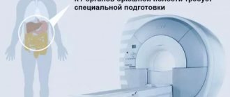
The process of preparing for a CT scan of the abdominal cavity
For this type of examination, doctors put forward a number of special requirements regarding nutrition, since during the process the organs of the digestive tract are scanned.

Already a few days before the planned tomography, you need to completely exclude from your diet foods that increase gas formation in the intestines:
- bread, buns, muffins;
- dairy and fermented milk products;
- carbonated drinks;
- foods rich in coarse fiber.
In addition, alcohol is prohibited.
On the day of a computed tomography scan of the abdominal organs, you should not eat or drink at least 3-4 hours before the procedure, so it is recommended to schedule it in the morning or first half of the day so that the patient does not have to go hungry all day.
Failure to comply with these rules can distort the results of the study - shadows and spots that are not related to the presence of diseases may appear in the images.
The night before, a cleansing enema or laxative is recommended to cleanse the intestines. At this time, you should drink plenty of still mineral water.
When planning an examination with contrast, you must first take a creatinine test.
If you are taking medications, you should inform your doctor about this. In some cases, the doctor prescribes the use of drugs that block x-rays - they, in some way, act as a contrast agent if the contrast itself is not planned during a CT scan.
If you have one, you need to take with you to the procedure:
- doctor's referral;
- extract from the medical record;
- results of previously performed examinations and tests (these may be prescribed by a doctor in preparation for a CT scan, or as part of a general diagnosis of the body).
Where to get a CT scan in Volgograd
Still don't know where to get diagnosed? has modern equipment for quick, accurate examination of any area of the body. Specialists are ready to provide diagnostic support any day of the week. Interpretation of the results and a detailed description of the images are carried out in a minimum period of time.
Detailed information about Diagnostic and contact numbers can be found on the website https://mrtplus.rf/. Call the number and get answers to all your questions. The administrator will explain in detail what the preparation for the CT procedure involves and will schedule an appointment for diagnostics at a time convenient for you. The center regularly holds promotions that allow diagnostics to be carried out on favorable terms for the patient.
What does a CT scan of the abdominal cavity without contrast show?
This type of tomography is used more often than CT with contrast, since it is more harmless, costs slightly less and is easier to perform, without double scanning and manipulations with the introduction of a contrast agent with special syringes or catheters.
CT without contrast makes it possible to assess the condition of the abdominal cavity and each organ separately, check for the presence of developmental anomalies, and also identify the presence of stones in the kidneys, gall bladder, bile ducts and ureter, an increase or decrease in the size of organs, and changes in their tissues.
This type of study helps to determine the presence and amount of calcifications in the arteries or pancreas, determine aneurysm, cirrhosis, the development of malignant tumors, and the spread of metastases.
Why do you need contrast injection?
The contrast agent contains iodine. Distributed throughout the tissues of organs, it makes it possible to most clearly visualize the internal structures of these tissues. The drug is administered intravenously and spreads through the vessels, staining them, after which it accumulates in the tissues, improving their display on photographs. Since it is transported in the blood, its highest concentration is created in tissues and organs richly supplied with blood. For this reason, the contrast method is used to identify pathological foci with increased blood flow. The method is effective for detecting and studying the texture of neoplasms in the abdominal organs; it can even identify those that CT without contrast is not capable of detecting. As a result of the introduction of contrast, the condition of the vessels, the presence of pathological narrowings and functional pathologies in them will be displayed on the image or monitor.
Within 24 hours, the substance is completely eliminated from the body through the kidneys.
How does a computed tomography scan of the abdominal organs work?
CT scans can usually be performed either in a hospital setting or in a clinic.
If an abdominal CT scan with contrast is planned, a venous catheter is inserted into the cubital vein before the scan begins. This mechanism, using an automatic injector, gradually delivers the contrast drug into the body. In this case, we are talking about CT with bolus contrast. It is also possible to use a syringe to administer contrast. To examine the upper gastrointestinal tract, contrast is administered orally; if it is necessary to examine the pancreas, kidneys or liver in detail, contrast is administered parenterally - into a vein.
The patient is placed on the tomograph table and asked to take the most comfortable position. It is especially important that the person does not move during the process, so sometimes it is necessary to secure him with belts. The doctor may ask you to hold your breath for a short time.
The duration of the tomography is no more than 30 minutes. All this time the patient will be under the supervision of medical personnel. The radiologist and x-ray technician are in the next room, but can see and hear the subject and communicate with him. After the procedure, the patient may be prescribed plenty of fluids to speed up the process of removing the contrast.
For those people who suffer from claustrophobia, it is recommended to undergo an examination with an open-type tomograph.
When is it prohibited to undergo CT and CT with contrast?
Despite its apparent simplicity and safety, any type of CT examination may be unacceptable in the following cases:
- The patient is in a general serious condition;
- The patient is prone to claustrophobia;
- The patient has serious cardiovascular diseases;
- The patient is overweight, which does not allow him to take the desired position in the tomograph;
- The patient is less than 15 years old;
- The patient had already undergone a recent CT examination. Repeated examination is permissible only after 3-4 months, after the last session;
- The patient has serious kidney problems, which prevents him from taking contrast agents.
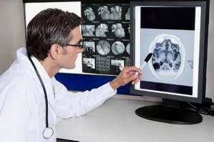
Study of the results of CT scan of the abdominal cavity
Best materials of the month
- Coronaviruses: SARS-CoV-2 (COVID-19)
- Antibiotics for the prevention and treatment of COVID-19: how effective are they?
- The most common "office" diseases
- Does vodka kill coronavirus?
- How to stay alive on our roads?
After the examination is completed, the patient is given photographs of the abdominal cavity, a digital media with saved tomography results, as well as a report from a radiologist. It is necessary to take into account the fact that the quality of the images directly depends on the sensitivity of the tomograph, on the patient’s thorough compliance with the preparation rules, and on whether he was able to remain motionless during the process.
If all the requirements have been met, in the image the doctor will be able to detect the location, size and number of foci of inflammation, stones, neoplasms, thrombosis, and other disorders, and the degree of their spread.