![]()
The most universal and painless methods for diagnosing the retroperitoneal space include ultrasound, which is often performed together with ultrasound of the abdominal organs. This happens due to the fact that both areas are located nearby, and it will not be difficult for a physician, while examining the abdominal cavity using an ultrasound probe, to “look” into the retroperitoneal area.
The ultrasound method is considered one of the safest ways to diagnose internal organs: it does not use x-ray radiation, so it has few contraindications. As for the degree of information content, doctors note that it directly depends on the quality of the patient’s preparation.
What is retroperitoneal ultrasound?
Diagnostics using ultrasound makes it possible to obtain images of organs, vessels and soft tissues in the retroperitoneum in real time. This occurs due to ultrasonic waves that emanate from the sensor and enter the human body through the skin to a certain depth. Then they are either completely or partially absorbed by the tissues and pass on, or they are repelled and returned to the sensor. The tissues of the human body have different densities, so with the help of ultrasound scanning, the diagnostician receives information on the device monitor in the form of a black and white image of areas of a round, blurry or clear shape, somewhere darker, and somewhere lighter. Ultrasound examination has three main advantages:
- painlessness and safety;
- general availability;
- non-invasiveness.
This method is considered painless because the sonologist during scanning does not introduce any instruments that injure the patient’s body or cause pain. Ultrasound is easily tolerated by people of all ages. If necessary, it is prescribed to pregnant women and infants. In addition, this technique, unlike CT, does not carry radiation exposure, so you don’t have to worry about your health.
What will an ultrasound of the retroperitoneal space show?
Ultrasound of the retroperitoneal space will clearly show the following pathological conditions:
- hematoma caused by mechanical injuries, where the cause may be accidents or sports;
- inflammation of purulent, putrefactive and serous type;
- paracolitis;
- paranephritis or inflammation of the retroperitoneal tissue;
- abscesses of the retroperitoneal space;
- cysts of the kidneys and adrenal glands;
- chronic kidney disease;
- nephropathy;
- necrosis of the renal cortex;
- nephrosclerosis;
- aplasia and malrotation of the kidneys;
- acute kidney injury;
- squamous cell carcinoma;
- metastases in the kidneys, adrenal glands and lymph nodes;
- autosomal recessive polycystic kidney disease;
- duplication and merger anomalies;
- multicystic kidney dysplasia;
- renal agenesis;
- dysplasia and hypoplasia of the kidneys;
- lymphadenopathy.
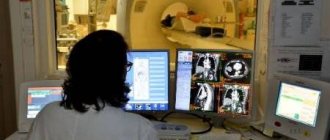
Ultrasound of the retroperitoneal zone plays an important role in the diagnosis of cancer. The fact is that at the initial stage the neoplasms do not manifest themselves in any way, but then they can grow and thereby put pressure on or displace nearby organs. As a rule, it is impossible to detect them by clinical symptoms at the initial stage, when treatment will be most effective. To do this, doctors need to resort to hardware diagnostics such as ultrasound, CT and MRI. Ultrasound of the retroperitoneal space during oncological search is usually prescribed in conjunction with an examination of the abdominal cavity. The organs of these two areas of the body are located so close that it is not difficult for a doctor to assess the condition of all structures at the same time. The information content obtained from a comprehensive ultrasound examination of the abdominal and retroperitoneal space is quite large if you follow all the recommendations when preparing for the procedure.
| Ultrasound service | Price according to Price, rub | Promotion price, rub |
| Ultrasound of the abdominal organs and retroperitoneal space (liver, gall bladder, pancreas, spleen, stomach) | 1500 rub. | |
| Ultrasound of one organ (liver, gall bladder, spleen, pancreas, bladder, adrenal glands) | 800 rub. | |
| Ultrasound of the abdominal organs and kidneys | 1700 rub. | |
| Ultrasound of the abdominal organs + ultrasound of the kidneys + ultrasound of the bladder | 2000 rub. | |
| Kidney ultrasound | 800 rub. | |
| Comprehensive ultrasound (ultrasound of the abdominal organs + ultrasound of the kidneys + ultrasound of the thyroid gland) | 2400 rub. | 1999 rub. |
| Comprehensive ultrasound (ultrasound of the abdominal organs + ultrasound of the kidney + ultrasound of the thyroid gland + pelvic ultrasound with an abdominal probe + ultrasound of the mammary glands) | 4200 rub. | 2999 rub. |
| Comprehensive ultrasound (ultrasound of the abdominal organs + ultrasound of the kidneys + ultrasound of the thyroid gland + ultrasound of the prostate gland with an abdominal probe) | 3300 rub. | 2499 rub. |
| Comprehensive body diagnostics (MRI of the thoracic spine, MRI of the lumbar spine, ultrasound of the abdominal organs, ultrasound of the kidneys, ultrasound of the bladder, consultation with a neurologist, consultation with a therapist) | 11700 rub. | 7000 rub. |
When is the examination scheduled?
You can do an ultrasound of the abdominal cavity at your own request. This is a good prevention of pathological processes. But in some cases, the doctor prescribes such a study.
Indications for prescribing an ultrasound procedure are:
- patient complaints of throbbing or pain in the abdomen;
- in case of suspected appendicitis (especially if the case involves small children);
- if the patient claims that he feels heaviness under the right rib, bitterness has appeared in the mouth, bitter belching has also appeared, and there is a yellow coating on the tongue;
- if a person develops an aversion to fatty foods, but does not take any medications;
- to monitor the condition of those patients who have been diagnosed with liver diseases (hepatitis, hepatosis), various types of jaundice, stones and sand in the gallbladder;
- ultrasound is prescribed for patients with an enlarged liver, spleen, for diseases such as malaria, mononucleosis, sepsis, anemia, etc.;
- indications for diagnosis by ultrasound are difficult or painful urination, changes in the color or amount of urine, especially in cases where the amount of fluid consumed has not changed;
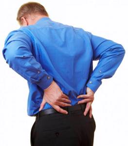
- The doctor may refer a patient complaining of pain in the lower back for an ultrasound after injuries to this area or the abdomen;
- before elective surgery planned on any of the abdominal organs or kidneys;
In addition, an examination of the abdominal organs may be prescribed to patients who have been taking medications for a long time or who like to drink alcohol in unlimited quantities.
An abdominal ultrasound is also prescribed when the following signs appear:
- if your belly begins to grow for no reason;
- with excessive gas formation;
- in cases of persistently elevated temperature, which is in no way related to colds;
- for dyspeptic syndrome, accompanied by nausea, vomiting, heartburn, and loss of appetite.
Important! Ultrasound has no contraindications.
Ultrasound examination is used not only as a diagnostic method, but also for the purpose of monitoring certain processes. The procedure may be performed in conjunction with a kidney biopsy, as well as removal of fluid from the abdominal cavity. This is required in order to control the movements of the needle.
If there are certain clinical manifestations, then standard ultrasound is supplemented with the necessary procedures. Thanks to them, it is possible to assess the motility and evacuation activity of the stomach, establish the functional type of the gallbladder, recognize signs of appendicitis that are visible to ultrasound, study the portal blood flow in detail, and assess the blood circulation in the kidneys.
Since ultrasound does not harm the body, it can be performed an unlimited number of times and can be used not only as a screening examination, but also if there are prerequisites.
How to prepare for an ultrasound of the retroperitoneum?
Ultrasound results can be distorted due to improper preparation of the patient for examination of the retroperitoneal space. The preparation is simple and does not differ from that for an ultrasound of the abdominal cavity. Diet. You should start a few days in advance with a diet that excludes ingredients that cause flatulence. For 2 days, it is worth removing from the diet all yeast-based products - bread, kvass, beer, as well as lactic acid products, legumes, sauerkraut, raw vegetables and fruits, carbonated drinks, alcohol. Gas-lowering drugs . During the diet, it is recommended to take activated carbon at the rate of 1 tablet per 10 kg of weight. Coal can be taken at once or divided into several doses. If the patient has increased flatulence, the day before the scan you should start taking medications such as Espumisan, Mezim-Forte. Colon cleansing. If there is a tendency to constipation, then the day before the procedure you will need to take a mild laxative to cleanse the intestines of excess toxins. Starvation. Ultrasound of the retroperitoneal space is performed on an empty stomach, so it is more convenient to do it in the morning. If, however, the procedure is scheduled for the afternoon, then a light breakfast in the form of crackers and tea is allowed. However, you need to remember that the fasting time before diagnosis should be at least 6 hours. Taking water . 30 minutes before the examination, you will need to drink 0.5-0.7 liters of water so that the sensor can better see the bladder, if an ultrasound of the retroperitoneal space and bladder is prescribed.
What are the indications and contraindications for the procedure?
Doctors, when receiving a patient, determine whether he has specific symptoms and manifestations, in which the patient needs to undergo an ultrasound procedure of the retroperitoneal space.
Indications for examination include:
- disturbances in the process of urination, cutting pain during the process, the appearance of foreign impurities in the urine, for example, blood;
- pain in the pelvic and lumbar region;
- abdominal injuries;
- chronic infectious lesions;
- significant jumps in body temperature for unknown reasons;
- renal failure;
- suspicion of the presence of neoplasms of any nature;
- the likelihood of urolithiasis;
- atherosclerosis of the abdominal aorta;
- glomeluronephritis, pyelonephritis, diabetes mellitus, hydronephrosis.
One of the major advantages of the ultrasound method is the small number of contraindications. In this case, the procedure is impossible if the patient has extensive wound lesions of the abdomen, large-area violations of the integrity of the skin on the abdomen. Ultrasound diagnostics is difficult if the skin of the patient is affected by pustular-inflammatory processes.
As for age restrictions, this diagnostic method does not have them: ultrasound of the retroperitoneal space is prescribed for both children and adults. It can also be performed on infants.
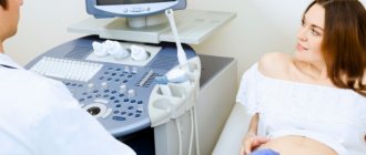
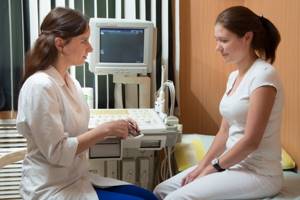
How is an ultrasound of the retroperitoneal organs performed?
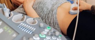
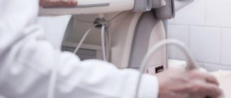
Ultrasound of the retroperitoneal space is quite simple and does not cause discomfort. Before the procedure, the patient must expose the abdominal area and lie down on the couch. The scan is usually done while lying on your back, but sometimes the doctor asks the patient to turn to one side or the other or stand up. Screening comes down to the fact that the diagnostician lubricates the area of study and the sensor of the device with water-based gel, places the sensor on the stomach, pressing it tightly against the skin closer to the area of study, and begins to move it slowly in different directions, sometimes stopping at one point and then moving to another. At this time, an image of the organs appears on the monitor in the form of a black and white picture of structures with different densities. In some cases, in order to monitor the functioning of blood vessels and the speed of blood flow, duplex scanning is additionally used as part of ultrasound of the vessels of the abdominal cavity and retroperitoneal space. The entire procedure takes approximately 30 minutes. The ultrasound results are delivered to the patient within 10 minutes.