Ultrasound of the abdominal cavity is the most common method of functional diagnosis of internal organs and soft tissues of the abdomen. This examination method is carried out quickly and painlessly, which is its significant advantage in comparison with other diagnostic techniques. We will discuss below what is included in an abdominal ultrasound and how to properly prepare for the procedure in order to obtain accurate and reliable results.
Indications for examination
It is recommended to perform an abdominal ultrasound if the following complaints are present:
- abdominal pain;
- bloating;
- bitterness in the mouth;
- jaundice;
- increased gas formation;
- tendency to constipation;
- feeling of heaviness and fullness in the stomach;
- bad breath;
- palpable organ changes;
- injuries;
- vomit;
- regurgitation.
Other indications for ultrasound include long-term use of medications, alcohol abuse, and suspicion of the presence of tumor diseases. An abdominal ultrasound is also performed to monitor the effectiveness of the treatment.
What organs are examined on an abdominal ultrasound?
During the examination, doctors evaluate the size, contours, location and structure of:
- gallbladder;
- liver;
- spleen;
- pancreas.
In addition, based on the results of an ultrasound of the abdominal organs, the presence or absence of retroperitoneal lymph nodes, fluid and neoplasms is determined.
General practitioners, gastroenterologists, pediatricians, oncologists, and family doctors can refer you for examinations. You can also get detailed information from specialists on how to prepare for the examination.
Abdominal examination modes
There are 4 modes of abdominal examination.
- A-mode (amplitude)
is the simplest mode, it allows you to determine the distance to the organ and its parameters; - B-mode or 2D
makes it possible to see an image of an organ in 2D format; - M-mode
creates a video recording of an image with clear boundaries; - Doppler mode
. It shows blood flow in color or black and white modes; - The harmonic mode
allows deep penetration of ultrasonic beams, creating a high-resolution image.
What can be revealed on an abdominal ultrasound
Using ultrasound diagnostics, a doctor can detect the following conditions:
- gallstones;
- hepatitis;
- liver dystrophy;
- cirrhosis;
- neoplasms;
- lymphadenopathy;
- congenital or acquired structural organ disorders;
- post-traumatic disorders.
In addition, the procedure allows you to study blood flow in the area under study. Circulatory disorders are diagnosed in a similar way.
Factors distorting the results
In order to obtain the most accurate ultrasound results, it is necessary to eliminate possible causes leading to distortion of the picture. Among them are:
- drinking alcohol, strong coffee, energy drinks, which contribute to muscle spasms;
- taking antispasmodics;
- presence of gases in the intestines;
- recent endoscopic examination;
- residual contrast agent in the body.
Excess body weight makes the examination difficult. This is due to the poor permeability of ultrasonic waves through the fat layer. Therefore, it is important to properly prepare for the examination.
Preparing for the examination
Preparation for ultrasound of the abdominal organs and kidneys begins 3-4 days before the study. Recommendations on how to prepare for the examination include the following:
- Following a diet aimed at reducing gas formation.
- Ultrasound of the abdominal organs is performed on an empty stomach. Therefore, the last meal should be 4-5 hours before the examination. In case of a removed gallbladder, food and water intake is allowed.
- In case of previous radiography with contrast injection, ultrasound should be performed after 3 days.
- Inform your doctor in advance about taking medications and agree on their discontinuation if necessary.
When preparing for an ultrasound of the abdominal organs, important importance is attached to the diet.
| Authorized Products | Prohibited Products |
| Cereal porridge | Milk and any dairy products |
| Chicken breast | Cabbage, radish, radish |
| Beef | Alcohol |
| Low-fat varieties of hard natural cheese | Legumes |
| River fish | Carbonated drinks |
| Boiled eggs | Pasta and flour products |
| Soups | Fresh apples, pears, grapes |
| Turkey meat | Coffee and caffeinated drinks |
| Stewed or steamed vegetables | Sweet desserts |
| Boiled potatoes | Chewing gum |
Adult patients are allowed to take certain medications a few days before the examination. The prescription must be carried out by the attending physician.
- If there is increased gas formation or bloating, medications may be taken.
- To normalize digestion, enzyme preparations are allowed.
- On the eve of the procedure, it is allowed to take an adsorbent.
Diet
The diet before an abdominal ultrasound requires a complete exclusion from the diet of dairy products, black bread, fresh vegetables and fruits, juices, legumes, sauerkraut, fatty meats, sweet confectionery, coffee, alcohol, and carbonated drinks.
The diet should be fractional, i.e. often (every 3-4 hours), but in small portions. The amount of liquid you drink is not limited.
On the recommendation of a doctor, as an addition to the diet, you can take medications that improve digestion and help reduce gas formation (Activated carbon, Espumisan, Festal, Pancreatin, Mezim-forte).
Note: an enema before an abdominal ultrasound is necessary for those people who suffer from chronic constipation.
12 hours before the start of the procedure, you can put a laxative suppository (Bisacodyl), and only if it is ineffective, cleanse the intestines with an enema. On the eve of the study, a light dinner is allowed (until 20.00), during which it is not advisable to eat meat and fish dishes (dietary ones too!). On the day of the procedure, if the ultrasound is scheduled for the morning, you cannot have breakfast. If the examination is carried out after 15.00, then you can eat something light in the morning (before 11.00).
Activated charcoal must be taken before an ultrasound of the abdominal cavity - 2 hours before diagnosis, 5-10 tablets per dose.
Important! Smoking before an abdominal ultrasound, as well as drinking water, sucking hard candies, and chewing gum is strictly prohibited. If you are taking vital medications or shortly before the ultrasound appointment you underwent irrigoscopy, FGDS, or colonoscopy, be sure to warn your doctor about this. Your doctor may give you additional recommendations before an abdominal ultrasound or reschedule the examination for another day.
Special preparation cases
In some cases, preparation recommendations may change. This applies to people with certain health problems, special physiological conditions and children.
- Pregnant
Metabolic processes in pregnant women may occur more intensely than in other people. Therefore, the last meal before the examination can be 1.5-2 hours. However, it is not recommended to exceed the standard serving size.
The serving size and timing of ultrasound may be revised for patients with multiple pregnancies.
- Patients suffering from diabetes mellitus
Patients with diabetes follow a special diet. Long breaks between meals can worsen the condition. Therefore, the preparation stage for this category of people has some peculiarities: immediately before the study it is permissible to eat a light breakfast.
- Children
For infants, the last feeding is allowed 3 hours before the procedure. Children under 15 years of age are allowed to eat 4 hours before the test. In cases where the child persistently asks to eat, you can give him a few sips of water. However, it is worth noting that to obtain the most accurate results, ultrasound should be performed on an empty stomach.
- Gallbladder function test
Involves performing an ultrasound with a test breakfast. Initially, the examination is carried out on an empty stomach, then 30 minutes after eating. The following products are allowed as a trial breakfast: yogurt, sour cream, 2 yolks, boiled egg.
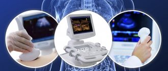
Why is the examination carried out with different sensors?
Modern devices have sets of sensors with different power, curvature and other parameters. The correct choice of sensor is very important and affects the effectiveness of ultrasound.
- Dimensions
. Sensors for abdominal examination are: 40 mm; 50 mm; 60 mm; 70 mm. - Scan angle
. Also, the sensors have different scanning angles: 500; 600; 700; 800. - Frequency
. The frequency ranges of the sensors differ as follows:
- 2-6 MHz (used when studying very obese people with a thick layer of fat on the abdomen (fat tissue interferes with the examination) or for studying deeply located organs up to 25 cm);
- 3-8 MHz (used for examining pregnant women and children 1-8 years old);
- 5-10 MHz (used in neonatal studies (infants up to 28 days of age) or for studying superficial organs).
There are microconvex abdominal sensors that are used for more detailed examination of organs in young children and in gynecology and urology.
Sensors with a surface curvature of 10-14 mm are suitable for vaginal examinations, and with a curvature diameter of 8-10 mm - rectal ones. Such details are important for a clear image of small areas of the organ (when considering fibroids, prostate adenoma, fetus at short term). In pediatrics, when studying small organs, sensors with a curvature of 20-40 mm are used (for example, to study the brain through the fontanel).
Microconvex sensors have a handle for ease of insertion, and can rotate around its axis to view the organ in different projections. Convex sensors may change during the examination. They are selected depending on the tasks assigned to the doctor, the characteristics of the patient (overweight, age) and other factors.
How is an abdominal ultrasound performed?
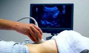
The essence of the method is to direct ultrasonic vibrations to various tissues, which are characterized by varying degrees of reflectivity and absorption. A special sensor detects reflected rays. Thanks to this, it is possible to obtain a visual image of the area under study on the monitor screen.
During the examination, the patient sits on a comfortable couch. A special gel is applied to the skin of the abdomen. Scanning is performed using a sensor. The doctor sees an image of the internal structures in real time. The examination procedure is absolutely painless and comfortable. The average duration of the study is up to 20 minutes. In some cases, the time may be increased at the discretion of the diagnostician. If necessary, the specialist may ask the patient to change his body position or hold his breath for a few seconds.
What do the indicators mean in the abdominal ultrasound protocol?
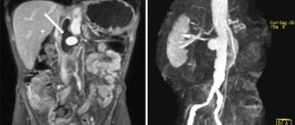
During the procedure, the doctor examines a certain organ and, based on the measurements taken, draws conclusions about how well it complies with the established norm. Depending on the echogenicity of the organ being examined, the homogeneity of its structure is assessed, as well as the presence of pathological inclusions in it: cysts, stones, tumors. During diagnosis, the anatomy of the internal organ must be assessed. If the ultrasound scanner used is equipped with a Doppler, then the blood flow through large vessels is checked.
Abdominal ultrasound is deservedly called a fairly wide-spectrum study, since it allows you to examine almost all internal organs of the pelvis and abdominal cavity.
The ultrasound protocol form indicates the results that relate to each internal organ separately. So, when examining the liver, the presence of fatty hepatosis is indicated by the echo densities of this organ, which appear in the form of small foci. With this disease, the edges of the liver are rounded, and in the last stages of the disease it is no longer even possible to see the portal vessels. With cirrhosis of the liver, the monitor screen clearly shows its increased size, as well as the expansion of the splenic and portal veins. The lower edge of this organ will be rounded, and its contours will be uneven. With this diagnosis, the increase in echo density will be large-focal. Congestion in the liver may be indicated by the expansion of the vena cava and the absence of its narrowing during inspiration. The presence of malignant and benign tumors is indicated in the presence of a violation of the normal echostructure.
Conclusions are drawn about the normal state of the gallbladder if its correct pear-shaped or cylindrical shape was confirmed during an ultrasound. Its width should not exceed three to five centimeters, and its length should not exceed six to ten centimeters. The thickness of the walls of the gallbladder can reach four millimeters. If there is a lumen on the gallbladder, there should not be any formations.
Normally, on ultrasound, the total bile flow should have a diameter of six to eight millimeters. The contours of the pancreas should be smooth, its echogenicity should not be reduced, and the size of the head should not exceed thirty-five millimeters.
If an abdominal ultrasound of the kidneys was performed, then their normal width should be five to six centimeters, length – about eleven centimeters, and thickness – four to five centimeters. The renal parenchyma has a normal thickness of twenty-three millimeters, its pelvis should not be dilated, and no additional structures should be found in the lumens of the ureters and pelvis.
Decoding the results
The results of the examination are delivered 10-15 minutes after its completion. If any changes are detected, the diagnostician will give detailed comments in an accessible form and recommend further observation by a specialized specialist. The conclusion reflects the following parameters:
- Size, shape, structure of the abdominal organs (liver, spleen, gall bladder, pancreas). Ultrasound also involves assessing the condition of the kidneys.
- Proliferation of organ tissue.
- The presence or absence of fluid in the abdominal cavity.
- The presence or absence of gallstones.
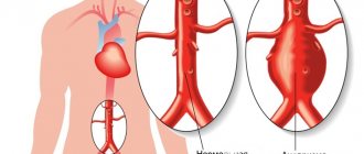
- Aortic diameter.
At the multidisciplinary clinic “Zdorovye”, ultrasound of the abdominal organs is performed using modern equipment. Expert class equipment has high resolution. This allows doctors to accurately determine the location of pathological changes in the area being examined.
Our team consists of the best professionals in the city. Doctors have many years of experience and regularly improve their skills in specialized courses. Diagnosticians detect diseases with high accuracy even in the early stages of development.
Contraindications for abdominal ultrasound
There are no obvious contraindications for this procedure. If an emergency diagnosis of the pelvic organs is carried out in women, then they must be asked the date of their last menstruation to obtain more complete information. In some cases, additional abdominal ultrasound is performed in the following days in order to correctly assess the clinical picture over time.
To diagnose inflammatory processes occurring in the uterine cavity, ultrasound is performed on any day of the menstrual cycle, and if dilation of the fallopian tubes is detected, the study must be repeated after the end of menstruation.
To identify endometriosis, ultrasound is prescribed in the second half of the menstrual cycle; for endometrial hyperplasia, the study is repeated immediately after the end of menstrual bleeding. In the first half of the cycle, an abdominal ultrasound is performed if uterine fibroids are suspected, but after an abortion or surgery, the procedure is prescribed at the end of the next menstruation.
In case of severe pain, bleeding and other emergency situations, an abdominal ultrasound is performed any day without special preparation or any restrictions. Internal organs are examined with special ultrasound devices, for example, the HD 15 ultrasound machine. High-quality and multifunctional equipment, as well as the high skill of ultrasound diagnostic doctors, allows us to identify serious diseases in the early stages and take appropriate measures for their timely and effective treatment.