Abdominal pain is an alarming sign that requires immediate medical attention. After all, it can signal both chronic and acute pathologies, when urgent surgical intervention is indicated. When making a diagnosis, the surgeon will focus on the location of the pain, but the symptomatic picture alone is not enough. Moreover, pain can be radiating, that is, projected to other parts of the abdomen. Then it is necessary to resort to other diagnostic options. One of these hardware methods is abdominal CT. This is a highly informative method that allows you to examine the tissues of organs, vessels and lymph nodes of the peritoneum.
CT helps to obtain a three-dimensional image of the area under study, thanks to the ability to take X-ray images layer by layer, with the thickness of each layer in this case being about several microns. X-rays are emitted in a ring-shaped circuit with sensors, due to which images are obtained in different planes. Entering the computer monitor, they are superimposed on each other, forming a three-dimensional model of the organ being studied.
An even more detailed picture can be recreated by a CT scan of the abdominal cavity with contrast. Iodine preparations move along with the bloodstream to the desired area. Thanks to the ability of this drug to increase the contrast of tissues, maximizing differences in the absorption of X-rays, it is possible to most accurately determine the location of pathologies, their structure, shape and diameter.
What is CT or computed tomography
Computed tomography (CT) or multislice computed tomography (MSCT) is a modern diagnostic method that consists of a layer-by-layer examination of the human body using X-rays to obtain a detailed image of internal organs and structures.
CT imaging is based on the varying densities of organs and tissues through which the X-rays pass. During the examination, an X-ray tube with a narrow beam of X-rays rotates around the patient. At the same time, many sensors record the slightest changes in density. Information from the sensors is processed by a powerful computer, after which it is sent to the monitor in the form of successive longitudinal and transverse sections (“tomography” literally means “image of a slice”). The thinner the section, the more accurate the diagnosis.
The University Clinical Hospital No. 2 has modern equipment - a high-class computed tomograph, which allows obtaining high-quality images with minimal radiation exposure.
Today, CT is one of the main diagnostic methods and is widely used both for making a diagnosis and for clarifying it. One of the main advantages of computed tomography is the speed of the study and the ability to scan several organs simultaneously. Modern tomographs can significantly reduce the duration of the examination and reduce patient exposure to a minimum, while obtaining hundreds of sections up to 0.5 mm thick. Thanks to high-precision three-dimensional projections, which make it possible to examine the position, shape, size and structure of various organs and structures of the body, the results obtained from CT examination are the key to success in making an accurate diagnosis, selecting the most effective treatment and planning surgery if necessary.
A significant advantage of this method is the ability to examine patients with pacemakers, artificial heart valves, metal clips in the body, etc., that is, those for whom MRI is contraindicated.
CT scanning is used to examine almost all parts of the body and organs: chest, abdomen, pelvis, limbs, liver, pancreas, intestines, kidneys and adrenal glands, bladder, lungs, heart, as well as blood vessels, bones and spine.
The range of diseases for which this research is necessary is extremely large. In each individual case, the need and feasibility of this study is assessed by your attending physician.
What is used to perform an MRI with contrast?
Due to the peculiarities of the magnetic resonance imaging method, drugs based on gadolinium perform best. This is a non-toxic component that dissolves well in water and is quickly eliminated from the body naturally.
There is also no chance that such a product will settle in your internal organs.
When using contrast, only those products that have been officially approved and registered in the Russian Federation are used. These include such as “Omniscan”, “Dotarem” and a number of others.
The drugs are created specifically to minimize the likelihood of allergies. But in a small number of cases, the patient may still experience an allergic reaction.
Carrying out a special analysis allows you to predict this probability in advance.
What is the difference between CT and MRI
This is the most common question asked to a radiologist. CT and MRI are diagnostic methods. They have similarities and differences. The similarity lies in the external similarity in the design of the devices themselves - tomographs. In their center there is a tunnel into which the table slides.
The main difference is a completely different principle for obtaining images: CT uses x-rays, MRI uses a magnetic field. Which method is right for you is determined by your doctor, taking into account the specifics of the disease and the purpose of the study.
Screening for coronary calcium. CT scan of the coronary arteries.
CT scan of the coronary arteries with determination of coronary calcium is performed to obtain diagnostic information about the presence, location and size of calcified atherosclerotic plaques in the coronary arteries. Patients with atherosclerotic plaques have a higher risk of developing coronary heart disease and myocardial infarction. Coronary calcium screening is a rapid, non-invasive diagnostic method that does not require the administration of a contrast agent. If it is necessary to visualize the lumen of the coronary vessel, intravenous contrast is used. This technique is called CT angiography of the coronary arteries. The study makes it possible to identify not only calcified, but also soft plaques, assess the diameter of the coronary arteries along their entire length, and also identify anomalies in the development and origin of heart vessels. It is possible to perform CT angiography to assess the patency of coronary stents, as well as aortocoronary and mammary coronary bypass grafts. Cost of examination Pre-registration and inquiries by phone (495) 942-40-20 . Registration hours: Monday-Friday from 8.00 to 20.00, Saturday - from 9.00. until 17.00.
Important!
Please prepare in advance and bring with you all extracts, protocols or recordings (disks) of previous studies. The more information the doctor has before the study, the clearer the task assigned to him. In addition, previous results will allow us to assess the dynamics of the disease.
Before the procedure, be sure to notify your doctor if you have:
- Pregnancy
- Are you allergic to medications, including iodine in the contrast agent?
- Cardiovascular disease (eg, heart failure)
- Diabetes mellitus or you are taking metformin (Glucophage). You may need to avoid taking this medication the day before and for the day after your procedure.
- Kidney diseases
- Bronchial asthma
- A pacemaker or insulin pump is installed
- Multiple myeloma.
- You have had an X-ray examination within the previous 4 days using a barium contrast agent (irrigoscopy) or you have used medications containing bismuth. Barium and bismuth, appearing on X-ray film, reduce image quality.
- There is a fear of closed spaces (claustrophobia). Because you will have to lie still inside the scanner during the procedure, you may need sedatives. In this case, you should ask someone to take you home after the procedure.
The Department of Radiation Diagnostics of the University Clinical Hospital No. 2 operates within a multifunctional clinic, therefore, if any possible complications are suspected, the decision on conducting the study, its type and methods of preparation is made together with medical specialists (nephrologist, endocrinologist, etc.).
Contraindications for MRI with contrast
The ability to administer contrast is not always provided. There are several main contraindications that may limit the use of a contrast agent:
- The likelihood of allergic reactions to some components of the drug.
- Diseases of the kidneys and cardiovascular system.
- Constant use of drugs from the group of beta blockers.
- Severe dehydration.
There are a number of other diseases for which the use of contrast is contraindicated. These include myeloma, sickle cell anemia, and polycythemia.
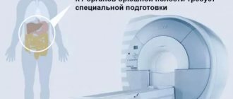
Preparing for an abdominal CT scan
If you are scheduled for a CT scan of the abdominal organs, refrain from eating solid food the evening before the examination. Before the procedure, you may be asked to drink a contrast agent, and in some cases, take a mild laxative or have a barium enema.
CT and MSCT of the abdominal organs are performed on an empty stomach or no earlier than 3-4 hours after the last meal. It is allowed to take medications (with a small amount of water). Immediately before the examination, the department may ask you to drink about 500-600 ml of water.
MSCT examination is performed after preliminary contrasting of the intestine with a diluted contrast agent. The gastrointestinal tract is filled either on the day of the study or the day before: On the eve of the study, drink 0.5 liters of solution in the evening, in the morning (06:00-07:00) drink the remaining 0.5 liters. The study is carried out with a full bladder. Women must have a sanitary tampon with them.
Attention! After an X-ray examination of the stomach, colon with barium (gastric fluoroscopy, irrigoscopy), CT examination of the abdominal organs is recommended no earlier than 5-7 days (at least 3 days). In cases of emergency - after cleansing enemas.
How the research works
We ask you to come to the radiology department (9th floor) 10-15 minutes before the scheduled start time of the study and, having contacted the office, inform the staff of your arrival. Given the possibility of emergency research, there is a possibility of waiting for the procedure for some time. Experienced doctors and x-ray technicians will do everything to make you feel comfortable and safe throughout the entire examination.
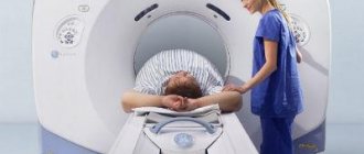
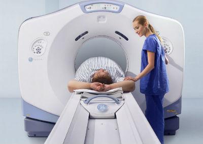
Computed tomography is an absolutely painless procedure that takes no more than 30 minutes (including changing clothes and, if necessary, intravenous contrast). The scanning itself lasts several minutes. If you need a CT scan with contrast, you will first be invited to the procedure room, and a nurse will install an intravenous catheter for you to administer a contrast agent during the examination.
Before the examination, you will be asked to remove all jewelry and metal objects that fall into the scanning area; the x-ray technician who will carry out the procedure will explain to you the rules of conduct during the examination and will help you sit comfortably on the tomograph table.
If the study is carried out using a contrast agent, it can be introduced into the patient’s body in various ways depending on the purpose of the study: intravenously - in CT scans of the chest, abdomen and pelvis.
- orally - for some abdominal examinations, the contrast agent must be drunk
- through a special catheter into the bladder or intestines
During the procedure, the patient lies on a special table connected to a CT scanner, which is a large ring-shaped machine. The scanner rotates around the area of the patient's body being examined, taking layer-by-layer images of the corresponding organ. A faint hum or clicking sound may be heard.
Try not to move during the procedure, as moving your body may reduce the quality of the image.
Given that a CT scanner emits X-rays, the machine is usually located in a special shielded (protected) room. The device is controlled automatically from the adjacent office, which houses the tomograph’s computer unit, monitors and equipment for monitoring the patient’s condition. The doctor performing the examination sees and hears you. You will be able to talk to him through a special intercom. The specialist may ask you to hold your breath for a few seconds to get a clearer picture.
Based on the images obtained, the doctor at the radiology department gives a medical opinion. In addition, your doctor or surgeon can comment on the results of the study.
What sensations does the patient experience during the CT scan procedure?
The procedure itself is absolutely painless. Some discomfort may be caused by the hard surface of the table, the inability to move, and the temperature in the CT room (the room may be cool).
Some patients feel nervous while inside the scanner. If contrast material enters a vein, you may experience a feeling of warmth, heat, or a metallic taste in your mouth. Sometimes patients experience nausea or headache. Be sure to tell your doctor or x-ray technician how you feel.
CT scan of the brain and skull bones
is used for:
- Diagnosis of the most dangerous changes after brain injury (bleeding, bruises and concussions, skull fractures).
- Detection of hemorrhage in the brain due to rupture of a cerebral aneurysm, hypertensive crisis or other cause.
- Visualization of aneurysms and arteriovenous malformations of cerebral vessels during MSCT angiography.
- Assessment of cerebral perfusion in acute stroke.
- Detection, preoperative planning and dynamic monitoring of brain tumors.
- Assessment of cerebral ventricles in hydrocephalus.
- Diagnosis of tumors and polyposis-inflammatory changes in the paranasal sinuses.