MSCT of the abdominal cavity and retroperitoneal space is one of the most modern methods for visualizing organs and tissues in this area. The use of multi-detector scanning allows not only to significantly reduce the dose of ionizing radiation (compared to X-ray or CT), but also to quickly obtain high-quality images of organs and tissues thanks to thin sections (less than 1 mm). Also, MSCT provides the opportunity to conduct 3D reconstruction of organs, which is very important both in diagnostic terms and in the choice of treatment tactics.
MSCT allows you to obtain images of soft tissues, vessels and organs of the abdominal cavity and retroperitoneal space such as:
Liver, gallbladder, biliary tract, pancreas, stomach, small intestine, large intestine, kidneys, adrenal glands, ureters, lymph nodes. The purpose of MSCT is to evaluate the position, size and anatomical structure (presence of pathological formations or diffuse changes), the presence of radiopaque stones in the gall bladder, kidneys or ureters, the presence of fluid in the abdominal cavity, changes in bone structures such as the spine or ribs (as a screening ).
MSCT of the abdominal cavity with contrast reveals
MSCT of the abdominal organs is used to identify the causes of pain in this area.
It shows organ damage, infection and disease. Liver diagnostics makes it possible to detect cysts, cirrhosis, injuries, tumors, and jaundice. When examining the gallbladder, stones, inflammatory processes, cholecystitis, cholangitis, and adenomyomatosis are clearly visible. MSCT of the intestine shows polyps, fistulas, tumors, damage due to trauma. By studying the pancreas, its contours and dimensions are determined. Pancreatitis, false cysts and aneurysms, tumors, and abscesses are often diagnosed. MSCT of the kidneys is performed to detect injuries, cysts, abscesses, hydronephrosis, tumors, and pyelonephritis. Often, kidney diagnostics are performed together with MSCT of the adrenal glands because both of these areas are often affected by the same pathological process.
Preparation
Preparation for a computed tomography scan of the abdominal organs begins the day before the main study.
From the evening before the start of CT diagnostics, the patient should exclude gas-forming foods and drinks from the diet (cabbage, legumes, fermented milk and fermented products, apples, yeast products), and under no circumstances drink alcohol. The procedure itself is carried out on an empty stomach, since active intestinal motility, which appears after eating food, can interfere with computer images. CT can be performed on an outpatient basis, without hospitalization of the patient.
When performing a computed tomography scan of the abdominal organs, contrast enhancement is used according to a special medical scheme. A contrast agent is an iodine-based substance that is used to visualize in detail the areas of interest. The iodinated drug is injected intravenously through a special catheter (Venflon) into the cubital vein and is carried by the blood, staining the vessels, with subsequent accumulation in organs and systems, improving their visualization on images.
Contrast penetrates especially well into pathological areas that are abundantly supplied with blood (areas of inflammation, tumor growths, etc.).
For each patient, a different volume of the substance may be needed to undergo a computed tomography scan with contrast of the abdominal cavity, which is individually selected depending on the person’s weight category. After the examination, the drug is completely eliminated from the body within 24 hours.
If there is an indication for scanning a specific parenchymal organ, oral forms of radiopaque contrast agent are used. The patient must drink several glasses of liquid within a certain amount of time. This method is indicated for studying the work and condition of parenchymal organs:
- liver;
- kidney;
- pancreas.
In rare cases, the patient will need contrast administration in the form of an enema.
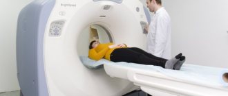
Then the whole procedure takes place directly on the table. Before the scan itself, the patient lies down on the table. He must not move so that the data obtained is reliable. According to the protocol, the X-ray specialist asks the patient to hold his breath for a couple of seconds. The entire scanning process takes from 15 minutes, in the case of contrast injection - 25 minutes. During the entire diagnostic process, the patient will be under the supervision of qualified medical personnel. After the study is completed, the intravenous catheter is removed. To speed up the removal of the contrast agent from the body, all patients are recommended to drink plenty of fluids.

Contraindications to MSCT of the abdominal cavity with contrast:
- a clear contraindication for MSCT is pregnancy, regardless of the stage of pregnancy
- Patients under 14 years of age are not recommended to undergo the study
- large patient weight, since most CT scanners have certain weight restrictions
- a relative contraindication is hyperkinesis (nervous disorder accompanied by involuntary movements, twitching of limbs) and other neurotic conditions
- allergy to iodine
- renal failure
- severe diabetes mellitus
- lactation period
- thyroid diseases
- bronchial asthma
- if the biochemical blood test shows creatinine content more than 1.5 mg/dl
- multiple myeloma – during angiography
Registration for examination
You can sign up for diagnostic procedures at JSC “Medicine” (clinic of Academician Roitberg) on the website - the interactive form allows you to select a doctor by specialization or search for an employee of any department by name and surname. Each doctor’s schedule contains information about visiting days and hours available for patient visits.
Clinic administrators are ready to accept requests for an appointment or call a doctor at home by calling +7 (495) 775-73-60.
Convenient location on the territory of the central administrative district of Moscow (CAO) - 2nd Tverskoy-Yamskaya lane, building 10 - allows you to quickly reach the clinic from the Mayakovskaya, Novoslobodskaya, Tverskaya, Chekhovskaya and Belorusskaya metro stations .
Preparation for MSCT of the abdominal cavity with contrast
Preparation for MSCT of the abdominal cavity is that before the procedure in the evening the patient is allowed to eat a light dinner without overeating, and on the day of the examination he cannot eat anything, not even breakfast. If the session takes place in the afternoon, you can drink juice in the morning or eat liquid porridge. If MSCT of the abdominal cavity with contrast is expected, then in the evening you need to dilute two ampoules of 75% urografin in 1.5 liters. Of this, 0.5 liters are drunk in the evening and 0.5 liters in the morning, and the remaining third of the solution is taken with you for testing and drunk half an hour before the procedure. Before undergoing the procedure, the doctor prescribes a blood test for biochemistry to the patient. A modern version of the method is MSCT angiography - a method for detailed examination of blood vessels. Before performing it, you need to drink plenty of fluids, but no later than 3 hours before the test.
Preparation for MSCT of the brain with contrast:
Patients with an increased risk of allergic reactions and reduced renal function should be pre-treated with antihistamines and should not take diuretics or nephrotoxic drugs on the day of the study.
3. Disadvantages and contraindications
The main problems, limitations and contraindications to MSCT are associated, oddly enough, not with the examination itself, but with the introduction of an auxiliary contrast agent: as a rule, the contrast enhancer contains iodine, which in some cases can cause an allergic reaction. Contrast studies may be contraindicated in certain conditions and diseases of the kidneys, heart, liver, etc.
It is also undesirable to have metal objects on your body (this is not a contraindication, but it can reduce the quality of the image). In addition, even minimal radiation exposure has to be taken into account, so pregnant women are prescribed MSCT only for health reasons and in the absence of diagnostic alternatives.
Depending on the geometric dimensions of the equipment, overweight and obesity of the patient may become a problem. A prerequisite is also that the patient understands and meaningfully follows the instructions.
Indications and contraindications, possible restrictions and precautions - all these points are relative; the benefit-risk ratio in each individual case is carefully weighed and assessed by the doctor based on a study of the medical history and illness.
About our clinic Chistye Prudy metro station Medintercom page!
Advantages
Abdominal CT with contrast in Moscow is the leading method for diagnosing many pathological processes. A computed tomograph qualitatively visualizes traumatic, inflammatory, cystic, and many other changes in the body, in particular neoplasms (both benign and malignant with metastasis). Using such images, it is possible to determine the true size, localization, boundaries of pathologies, the presence of metastases, the degree of invasion into organs and other important parameters that determine the stage of the disease and further tactics of patient management.
For example, computed tomography of the pancreas shows in detail the presence of neoplasms of various structures, and also perfectly differentiates them due to the difference in the density of the examined tissues. During a study with a contrast agent, the drug that passes through the splenic artery shows signs of tumor growth into the vessel.
Contrast-enhanced computed tomography also has a number of other advantages:
- Low radiation exposure;
- High scanning speed;
- Obtaining slices of minimal thickness;
- High image quality;
- Identification of minimal changes in internal organs that are not available for visualization in other studies;
- After the completion of the CT scan, the patient returns to his usual lifestyle;
- Construction of highly informative three-dimensional images;
- The results obtained are presented on film and CD, which the patient can provide to the attending physician for further therapy, diagnosis and development of further diagnostic tactics.
Features in children
If a child needs to undergo MSCT, it should also be prepared in advance. He is also advised to eat a diet that excludes foods that increase gas formation, inhibit motor skills, or complicate digestion. Additionally, medications may be prescribed.
Breastfed 
children should not be introduced to new foods before the study .
Since they eat much more often than an adult, the last feeding is carried out 3-4 hours before the procedure. The same goes for drinking. It is important that MSCT is usually performed under sedation. Therefore, at the planning stage, the doctor prescribes additional examinations (blood and urine tests, electrocardiogram).
When performing the study, older children can do without general anesthesia. Provided proper psychological preparation, the child clearly follows all the doctor’s recommendations and reacts normally to the procedure.
CT scan of the abdomen and retroperitoneum
Indications for a CT scan of the abdominal organs may include:
- suspicion of primary and secondary (metastasis) liver tumor;
- “vague” lesions in the liver and spleen detected by ultrasound;
- suspicion of pancreatic diseases (tumor, cyst, acute pancreatitis, etc.);
- suspicion of neoplasm of the kidneys and adrenal glands;
- hematuria of unknown etiology;
- non-functioning kidney of unknown etiology;
- suspected kidney abscess;
- detection and identification of suspected perinephric pathology;
- with a clinical picture of urolithiasis that is not confirmed by traditional X-ray methods and ultrasound;
- kidney cancer after nephrectomy to exclude tumor recurrence;
- tumors of the gastrointestinal tract (esophagus, stomach, intestines) for staging the malignant process;
- tumors of the gastrointestinal tract (esophagus, stomach, intestines) to evaluate the treatment;
- impossibility of performing fibrocolonoscopy (virtual CT colonoscopy is performed to identify polyps, diverticula and other pathologies);
- hemoblastosis (Hodgkin's lymphoma, lymphosarcoma, etc.) (to assess the degree of damage to the abdominal and retroperitoneal lymph nodes);
- metastases of a malignant tumor to the retroperitoneal lymph nodes;
- suspicion of a retroperitoneal tumor;
- focal inflammatory changes in the liver or spleen;
- gallbladder pathology;
- traumatic damage to parenchymal organs (liver, spleen) and blood vessels;
- developmental abnormalities of the abdominal organs.
Carrying out computed tomography of the abdominal cavity and retroperitoneal space.
The information content of computed tomography of the abdominal cavity and retroperitoneal space depends not only on the technical perfection of the tomograph on which the patient is examined, but must also be carried out methodically correctly.
To answer the questions listed above, CT scans of the abdomen and retroperitoneum should be performed with bolus intravenous contrast. A catheter is inserted into the patient's elbow vein, and during the examination, an automatic injector injects an iodine-containing contrast agent. Scanning in automatic mode is carried out in the arterial, venous, parenchymal phases of the contrast agent entering the organs. Why is this being done? Healthy organ tissue and pathological formations (primarily tumors) accumulate the contrast agent in different ways. This makes it possible to detect minimal structural changes in the organs under study (for example, tumors up to 0.5 mm in size in the adrenal glands, liver, pancreas, kidneys). The examination is painless for the patient, the total duration of the examination is about 30 minutes. The contrast agent is excreted by the kidneys within several hours.
Preparing for a CT scan of the abdominal cavity
An abdominal CT scan does not require special preparation. The patient arrives for the study on an empty stomach. To conduct a study with intravenous bolus ascertainment, the patient must have the results of biochemical blood parameters - urea and creatinine.
Advantages of CT scan of the abdominal cavity and retroperitoneum at the Scandinavian Health Center
At the Scandinavian Health Center, CT scans of the abdominal organs are performed by highly qualified and certified radiologists using a TOSHIBA Prime Aquilion 160 multi-slice spiral tomograph (160 slices). Thanks to the latest technologies and software, the device allows you to obtain high-resolution scanning results, and the examination is completed quickly and with minimal radiation exposure.
In total, the test lasts about 30 minutes, although the scan itself lasts a few seconds. The rest of the time is spent processing the received data and preparing the research results (protocol or conclusion, images, DVD).
In addition to computed tomography of the abdominal organs and retroperitoneal space, the Scandinavian Health Center can perform multislice CT:
- chest and pelvic organs (with and without contrast);
- lower and upper extremities;
- spine;
- joints (using a contrast agent);
- examination of the vessels of the head and neck;
- examination of all parts of the aorta and its branches;
- MSCT coronary angiography;
- MSCT of the arteries of the lower and upper extremities;
- MSCT examination of the pulmonary artery and its branches;
- and all other CT scans used today.
Request a call back
How is it carried out?
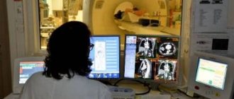
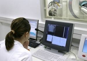
Multispiral computed tomography of the abdominal cavity involves the study of its organs and other anatomical structures using x-ray radiation , which passes through a certain area of tissue in a narrow beam. As a result, the specialist receives a layer-by-layer image of the object under study on the monitor screen, which allows a detailed study of its structure.
The procedure is carried out on an empty stomach after preliminary preparation.
- During the study, contrasting of the intestine is usually performed, since without this, its loops can imitate a space-occupying formation.
- Immediately before the procedure begins, the patient is placed on the conveyor table of the device and is asked to lie more comfortably on his back. To reduce artifacts, the hands must be removed from the scanning area (usually they are moved to the sides).
- Highly radiosensitive areas ( genital organs, mammary glands) are covered with lead screens , provided that they are outside the scanned field.
- The x-ray technician places the light beam at the level of the anatomical area being examined. And he goes into a separate room, from where he will contact the patient through a microphone.
- The patient is advised not to move and hold his breath . At this time, a scan is performed, during which the X-ray tube moves around the object in a spiral and performs a series of tomograms in different planes. The research process takes only a few minutes and is well tolerated by patients.
Certain difficulties arise when performing MSCT in children . After all, during the procedure you need to lie still and not make unnecessary movements. In young children (under 3 years old) this is almost impossible to achieve, so the procedure is often performed under anesthesia or sedation. You can learn more about medicinal sleep from this article.