Ultrasound of lymph nodes is one of the main modern diagnostic methods that can be used to assess the condition of lymphoid tissue. The main indication for this study is lymphadenopathy - an enlargement of the lymph nodes of any group, accompanied by changes in the skin in the form of redness and a local increase in temperature. Enlarged lymph nodes may be painful on palpation. Often, inflammation of the lymphatic vessels associated with this group of nodes is simultaneously observed - lymphangitis.
In the PreAmbula network of clinics, the latest digital ultrasound diagnostic devices are used to examine lymph nodes, guaranteeing the receipt of highly accurate information. If the examination reveals any pathologies, the patient is referred for treatment to an appropriate specialist.
What are the causes of enlarged lymph nodes?
The immune system includes the spleen, thymus (or thymus), lymphatic vessels and nodes. Lymph nodes are a peripheral organ of the immune system and are located in different areas of the body; the largest groups of lymph nodes are the axillary, inguinal and submandibular. The main purpose of the lymph nodes is to protect the body from the spread of infection. Special cells contained in lymphoid tissue kill pathogenic bacteria and viruses that enter the body.
Lymphadenopathy is almost never an independent disease. Enlargement and inflammation of any group of lymph nodes may indicate a pathology of nearby organs or an infectious disease. Thus, the lymph nodes in the neck area can react to a sore throat, the mental nodes often become inflamed due to carious lesions of the teeth, and an increase in several distant groups of lymph nodes at once occurs during acute viral infections, for example, with mononucleosis.
In addition, lymphadenopathy is one of the symptoms of cancerous tumors and leukemia, some types of immunodeficiency. Therefore, if your child has symptoms of lymphadenopathy, you should immediately make an appointment for an ultrasound of the lymph nodes. After all, an increase in the size of these immune organs always indicates some kind of problem in the body.
Decoding the results
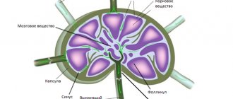
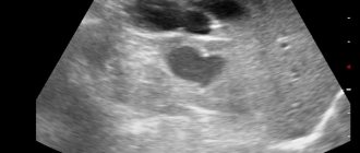
During the study, the density, structure, size, and contours of the lymph nodes are assessed. Normally, they are small in size, oval in shape, and not fused to each other. The contours are clear and even. The structure is homogeneous, without areas of decreased or increased echogenicity. The sizes are small, do not exceed 1-3 cm. An increase in the size of the lymph nodes can be a sign of infection, autoimmune pathology, inflammation, cancer or hematological disease.
Conclusion Ultrasound of the retroperitoneal lymph nodes is not a final diagnosis. It can only be determined by a doctor based on all the results of tests and studies!
How is the ultrasound examination procedure carried out?
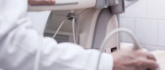
The ultrasound diagnostic procedure is carried out quite quickly, it is safe and completely painless. The child is placed on the couch. The skin in the area being examined is lubricated with gel, which is necessary to facilitate the penetration of ultrasonic waves to the surface of the organ. The ultrasound specialist then presses the device's sensor to the skin and uses it to obtain an image displayed on the monitor. From time to time, the doctor may press on certain areas of the body to assess the degree of mobility of the lymph nodes. Examination of the abdominal lymph nodes is carried out in a state of deep inspiration. Therefore, before visiting the clinic, you should practice holding your breath with your child.
Examination of each group of lymph glands usually takes about 10-15 minutes. Most often, lymph nodes located in the neck and abdomen are examined using ultrasound. Changes in the cervical lymph nodes are observed in pathologies of the tonsils, salivary glands, hard palate, hearing organ and thyroid gland. If lymphadenopathy is found in the abdomen or retroperitoneum, this may indicate diseases of the stomach, liver, pancreas or intestines. Lymph nodes are located at different depths from the surface of the body, so it is not always possible to clearly diagnose pathology using ultrasound alone. To clarify the diagnosis, the doctor may prescribe several studies at once; for example, traditional ultrasound diagnostics can be supplemented with MRI, duplex echosonography and biopsy with histological analysis of lymphoid tissue.
If a pathology of any group of lymph nodes is detected, the specialist also evaluates the condition of the organs located next to them in order to find out the cause of lymphadenopathy. If necessary, other laboratory and instrumental studies are prescribed for a more accurate diagnosis.
What will an ultrasound of the retroperitoneal space show?
Ultrasound of the retroperitoneal space will clearly show the following pathological conditions:
- hematoma caused by mechanical injuries, where the cause may be accidents or sports;
- inflammation of purulent, putrefactive and serous type;
- paracolitis;
- paranephritis or inflammation of the retroperitoneal tissue;
- abscesses of the retroperitoneal space;
- cysts of the kidneys and adrenal glands;
- chronic kidney disease;
- nephropathy;
- necrosis of the renal cortex;
- nephrosclerosis;
- aplasia and malrotation of the kidneys;
- acute kidney injury;
- squamous cell carcinoma;
- metastases in the kidneys, adrenal glands and lymph nodes;
- autosomal recessive polycystic kidney disease;
- duplication and merger anomalies;
- multicystic kidney dysplasia;
- renal agenesis;
- dysplasia and hypoplasia of the kidneys;
- lymphadenopathy.
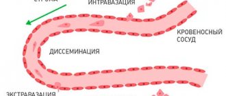
Ultrasound of the retroperitoneal zone plays an important role in the diagnosis of cancer. The fact is that at the initial stage the neoplasms do not manifest themselves in any way, but then they can grow and thereby put pressure on or displace nearby organs. As a rule, it is impossible to detect them by clinical symptoms at the initial stage, when treatment will be most effective. To do this, doctors need to resort to hardware diagnostics such as ultrasound, CT and MRI. Ultrasound of the retroperitoneal space during oncological search is usually prescribed in conjunction with an examination of the abdominal cavity. The organs of these two areas of the body are located so close that it is not difficult for a doctor to assess the condition of all structures at the same time. The information content obtained from a comprehensive ultrasound examination of the abdominal and retroperitoneal space is quite large if you follow all the recommendations when preparing for the procedure.
| Ultrasound service | Price according to Price, rub | Promotion price, rub |
| Ultrasound of the abdominal organs and retroperitoneal space (liver, gall bladder, pancreas, spleen, stomach) | 1500 rub. | |
| Ultrasound of one organ (liver, gall bladder, spleen, pancreas, bladder, adrenal glands) | 800 rub. | |
| Ultrasound of the abdominal organs and kidneys | 1700 rub. | |
| Ultrasound of the abdominal organs + ultrasound of the kidneys + ultrasound of the bladder | 2000 rub. | |
| Kidney ultrasound | 800 rub. | |
| Comprehensive ultrasound (ultrasound of the abdominal organs + ultrasound of the kidneys + ultrasound of the thyroid gland) | 2400 rub. | 1999 rub. |
| Comprehensive ultrasound (ultrasound of the abdominal organs + ultrasound of the kidney + ultrasound of the thyroid gland + pelvic ultrasound with an abdominal probe + ultrasound of the mammary glands) | 4200 rub. | 2999 rub. |
| Comprehensive ultrasound (ultrasound of the abdominal organs + ultrasound of the kidneys + ultrasound of the thyroid gland + ultrasound of the prostate gland with an abdominal probe) | 3300 rub. | 2499 rub. |
| Comprehensive body diagnostics (MRI of the thoracic spine, MRI of the lumbar spine, ultrasound of the abdominal organs, ultrasound of the kidneys, ultrasound of the bladder, consultation with a neurologist, consultation with a therapist) | 11700 rub. | 7000 rub. |
How to prepare for the lymph node ultrasound procedure?
For most lymph node groups, such as ultrasound of the neck lymph nodes, no special preparation is required. But if it is necessary to assess the condition of the lymph nodes of the abdominal cavity or retroperitoneal space, then a little preparation should be done to increase the information content of the study. The presence of gas in a child's intestines can interfere with a correct diagnosis. Therefore, for 2-3 days before the procedure, the child must follow a diet that involves excluding foods that promote gas formation from food. Such products include:
- legumes (peas, beans, soybeans, beans);
- cabbage;
- Rye bread;
- dairy products;
- carbonated drinks;
- sweets;
- yeast baked goods.
If a child has bloating, then on the eve of the test it is recommended to give him activated charcoal or any other remedy approved for children that relieves gases in the intestines. You need to come for an ultrasound on an empty stomach; you should not drink tea, water, juice or any other drinks immediately 2-3 hours before the procedure.
How the research is carried out
Ultrasound examination of lymph nodes in the retroperitoneal space is carried out in the following order:
- The patient undresses and sits on the couch.
- The doctor lubricates the skin with ultrasound gel.
- An external sensor is applied to the abdomen, which is slowly moved from side to side.
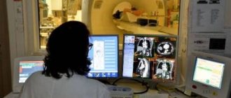
- The doctor monitors the scan result on a computer monitor and records all important data in the study protocol.
- After the examination is completed, the patient wipes the hydrogel from the skin and gets dressed.
The whole procedure takes 15 minutes, and the result is given to the patient.
How to sign up for an ultrasound of lymph nodes?
To make an appointment for an ultrasound examination of the lymph nodes, simply call the single referral network of PreAmbula clinics. Our staff will schedule a study at a time convenient for you and, if necessary, explain the information you are interested in.
Ultrasound diagnostic specialists at Preambula are highly qualified and have extensive practical experience in this area of medical research. In combination with high-quality equipment, this guarantees accurate diagnosis.
Ultrasound of lymph nodes of the 1st-2nd regions - 1100 rubles.
How to reduce the cost? MAKE AN APPOINTMENT
Examination of lymph nodes in cancer diagnosis
Ultrasound of the subclavian lymph nodes is used in the diagnosis of cancer. According to statistics, they account for about 5-10% of the reasons that cause changes in the lymphatic system. At the initial stages of the development of a malignant tumor, the supraclavicular lymph node increases in size, but it does not hurt when palpating or moving the neck.
Important! If the supraclavicular lymph node is enlarged, palpable, but there are no symptoms of inflammation, you should immediately undergo an examination, as this may be associated with the development of oncological pathologies.
The node can be affected by melanoma, neuroblastoma, seminoma, Kaposi's sarcoma. With these oncopathologies, it is painless and elastic to the touch. To make an accurate diagnosis, a comprehensive examination is required, in which ultrasound is only one of the methods.
Prices for ultrasound and alternative diagnostics
In different clinics, prices for ultrasound may differ slightly, but remain within the range of affordability for most patients. For special indications, screenings are carried out in regular clinics by appointment and free of charge. But if you don’t have time to wait in line, or you want to undergo diagnostics in comfort, you can choose a center with the necessary services using our free service. The table contains the minimum prices for different types of scanning; more complete data is posted in the price lists via links to each medical institution.
| Services | Prices for procedures, rub. |
| Ultrasound of the pelvic organs | 1300 |
| Ultrasound of the pelvic organs in women transvaginally | 1000 |
| Ultrasound of the pelvic organs in women (transabdominal) | 900 |
| Ultrasound of the pelvic organs in men | 1300 |
| Ultrasound of the pelvic organs and kidneys | 2000 |
| CT scan of the pelvis | 3150 |
| MRI of the pelvis | 3600 |
Ultrasound of the abdominal cavity at the MedicCity clinic
Did your attending physician prescribe an ultrasound scan of your liver, gallbladder, or pancreas, or did you decide to undergo this study yourself? We need a clinic with reliable equipment and qualified specialists!
At the MedikCity clinic, ultrasound diagnostics are carried out using expert-class equipment; our ultrasound diagnostic doctors know all the professional secrets and have extensive practical experience. Within half an hour after the examination, you will receive a clear image and its high-quality description. Ultrasound at MedicCity is carried out by appointment, just like other studies. Take care of your health right now!
Consequences and harm from the procedure
Ultrasound diagnostics is one of the few technologies that are recognized as absolutely safe for humans. Doctors prescribe ultrasound for both relatively healthy young people and frail elderly patients, pregnant women and newborn children (or those still in the womb). The frequency of application may be as necessary to fully assess the clinical situation. However, doctors do not advise doing ultrasounds too often just for fun.
The fact is that intense ultrasonic waves slightly increase the temperature of the tissues being examined. This causes microscopic gas bubbles in body fluids to expand. This process is scientifically called cavitation. When expanding, the bubbles merge, grow and can damage adjacent cells (theoretically). To prevent this from happening, during each ultrasound session the specialist monitors its time and the intensity of the ultrasound field.
If an ultrasound is prescribed by an expert, there is no need to fear adverse consequences. According to the World Health Organization, routine screenings of the mother during pregnancy do not pose a risk to the health of the unborn baby.
Who is indicated for ultrasound of the abdominal organs?
The study can be prescribed to patients with complaints of symptoms and conditions such as:
- abdominal trauma;
- stomach ache;
- abdominal and back pain;
- pain in the hypochondrium;
- pain in the side;
- frequent attacks of nausea, heartburn, vomiting;
- coating on the tongue, bad breath;
- flatulence, constipation/diarrhea;
- blood in stool;
- change in the color of urine, feces (darkening, discoloration);
- signs of fluid accumulation in the abdomen; presence of sand and/or stones;
- suspicion of the presence of a foreign object;
- suspicion of appendicitis; preparation for surgery; suspicion of a tumor process, etc.
Routinely, ultrasound of the abdominal cavity is performed to monitor the results of treatment, including for oncological pathologies.
There are practically no contraindications to abdominal ultrasound. Diagnosis is not carried out if the patient has a skin disease or skin damage that makes it difficult for the sensor to slide. The examination can also be complicated by failure to follow the recommended diet on the eve of the examination, or the patient’s excess fat deposits in the abdominal area (gases in the gastrointestinal tract and dense fatty tissue can impede the propagation of ultrasonic waves and affect the accuracy of the result). A relative contraindication to the procedure may be a gastroscopy performed on the same day - due to the fact that during this examination a certain amount of air enters the body.
Types of ultrasound
Most people have encountered this type of scanning at least once, but not everyone is familiar with its varieties. Depending on the gender of the patient, as well as the disease being sought, the following types of examination are distinguished:
- Transabdominal. The most common type of screening involves the external use of a hand-held probe. The patient needs to free his stomach from clothes and lie down on the couch. If necessary, the doctor will ask you not to breathe for a few seconds, turn on your side, turn your back to him or stand up.
- Transvaginal. This ultrasound requires a special sensor, reduced in diameter and elongated, which is painlessly inserted into the vagina. In this case, the ultrasonic field generation sector propagates from the inside. The intestines and bladder are not obstacles to a targeted study of the female reproductive system.
- Transrectal. Used primarily for male ultrasound. You will need to remove all the things from below, lie on your side and relax. An even narrower sensor is inserted into the rectum. The doctor insists on this method when checking the prostate gland. It can hardly be called comfortable, but there will be no pain during the ultrasound, and the session itself lasts several minutes.
As soon as the pathology or its absence is confirmed, a picture is taken and a description is prepared. The received documents must be provided to the doctor.
Features of the procedure
This type of patient examination is carried out using special equipment, which is available in almost every medical institution. Ultrasound is one of the primary procedures for suspected pathologies affecting internal organs. After examining the patient and collecting the necessary medical history, the attending physician must send for an ultrasound to obtain the first data on the clinical situation. In most cases, this is enough to establish the disease and begin restorative measures.
In addition to its high efficiency, ultrasound has a number of other advantages, due to which the examination is used more often than other types of scanning:
- Low cost per session, affordable for most patients.
- Visualizes all organs in the studied area.
- Screening is painless, there is no noticeable discomfort from touching the sensor.
- The specialist performing the procedure sees the results immediately on the device screen, records deviations with the ability to print images. Decoding also occurs at an accelerated pace; it is enough to wait 10-20 minutes to prepare a conclusion.
- The examination takes a little time, no more than half an hour.
Preparing for diagnosis
There are no too strict rules for preparing for an ultrasound, but in order to obtain the most informative data, you will have to devote the necessary time to some manipulations. So, for the examination to be complete, you need to follow dietary recommendations. To prevent the intestine and its loops from becoming a dynamic obstacle to the study of pelvic structures, it must first be “calmed” and cleaned. A couple of days before the session, it is better to exclude heavy foods, which take a long time to digest and cause increased gas formation. You need to give preference to boiled products with a low-fat composition. Sweet, carbonated and alcoholic drinks should be completely removed from the diet. You can replace them with weak tea or clean water.
If a doctor has prescribed a transvaginal ultrasound for a woman, the rule of a full bladder is excluded. In this case, you need to come for a scan immediately after visiting the toilet so that the bladder is completely emptied. For men, when using the transrectal method of inserting an ultrasound sensor, it is necessary to first cleanse the intestines. This can be done with regular enemas or laxatives on the eve of the procedure. The doctor independently chooses which technology to prefer, so he will warn you in advance how to prepare for the study.
Operating principle of ultrasonic equipment
As is clear from the name of the technology, the operation of ultrasound is based on the use of sound waves in an ultra-high range. They pass safely through biological tissue at varying intensities and speeds. The denser the fibers of the organs, the faster the ultrasonic field penetrates through them. This occurs most slowly in cavities, so the doctor always asks you to come for a scan with a full bladder in order to better examine its internal relief and the neighboring structures covered by it.
During operation, a handheld ultrasound sensor simultaneously acts as an ultrasound generator and a receiver of its reflected waves. Depending on the density of obstacles in the path of the wave, the speed of its movement through the body changes. This is recorded by an ultrasound machine and converted into images on a monitor, which are immediately examined and assessed by the doctor. Not only the general outlines of the objects under study are visible on the screen. The program is capable of assessing the spatial location of structures relative to each other, as well as accurately measuring the parameters of the desired object.
Which specialist should you trust to perform an ultrasound?
From the patient's point of view, it is very convenient when the diagnosis is under the jurisdiction of one selected doctor. They collect anamnesis, take into account existing chronic diseases, and perform an ultrasound. If the doctor is well versed in this and has a second degree in ultrasound research, he knows exactly what to look for when scanning. Experienced gynecologists with extensive experience using ultrasound can be trusted. However, in case of complex diseases, in controversial and life-threatening situations, it is better to trust specialized uzologists.