X-ray of the sinuses at the German Family Clinic
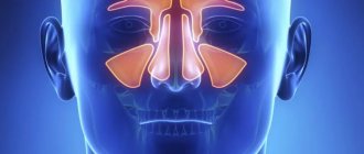
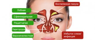
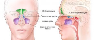
: conducting research and decoding images. Make an appointment for an x-ray by phone, (812) 337-12-12. The nose is a complex organ that is part of the upper respiratory tract. Performs respiratory, olfactory, protective and resonator functions. Each half includes four walls. The turbinates, which are cartilaginous projections of the nasal septum, divide both nasal cavities into the upper, lower and middle passages. The nasal cavity communicates with the paranasal sinuses (cavities in the bones of the skull filled with air) through the openings of the upper and middle nasal passages. According to localization, the maxillary, frontal, sphenoid sinuses and ethmoidal labyrinth are distinguished (the names correspond to the names of the bones). At birth, only the maxillary (maxillary) sinus and the ethmoid labyrinth are formed in the child. But the rest begin to form at the age of 3-4, finishing at about 25 years.
The complexity of the structure and functions performed, the location of the nose makes it susceptible to various injuries, as well as the development of various diseases. An effective method for diagnosing the paranasal sinuses is radiography.
Interpretation of results
The interpretation of the results of an x-ray examination may be as follows:
- normal (taking into account the patient’s age);
- the presence of dark spots in the paranasal sinuses;
- thickening;
- visible trauma or the presence of a foreign body.
Norm
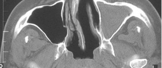
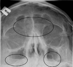
An X-ray of the sinuses of a healthy person looks like this:
- The nasal septum divides the nasal cavity into symmetrical sides of a triangle.
- The white stripes running to the right and left of the divided area are the nasal passages.
- The triangular cavities on the sides of the nose are the maxillary sinuses.
- Between the eye sockets there is an ethmoid sinus with thin walls, the cells of which should be clearly visible.
- Above the orbits, the frontal sinuses are defined, which can have different shapes. Their separation by bone partitions is allowed.
- There must be air in the sinuses. Their edges, like the contours of the bones, should be clear and even.
Photo of a healthy person
Darkening, cavities and thickening in the image
We can talk about diagnosing a pathology if the images show the following:
- darkening the area;
- thickening;
- the presence of a cavity of different shapes;
- deformation of bone tissue.
Acute inflammation is characterized by thickening of the mucosa and deformation of its borders (with purulent contents). The presence and level of fluid are determined by horizontal demarcation in the sinuses. An x-ray will not tell you about the composition of the contents; for this you will need to make a puncture (puncture). If the inflammation is chronic, the mucous membrane thickens and the lumen in the sinus becomes smaller.
Sinusitis
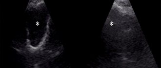
Sinusitis on an x-ray looks like a darkening, which has characteristic differences in various pathologies.
The pictures should be decrypted like this:
- With hyperplastic sinusitis, there is thickening of the mucosa directly next to the bone. In this case, the internal contour becomes wavy and blurry.
- With catarrhal sinusitis, thickening of the mucosal walls with complete or partial darkening is noted. The presence of a light cavity in the center of the sinus indicates a chronic process.
- With exudative sinusitis, darkening of the paranasal sinuses occurs with a horizontal delimitation, reflecting the level of fluid filling.
- With vasomotor and allergic sinusitis, pronounced swelling of the mucous membrane is noted.
Sinusitis
Sinusitis is an inflammation of the lining of the maxillary sinuses.
The following types of sinusitis are diagnosed:
- Exudative. The presence of fluid in the upper sinuses on one or both sides.
- Parietal. Localization of inflammation near the bone walls. The edges of the mucous membrane are deformed and directed inside the sinus.
- Polypous. There is bulging of areas of the mucous membrane, which can be either single or multiple.
Neoplasms and cysts in the sinuses
If photographs of the paranasal sinuses reveal a cavity with dense contents, this indicates the presence of a benign or malignant formation. In most cases, neoplasms are found by chance during the diagnosis of other pathologies.
- Sinusitis - what is it and how to treat it at home Symptoms of sinusitis and medications for children and adults
A sinus cyst is defined as a light, rounded area located outside the sinus mucosa. Its edges are clear and even.
Bone injuries
If a bone is broken or displaced, it will be visible on an x-ray. Bone injuries may appear on x-rays as dense fragments in the sinuses. The image helps to establish the location of the fracture and the displacement of bone fragments, if present. Severe fractures are accompanied by bleeding, which will appear as fluid in the sinuses. The doctor may also detect an old injury that appears as a callus on the image.
Foreign bodies
The foreign body in the image has contours in accordance with what exactly got into the nasal cavity. Typically, these are beads or other round objects that children place in their noses. On x-rays they appear as areas of varying darkness.
Photo gallery
Right-sided sinusitis Cyst in the left maxillary sinus Sinusitis. Frontal and maxillary sinuses Trauma to the nasal cavity
Advantages of performing a nasal x-ray in our centers
Experienced staff
X-rays of the sinuses in Moscow are performed by experienced radiologists of the highest category with extensive experience in radiology diagnostics.
Radiologists have an excellent knowledge of topographic anatomy, competently interpret images, and work closely with therapists, otolaryngologists, surgeons and other specialized specialists. The latest safe digital X-ray equipment
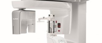
The equipment of our centers is regularly updated with more advanced models of the latest generation. Low-dose X-ray machines allow you to obtain highly informative, accurate results of diagnostic procedures and ensure high safety of the studies performed.
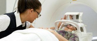
How does the procedure work and how long does it last?
The duration of the study is several minutes.
X-rays are taken depending on the indications as follows:
- The patient enters a special room. Children are accompanied by adults.
- In a standing or sitting position, the radiologist fixes the head in the position required to take the image. This can be a straight or lateral plane, a projection of the chin, the occipito-frontal part or the occipito-mental part. One of the parents holds the child's head.
- You need to take a deep breath and hold your breath for a few seconds.
- The doctor will inform you that the procedure is complete.
- The picture develops within 15-20 minutes.
Is X-ray dangerous? This is explained in the video from the Live Healthy channel.
X-ray of the sinuses: preparation and procedure
No special preparation is required for a nasal x-ray. The X-ray room laboratory technician will check to see if there are any metal objects on the body, and then help the patient to take the correct position for the procedure. It will be necessary to approach the vertigrapher (digital radiographic mobile vertical stand), take a position in which the nose and chin are in the same plane (nose-chin position), or the nose and chin are at an angle (chin position). The procedure is often performed sitting or standing, but a horizontal position of the subject is also possible (for example, nasofrontal projection). The subject will be asked to hold their breath and remain motionless.
It should be noted that there are many nuances when installing. Therefore, it happens that a radiologist, having analyzed the appointment of the attending physician, can cancel his appointment and perform x-ray diagnostics in a slightly different way.
In addition to the main two projections above (nasomental and mental), there is a frontal option, which will show only the ethmoid labyrinth, the right and left halves of the sphenoid sinuses. Therefore, this method of obtaining radiographs is rarely used to determine pneumatization of the paranasal sinuses. A targeted photograph of one of the sinuses may be taken.
For a detailed study of the structure of the paranasal sinuses, it is practiced to obtain an X-ray image using an X-ray contrast agent (usually an iodolipol solution). This method is used in particular to identify various types of intra-axillary formations, polyps, and cysts. The procedure involves puncturing the patient's sinus wall under anesthesia; rinsing the sinuses with furatsilin and administering iodlipol heated to body temperature through a needle. Nasofrontal, lateral and nasomental projections are used. However, due to the high probability of deterioration in the clarity of the images due to the mutual overlap of exposure between the nasal sinuses, images of both maxillary sinuses are not taken simultaneously.
Contraindications to X-ray examination
X-ray of the paranasal sinuses has several contraindications. This method is prohibited from doing:
- Pregnant women. Despite the fact that the study has low radiation exposure, it is contraindicated for expectant mothers due to the strong sensitivity of the fetus to radiation exposure. The use of X-ray machines during pregnancy can cause birth defects in the child.
- Children under 15 years old. For patients younger than this age, sinus x-rays may be rarely ordered because gamma rays can negatively affect the child's bone growth.
- People suffering from mental illness. In such cases, patients are unable to fix their head and not move for several minutes.
- A month before IVF. The cells of the reproductive system are considered the most vulnerable to radiation. Irradiated sperm in men and damaged eggs in women are unable to conceive. Therefore, gynecologists recommend undergoing an X-ray examination at least a month before in vitro fertilization.
In very rare cases, a doctor may issue a referral for x-rays for children under 15 years of age and pregnant women. But this happens if the research justifies the risk of probable harm that the diagnosed disease can lead to.
Preparing for an X-ray
Preparation for the procedure comes down to two rules:
- What does pneumatization of the sinuses indicate?
- remove jewelry and other metal objects located in the head area (chain, earrings, hairpins, hoop);
- wear a special apron (lead), which is issued immediately before the x-ray.
There are no restrictions related to food or liquid intake.
It is necessary to clarify when and how to take an x-ray when observing the disease over time. For example, if a patient is having a cuckoo sinus irrigate, x-rays are taken before the sinuses are cleared of fluid.
Description / what the TRG shows
The television film depicts a human skull in X-rays. Hard and soft tissues, their relationship and position are visible. TRG in dentistry is considered an important stage in preparation for orthodontic treatment, interventions in maxillofacial surgery and traumatology.
What the TRG image shows:
- Position, inclination, displacement of teeth.
- The ratio of the sizes of bones and soft tissues.
- Inflammation of the periodontium, sinuses, jaw joints.
- Anomalies in the development of the dentofacial apparatus.
- Asymmetry of the facial part of the skull.
- The state of the bite and the causes of its violation.
- Bone defects, their structure and location.
The image obtained during teleradiography provides the dentist with information about the structural features of the facial and brain parts of the skull, the condition of the bones, and the ability to build a treatment plan and prognosis. In the process of correcting the bite with orthodontic structures, the procedure is repeated. This makes it possible to track the progress of changes and check the correct progression of teeth.
Answering the question about what a TRG image is, we can say that this is the second most popular method of dental diagnostics after an orthopantomogram.
What can a doctor detect on x-rays?
This procedure provides ample opportunities for diagnosing and confirming various diseases of the paranasal sinuses. The resulting images (x-rays) help doctors visualize and examine the following anatomical parts: frontal, maxillary (maxillary) sinuses, ethmoidal labyrinth, orbits and facial bones.
Inflammation of the maxillary and frontal sinuses
An x-ray of the paranasal sinuses can reveal:
- defects in the structure of the organ and adjacent soft tissues;
- the presence of certain inflammatory diseases: acute or chronic sinusitis, sinusitis, sinusitis, pansinusitis;
- traumatic injury to the facial skeleton,
- foreign objects;
- tumor formations (benign and malignant), cysts of the nasal cavity;
- deviated nasal septum;
- developmental defects, congenital and acquired anomalies of PPN.
The method is used everywhere in the practical work of otolaryngologists and is the basis for making a diagnosis.
With the help of radiography, diseases of the ENT organs, injuries, and the presence of foreign objects are well diagnosed. This technique allows you to visualize pathological contents in the paranasal sinuses, changes in the mucous membrane lining the sinuses (swelling, polyps, etc.). Normally, the paranasal sinuses are filled with air and do not differ from the eye sockets. If they contain liquid (pus, blood, etc.), darkenings are formed (reduced pneumatization) with a horizontal upper border.
By taking a timely picture of the paranasal sinuses, it is possible to avoid complications fraught with irreversible consequences, for example, osteomyelitis, meningitis.
- Consultation with a specialist: how to remove pus from the maxillary sinuses?
Types and signs of sinusitis.
Based on the localization of inflammation, the following types of sinusitis are distinguished: sinusitis (inflammation of the maxillary sinuses), frontal sinusitis (frontal sinusitis), ethmoiditis (ethmoid labyrinth cells), sphenoiditis (sphenoid). The disease develops against the background of other infectious diseases: rhinitis, scarlet fever, measles, etc.
The main symptoms of the disease include:
- feeling of nasal congestion;
- thick discharge from the nasal passages, usually green or yellow in color;
- pain in the cheeks and eyes, headaches, pain between the eyes;
- puffiness under the eyes;
- swelling of the nose;
- cough due to mucus constantly flowing down the nasopharynx;
- impaired sense of smell;
- increased body temperature;
- weakness, decreased performance, high fatigue.
All these signs are a good reason to contact an otolaryngologist, conduct a diagnosis and prescribe effective treatment for sinusitis, sinusitis or any other form of sinusitis.