When is an X-ray of the nasal bones necessary?
Among the main indications for radiography of the nasal bones
highlight:
- suspicion of the presence of foreign bodies in the nasal passages;
- fracture of the nasal bones (an x-ray is required, as it can provide prompt and accurate information about the complexity of the fracture and its location);
- suspicion of neoplasms in the nasal paranasal sinuses;
- curvature, displacement of the nasal septum;
- suspicion of osteomyelitis (a purulent disease affecting bone tissue);
- suspicion of the presence of cysts and polyps.
X-rays can also be prescribed as a routine diagnostic procedure when it is necessary to draw up a strategy for an upcoming operation (for example, to correct a septum, remove polyps, and so on).
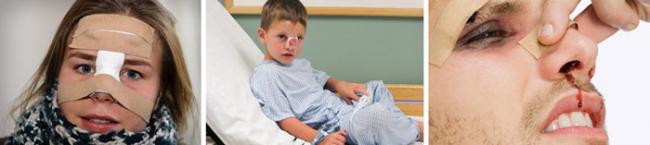
X-ray of nasal bones for injuries
Injuries to the nose, such as a fractured septum, are quite common. These could be injuries of a sports or household nature - in any case, an x-ray will allow you to see exactly where the fracture is located and how damaged the structure of the nasal bone is. Also, the fragments will be visible on the x-ray, it will be possible to predict complications and plan an operation to restore the bone. Using an x-ray, you can determine the type of nasal fracture:
- transverse;
- oblique;
- straight;
- no offset;
- in the form of a bird's beak;
- multi-fragmented, without noticeable displacement of fragments;
- destruction of the nasal septum.
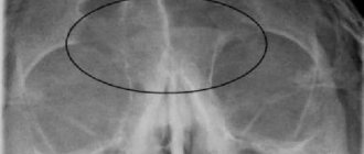
Severe types of destruction of the bone tissue of the nose are accompanied by hemorrhage into the paranasal sinuses, as can be judged by a nasochin photograph.
The formation of ossified callus in this area is slow due to the osteochondral structure. Some time after all the reconstructive operations have been carried out, x-rays may be ordered again, for example after 2-4 weeks. This is necessary in order to assess how quickly and correctly the fusion of bone tissue occurs. It is important to take X-rays of the facial bones of the nose in case of injuries for the timely diagnosis of periostitis. The main symptoms of this pathology: pain when making even small movements, discomfort and slight swelling in areas of inflammation. The color of the skin at the affected sites most often does not change. If periostitis of the nasal bone is the result of a blow, fracture, or bruise, then after 2-3 weeks all its symptoms, as well as the inflammation itself, disappear. If fibrous growth of bone tissue worsens, the disease becomes chronic. At this stage, redness of the skin in the nose area may already be noted. In this case, it is worth starting treatment immediately, since delay will lead to such a serious complication as purulent periostitis.
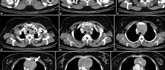
What does x-ray of the nasal bones show?
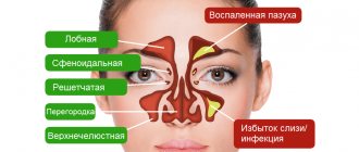
On x-rays, the doctor can see:
- Nasal bones in various projections. The images are used to evaluate their structure, location, and integrity.
- Frontal and maxillary sinuses. It is important to assess the size of the sinuses, detect foreign bodies (if any) and neoplasms in them.
- The main paranasal sinus and cells in the temporal bones.
By choosing a special head position, the ethmoid sinuses can also be captured in the photographs.
What will an x-ray show?
Radiography is the main method for diagnosing the nasal and other facial bones of the skull, and the paranasal sinuses. The images will show the nasal bones, paranasal sinuses, upper jaw, frontal and ethmoid bones.
The survey is informative for:
- Definitions of bone damage and trauma.
- Diagnosis of paranasal and maxillary sinuses.
- Detection of cysts, tumors, inflammation of bone tissue or cartilage.
- Searching for foreign objects in the nose.
- Assessment of deviated septum.
- Diagnosis of sinusitis, sinusitis and sinusitis.
- Definitions of bone loss – osteoporosis.
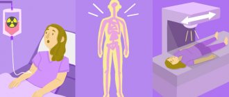
How is the procedure done?
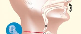
X-rays can be taken in different positions of the patient. Most often, he is seated in front of an X-ray machine or placed near an X-ray stand. Such positions allow you to obtain the most accurate images. X-ray is carried out as follows:
- the patient takes a comfortable position in the desired position;
- the chin is placed on a special stand;
- the midline of the head is arrow-shaped;
- a photo is taken.
How is radiography done in 2 projections (frontal and lateral)?
To obtain the most detailed and complete information about the condition of the nasal bones, photographs can be taken in two projections - frontal and lateral. In the first case, the patient faces the X-ray machine, in the second - sideways (left or right). Direct radiography shows only displaced destruction. To determine the side of the injury, photographs of the nose are taken in the left and right lateral projections. Another type of projection is also used in practice - nasomental. This image clearly visualizes the nasal bones, as well as the processes of the upper jaw. Due to the axial path of X-ray radiation, it is in this projection that it is easy to detect the displacement of fragments when taking pictures of a broken nose.
Advantages
Compared to other examination methods, digital Rg-graphy of the nasal bones has its advantages:
- Images are shown on the device screen immediately.
- The results are stored on disks in digital form - they are convenient to analyze, store, forward, and compare with previous and subsequent images.
- Fast – an image is taken in 2 seconds, while an MRI takes longer.
- Minimum level of radiation exposure, in contrast to a study such as CT.
- X-rays of the nose can be taken in a sitting position, which may be important for patients with severe bleeding.
- Everything goes without pain and discomfort.
Decoding the results obtained
When the X-ray images obtained are deciphered, the doctor analyzes the following indicators:
- Walls and edges of the nasal bones. It is necessary to evaluate how clear their boundaries are and whether their thickness is normal.
- Air space in the sinuses. It is important to evaluate the size of this space and its symmetry.
- Cells on the ethmoid bone. It is necessary to evaluate how well they are visualized, what their structure and size are.
A written medical report is issued to the patient, who passes it on to the attending physician.
Preparation
Depends on the situation and the reason why an x-ray of the nose is taken. If this is a study for diagnosing diseases (acute or chronic), then no preparation is required. If we are talking about injuries, fractures, severe bleeding, then, first of all, it is necessary to stabilize the general condition of the subject, stop the bleeding, and use painkillers. Before the x-ray, all jewelry and other metal objects that can reduce the information content of the images are removed from the head. To reduce potential harm from X-rays, the thyroid, chest, and abdomen areas are covered with a protective blanket.
Symptoms
Clinical signs of a nasal fracture:
- A nasal fracture causes severe pain. The patient is in shock or even fainting.
- Slight bleeding from the nasal slits occurs, which is blocked quickly and independently. In quite rare cases, blood flows in a stream and requires nasal packing. Such bleeding can lead to significant blood loss.
- The skin and mucous membranes are damaged, which can be manifested by the accumulation of air in the superficial fat layer. During the examination, the doctor determines whether the wound communicates with the nasal cavity.
- Then swelling of the soft tissues of the nose, eyes and cheekbones develops, bruising appears in the area of the cheekbones and eyelids. Hemorrhage under the skin occurs as a result of damage to blood vessels. After a day, swelling of the nose and surrounding tissues becomes extensive. Bruises form around the nose and under the eyes. Noticeable swelling in the impact area allows you to know whether the nose is broken. This symptom, combined with pain, deformity and bleeding, gives the characteristic clinical picture of a nasal fracture.
- Difficulty in nasal breathing is caused by displacement of bone fragments, deformation of the nasal septum, and increasing swelling.
- In case of a fracture of the bone and cartilaginous structures of the nose, the crunching of the fragments is determined by palpation - a clear sign that allows the traumatologist to make the correct diagnosis and prescribe adequate treatment.
- If the nose is broken, mucus continuously leaks from it.
- In the future, infection may occur and fever, pain and redness may appear at the site of the lesion. Softening of the tissue indicates the beginning of the process of abscess formation.
Where to get an x-ray of the nose in Krasnoyarsk?
The Medunion private medical clinic has installed the latest digital diagnostic apparatus, which is equipped with a device for digital X-ray image processing. It is displayed on the monitor and provides the doctor with the opportunity to assess the average level of shadow density and the total range between the light and dark parts of the image.
After the procedure, the printed image is given to the patient, and the digitized image is stored in the clinic’s archive. If necessary, it can be raised to monitor the patient’s condition over time. But the most important thing is that when using a digital X-ray machine, the patient is exposed to much less radiation exposure than when using an analog one.
You can make an appointment for diagnostics at the Medunion clinic by calling or filling out an online form on the website.
Features of x-rays of the nasal bones
The procedure does not require any special preparation from the patient. If it is performed after an injury, it is important to stop the bleeding and administer local anesthesia. The patient is asked to remove all metal objects, as well as removable dentures if they are made using metal. The radiographic anatomy of the nasal bones is such that it requires taking pictures in different projections:
- frontal;
- nasomental;
- lateral left;
- lateral right.
During these procedures, the patient can sit or lie down, depending on his condition. The procedure lasts a few seconds.
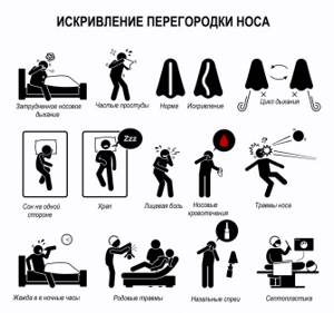
Deviated nasal septum
7881 May 28
IMPORTANT!
The information in this section cannot be used for self-diagnosis and self-treatment.
In case of pain or other exacerbation of the disease, diagnostic tests should be prescribed only by the attending physician. To make a diagnosis and properly prescribe treatment, you should contact your doctor. Deviated nasal septum: causes of occurrence, what diseases it occurs with, diagnosis and treatment methods.
Definition
The nasal septum is a plate that divides the nasal cavity in half; its main function is to adequately distribute the air flow between the right and left halves of the nasal cavity. A deviated septum can be either congenital or acquired. The consequence of this curvature is a narrowing of one of the nasal cavities. People with such a defect may experience impaired nasal breathing and sense of smell, mucous discharge, decreased hearing, nosebleeds, frequent respiratory infections and snoring.
Types of deviated nasal septum
A deviated nasal septum is determined by its severity:
I degree. There is a slight deviation of the septum from the midline, which usually does not lead to impairment of respiratory function.
II degree. The most prominent part of the septum is equidistant from the midline and from the wall of the nasal cavity, as a result of which the air flow is distributed unevenly.
III degree. In this case, the septum is in contact with the wall of one side of the nasal cavity, so breathing is carried out primarily through one nostril.
According to clinical manifestations:
- A small one-sided vertical ridge in the anterior sections of the nasal septum.
- A pronounced vertical ridge in the anterior part of the nasal septum with displacement of the cartilage in the opposite direction.
- One-sided vertical ridge in the deep parts of the nasal cavity.
- Two vertical ridges located one behind the other on opposite surfaces of the nasal septum.
- A one-sided horizontal ascending ridge in the posterior parts of the septum, resembling a Turkish saber in shape.
- Two horizontal ridges in the anterior and middle sections of the nasal septum on opposite surfaces.
- “Crumpled” partition with multiple fracture lines.
Possible causes of a deviated nasal septum
Among the causes of changes in the position and structure of the septum are developmental disorders in the embryonic period, birth injuries, and nasal fractures, polyps or tumors.
- Physiological curvatures are caused by defects during the growth and ossification of the child’s nasal septum, starting from the age of three months. The curvature is formed due to a discrepancy between the child’s growth rate and the development of bone structures.
- Traumatic curvatures are caused by any mechanical impact.
- Compensatory curvatures occur due to the formation of tumors or polyps in the nose.
Which doctors should you contact if you have a deviated nasal septum
? The choice of the doctor you should consult with depends on the cause of the deformity. If this is a recent injury, you should immediately contact a traumatologist. If a patient notices periodic nasal congestion, is bothered by snoring, hearing loss, or frequent infectious diseases, then he can routinely consult an otolaryngologist.
If you have breathing problems that lead to panic attacks or severe headaches, you may need the help of a neuropsychiatrist.
Diagnosis and examinations for deviated nasal septum
- If a bacterial or viral infection is suspected, a clinical blood test is required.
How to distinguish a fracture from a bruise?

What do you need to know about this? Many people are interested in how to identify a broken nose or bruise. In this case, the signs are quite similar to each other. However, there are some distinctive features. For example, if the nose is only injured and there is no fracture, then swelling will appear only along the edge of the damaged organ. Painful sensations from bruises are quite tolerable. If the bone is damaged, the pain becomes unbearable, and the patient may even go into shock. Bleeding from a bruise is not so severe and can be easily stopped using special compresses and vasoconstrictor drops. The symptoms of a broken nose and a bruised nose are similar. However, in case of bone damage, the clinical picture will be more pronounced.
- Nose fracture: symptoms, how to find out if the nose is broken, treatment, consequences