Computed tomography of the maxillary sinuses is a detailed examination of this area using x-rays.
Author:
- Sadykhov Rahim Agalarovich
ENT pathology expert
5.00 (Votes: 2)
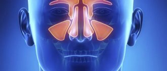
Computed tomography of the maxillary sinuses is a detailed examination of this area using x-rays. The maxillary sinuses are paired air cavities on either side of the nose, adjacent to the teeth of the upper jaw.
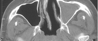
The main purpose of CT is to study the maxillary sinuses and their anastomoses (communications with the nasal cavity) in chronic, recurrent and advanced sinusitis. Computed tomography provides greater accuracy when examining hollow and bone structures, which is why it is prescribed for studying sinuses. In comparison, x-rays show only general signs of sinusitis (darkening in the area of inflammation). Computed tomography, on the other hand, provides a more thorough examination, which shows not only the pathological process in the sinus itself, but also allows one to determine its structure by X-ray density (cystic fluid, pus, blood, polypous tissue, mycetoma). Also, using a series of CT images, you can assess the condition of the anastomosis - important anatomical structures that ensure the normal functioning of the maxillary sinuses. Thanks to the high resolution of the tomograph, even minor abnormal formations are detected. If we compare computed tomography with MRI, then computed tomography is more effective for examining bone structures, and MRI is more effective for examining soft tissues.
The high degree of information content of the computed tomography method makes it possible to make a diagnosis and prescribe the most effective therapy for the treatment of pathologies of the maxillary sinuses. Based on the results of the examination, the doctor has in his hands photographs that allow him to study the scanned area, in the form of a single volumetric photograph or thin sections.
Indications for CT scanning
Computed tomography of the maxillary sinuses is prescribed for:
- sinusitis;
- problems breathing through the nose;
- pain in the nose;
- headaches of unknown etiology;
- toothache of unknown etiology;
- serious dental problems;
- complications of infectious diseases of the ENT organs;
- cysts and granulomas at the tips of the roots of the teeth of the upper jaw;
- chronic rhinosinusitis;
- recurrent otitis;
- suspected neoplasms of the sinuses and upper jaw;
- suspected injuries to bones or other tissue near the maxillary sinuses;
- suspicion of the presence of a foreign body in the maxillary sinus.
Preparatory stage for CT scanning of the paranasal sinuses
If a CT scan of the paranasal sinuses is performed without the use of a final drug, the patient does not need any preparation. All you have to do is arrive at the appointed time, change clothes and lie down on the machine’s table.
The use of contrast complicates the procedure. In this case, you need to prepare in advance for the examination. A few days before the test, the patient undergoes blood tests. This is necessary in order to identify the presence of allergies to the components of the drug and determine the condition of the kidneys. In addition, the patient must refrain from eating five hours before the examination.
What will a CT scan show?
The examination will display the following information on the images:
- inflammatory processes;
- foreign bodies in the nasal sinuses;
- bruises, violations of the integrity of the nasal bones;
- the location of the nasal septum and bones forming the maxillary cavities;
- oroantral fistulas (pathological communication between the oral cavity and the maxillary sinus);
- polyps, granulomas, cysts, pyoceles, exudate;
- symmetry of the right and left sides of the nose and sinuses;
- additional voids (ostia);
- degree of pneumatization of the sinuses;
- sinus drainage pathways;
- tumors of the mucous membranes;
- condition of the tear ducts.
Modern methods of x-ray examinations
Radiovisiography (intraoral x-ray of the tooth)
This is obtaining an X-ray image of one or more teeth and adjacent tissues on a digital matrix. A visiograph is a small X-ray sensitive sensor, an X-ray generator, and a computer program for recording and visualizing the resulting image.
The sensor is placed in the oral cavity behind the subject of photography. An X-ray tube is directed from the outside of the tooth towards the sensor. In a fraction of seconds, the photo is already displayed on the computer screen. Speed is not the only advantage of radiovisiography. The miniature dimensions of the sensor, high sensitivity and the minimum distance that X-rays must travel make it possible to reduce the radiation dose by hundreds of times compared to film devices. Modern sensors have high resolution, which gives the doctor the opportunity to see the smallest details of the tooth and bones.
X-ray of the temporomandibular joint (TMJ) allows us to study the relationship between the head of the articular process and the glenoid cavity, as well as the structure of the bone tissue of the bones that form the joint. As a rule, the doctor takes two pictures - in the usual occlusion (teeth closing) and with a functional load, that is, with the mouth wide open.
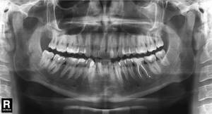
Panoramic tomography (orthopantomogram, OPTG)
This is an X-ray method that allows you to obtain a panoramic image of all teeth, jaws and adjacent parts of the facial skeleton, unfolded on a plane. Using it, the doctor can assess with a sufficient degree of accuracy the condition of each tooth, jaw bone tissue, temporomandibular joints, and maxillary sinuses.
Preparation and performance of computed tomography of the maxillary sinuses
A CT scan without contrast does not require any preparation. If a contrast agent is used, it is necessary to take a creatinine test and receive a referral from an otolaryngologist stating that the patient has no contraindications.
Before starting a CT scan, you need to remove all metal objects that could spoil the image. Tomographs have different designs, and depending on it, the patient can either lie on a couch that is immersed in the device, or stand, or sit. Regardless of your body position, you must remain motionless throughout the entire session.
Key contraindications
CT scanning of the maxillary sinuses also has its contraindications. The procedure is strictly prohibited for pregnant women. Because even minor radiation exposure can harm the fetus. The use of contrast agents has additional contraindications. In this case, the procedure is not recommended for people suffering from:
- kidney failure;
- allergic reactions to the components of the contrast agent;
- dysfunctions of the endocrine system.
Nursing mothers are advised to refrain from lactation for two days after the procedure. It is very important to express the existing milk. Note that the study is difficult in patients suffering from hyperkinesis.
Indications and contraindications for MRI of the sinuses
Magnetic resonance imaging is necessary in the following cases:
- prolonged headaches, especially pain in the face;
- repeated nosebleeds;
- treatment-resistant nasal congestion;
- impaired sense of smell;
- suspicion of a neoplasm;
- injuries to the facial area;
- congenital anomalies of the nasal cavity and sinuses;
- pathologies identified by X-ray or ultrasound results;
- postoperative observation.
All sinuses communicate with each other, so infectious processes (the most common pathology in this area) can pass from one sinus to another.
Acute sinusitis
Its main signs on MRI are a decrease or increase in signal intensity from the mucous membrane. Changes in the bone walls of the sinuses are possible in the form of thickening or destruction.
Signs of sinusitis and ethmoiditis on MRI
Chronic rhinosinusitis
MRI is of particular importance for inflammation of a fungal nature, which is difficult to diagnose by other methods. It is more common in young people with concomitant bronchial asthma, who are forced to constantly use inhaled corticosteroids, suppressing local immunity. Fungal sinusitis can also result from chronic infection in people with weakened immune systems. This case is characterized by severe difficulty in nasal breathing, thick, difficult to separate secretions in the nasal passages and sinuses, and rapid progression.
On MRI, the inflamed mucous membrane and the exudate contained in the sinus give an intense signal, in the center of which a low-intensity formation is determined, surrounded by a layer of fluid (the so-called “fungus ball”). With other research methods, it can be mistaken for air. MRI is the most informative method of differential diagnosis in this case.
Fungal sinusitis on MRI
Polypous rhinosinusitis
This is a disease of the mucous membrane, which is manifested by the progressive growth of polyps. The local process is often associated with chronic purulent inflammation. Diffuse polyposis, spreading to all paranasal sinuses, is most likely already caused by a decrease in the immunity of the whole organism.
MRI reveals formations of soft tissue density, heterogeneous in structure, sometimes with areas of cartilaginous density. Magnetic resonance imaging in this case helps to differentiate polyps from a tumor process.
Mucocele
This is a benign cyst-like formation of the sinuses. Most often it is a consequence of chronic inflammation and affects the frontal sinus.
On MRI, the sinus appears dilated, but with preserved airiness, and the image intensity is reduced. Magnetic resonance imaging makes it possible to distinguish it from cysts, sinusitis and polyposis. In the case where there is bone damage, it is differentiated from malignant formations. The prognosis is most favorable with early diagnosis and timely treatment.
Tumor diseases
Among benign neoplasms, the greatest clinical significance is:
- papillomas (from respiratory epithelium);
- adenomas (from glandular structures of the mucous membrane);
- vascular tumors;
- osteomas (from bone tissue);
- chondromas (made of cartilage tissue).
Manifestations of nasal and sinus tumors in the early stages are usually minimal and nonspecific. Some types may not produce clinical symptoms for years and are accidentally detected on MRI performed for other indications. Complaints may appear even when the tumor grows beyond the nasal sinuses. May appear:
- deformation of the nose or face;
- double vision;
- lacrimation (with compression of the nasolacrimal duct);
- decreased vision (with compression of the optic nerve);
- meningitis (if it spreads into the cranial cavity).
Often the only symptom may be a headache. This is due to the peculiarities of the innervation of this area. If a benign sinus disease has already been diagnosed, then the appearance of pain may indicate the addition of a secondary infection or malignant degeneration; a mandatory MRI is required.
Treatment of all tumors of the nasal sinuses is surgical and timely accurate diagnosis plays a great role in the prognosis of the disease.
Magnetic resonance imaging is the main diagnostic method for malignant processes in the sinuses and makes it possible to determine the nature, stage, boundaries, choose the right method of surgical treatment and carry out postoperative monitoring.
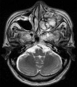
Hemangioma of the maxillary sinus on MRI (indicated by arrows)
MRI is contraindicated:
- pregnant women in the first trimester;
- if there are implants or foreign bodies containing metals with ferromagnetic properties in the patient’s body;
- if the patient has electronic devices installed (pacemaker, insulin pump, cochlear implant or others)
- if the patient suffers from claustrophobia or has an unstable mental state;
- in the presence of vascular clips on the arteries.
Children under 5 years of age and adults with a body weight of more than 130 kg or a waist circumference of more than 150 cm are not examined in our clinic.
What is the consent process before a procedure?
The physician will request consent for the CT - sinus procedure using an informed consent form.
Consent to the procedure. “Consent” is the patient’s agreement to undergo a procedure. The consent form is signed after discussing the risks and benefits of the procedure, as well as alternative testing options. This process is called informed consent.
The patient should sign the forms only after you are completely satisfied with the answers to your questions. In the event that the patient is a minor or an individual who is unable to give consent in person, the person's legal guardian or next of kin must give their consent to the procedure.
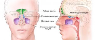
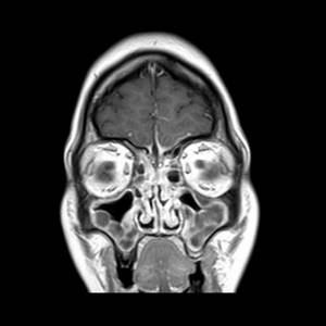
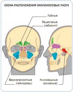
Structure of the sinuses
The paranasal sinuses are air-filled spaces located in the bones of the skull and are an extension of the nasal cavity. Their name corresponds to the name of the bone located nearby.
They are divided into:
- Lower. This is a pair of maxillary (maxillary) sinuses.
- Upper:
- anterior - paired frontal and anterior cells of the ethmoidal labyrinth;
- posterior – unpaired sphenoid sinus and posterior cells of the ethmoidal labyrinth.
Structure of the sinuses
Their main functions:
- Protective. Due to the fact that the inside of the sinuses is covered with a mucous membrane, the air in them is additionally warmed, moistened, and purified. The sinuses protect deeper and more vital structures from external damage.
- Resonator. The sinuses are involved in the formation of individual voice characteristics. Small cavities, such as the sphenoid and ethmoid cavities, create high-pitched sounds, while large cavities (maxillary and frontal) create low sounds. Therefore, during inflammatory processes, the timbre of the voice often changes.
Benefits and risks of this procedure
Advantages of the CT scanning procedure - sinuses:
- CT scan is a painless, non-invasive procedure; has no side effects
- CT scan can take pictures of soft tissue, bone and blood vessels at the same time
- CT scanning can be used even if the patient has an implanted medical device.
- CT scanning is a very accurate method and therefore eliminates the need for other diagnostic procedures.
- This method is also very useful in emergency situations, helping to quickly identify any internal injuries and bleeding
- unlike MRI, it is less sensitive to movement (usually patients are asked to remain still while the examination is in progress, for both CT and MRI scans)
- CT scans provide real-time images. Can be used to guide biopsy or aspiration needle insertion
- This procedure is very fast and accurate. Thus, it can be used to diagnose pain caused by infections and inflammations
Risks of the procedure when performing a CT scan of the sinuses:
- Excessive radiation from a CT scan procedure can cause cancer. But, as a rule, the likelihood of developing cancer is very small
- The CT scan procedure is not recommended for pregnant women.
- It is recommended that mothers breastfeed their babies (at least) 24 hours after the contrast agent is administered for a CT scan.
- There is a rare possibility of developing an allergic reaction due to the iodine-containing contrast agent used in CT scans
- CT scanning is recommended for children only if absolutely necessary, as children are very sensitive to radiation
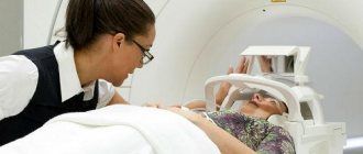
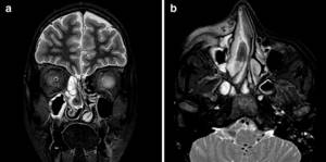
MRI of the sinuses, how a scan is done
The patient is placed on a table that moves inside the tomograph. There is no radiation exposure during this examination. MRI shows sections in different planes, without the need to change the position of the body. The procedure takes about 20 minutes and is very comfortable. There is good air ventilation inside the scanner and soft lighting.
In cases where the use of intravenous contrast is recommended, the study will take a little longer (about 15 minutes additional). After completing the tomography, you can immediately return to your normal lifestyle.
How is the research going?
It is necessary to make sure that there are no metal elements on clothing and remove jewelry. This will avoid distorting the results. The contrast agent is administered by injection. During the study, the person must sit with his chin and nose resting on a certain platform.
After some time, the scanner starts working. During this time you need to be still. Otherwise, the received frames will be unclear. A CT scan of the nose and sinuses without using a contrast agent takes about five minutes. If the specified substance is used, the study takes no more than a quarter of an hour.
The patient is alone in the control room. Medical personnel are located in an adjacent room. Doctors monitor the patient through a special viewing glass. Communication with staff occurs through speakers and a microphone.
If a child undergoes the procedure, parents have the opportunity to be nearby. But they must wear a special lead apron that protects them from exposure to radiation. After the results of a CT scan of the sinuses appear, they should be transferred to the attending physician.
CT or MRI of the sinuses, which is better?
In rhinology, MRI is most effective for identifying pathologies such as fungal diseases of the mucous membrane, mucoceles, polyps and cysts, and, if necessary, distinguishing tumors from inflammation. Magnetic resonance imaging is informative in case of intracranial and intraorbital spread of a tumor, determines tumor growth beyond the nasal sinuses, while bone tissue and filling materials do not create significant interference with visualization.
CT is more often used for facial injuries, especially in the emergency phase.