Price: 800 rub.
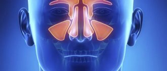
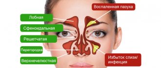
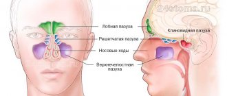
Paranasal sinuses are cavities in the facial bones of the skull filled with air. They communicate with the nasal cavity, which is why they can become infected during a cold. When the mucous membranes of the paranasal sinuses become inflamed, sinusitis is diagnosed. Depending on the location of the inflammatory process, the doctor may make a diagnosis:
- sinusitis - inflammation of the maxillary sinus,
- frontal sinusitis - frontal,
- ethmoiditis - ethmoiditis,
- sphenoiditis - wedge-shaped.
The main symptoms of sinusitis are pain in the forehead and bridge of the nose, nasal congestion, and high fever. Instrumental studies help diagnose the disease: ultrasound of the paranasal sinuses, radiography or computed tomography. To determine the causative agent of the infection and select effective treatment, the patient may be referred for a puncture.
Indications
An ultrasound of the paranasal sinuses is necessary when:
- severe runny nose, which does not go away for more than a week and is accompanied by high fever,
- persistent headaches that radiate to the teeth, ears,
- purulent nasal discharge with an unpleasant odor,
- loss or distortion of the sense of smell,
- constant nasal congestion, which does not help with vasoconstrictors,
- voice change.
Acute sinusitis without treatment can become chronic. Its symptoms include persistent nasal discharge, mucus running down the back of the throat, and general weakness. There is usually no temperature.
Patients diagnosed with chronic sinusitis are advised to undergo regular ultrasound scans, even if there are no complaints. It is also important to do a preventive ultrasound after each cold or flu. Regular examinations will allow you to exclude exacerbation of the infection or begin its treatment at an early stage.
An ultrasound of the paranasal sinuses is also recommended after a nasal injury. The study will help exclude damage to the mucous membranes and hidden inflammation.
X-rays and ultrasound of the paranasal sinuses are prescribed in preparation for implantation and other dental surgeries on the upper jaw. Diagnostics will allow you to determine the exact location of the maxillary sinus and exclude hidden infections that can cause dangerous complications after surgery.
What deviations are detected
Ultrasound is a universal procedure that allows you to identify many pathological processes. Examination of the nose confirms the presence of:
- sinusitis;
- frontitis;
- ethmoiditis;
- sphenoiditis;
- cystic formations;
- polypous neoplasms;
- swelling of the mucous membrane;
- boils;
- lipomas;
Ultrasound can help diagnose nasal polyps
- deviated nasal septum;
- allergic rhinitis.
The data obtained from ultrasound are highly accurate. Rarely there is a need for radiography.
Ultrasound or X-ray?
X-ray examination is considered the gold standard in diagnosing sinusitis. However, this examination method is associated with dangerous radiation and has a number of contraindications. If there is a suspicion of inflammatory processes or malignancy of tissue in the paranasal sinuses, the doctor will refer pregnant women and nursing mothers, small children for an ultrasound scan. Ultrasound scanning allows you to see tumors and cysts, accumulations of fluid and pus in the cavity of one or more sinuses, and evaluate the structure and condition of the tissues that form them. Based on these data, the doctor will rule out or confirm the presence of inflammation and neoplasms.
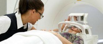
A comprehensive diagnosis of sinusitis usually includes a general examination, ultrasound, x-ray or CT scan of the paranasal sinuses. Based on the results of several studies, it will be easier for the doctor to determine the stage of development of the disease and select effective treatment.
Interpretation of examination results
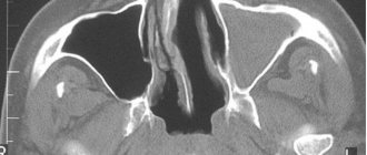
The protocol is deciphered by the treating ENT specialist. Normally, ultrasound images do not show the posterior and lateral walls of the paranasal sinuses. The echogenicity of the internal space of the cavity should be homogeneous, light gray, the mucosal tissue is layered. Behind the front white line, the echo is initially reflected frequently and then less frequently (reverberation effect).
In the sinuses, ultrasound can reveal a cyst, polyp, hematoma, or tumor of unknown origin. The monitor will show areas with shading if there is inflammation (sinusitis, sinusitis, sinusitis), mucus, pus, or blood accumulation. With tumors, the outline of the cavity will change. In case of disease, the side or back walls also become noticeable.
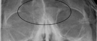
What pathologies look like on a sinusogram:
- inflammation, allergic reaction - the mucous membrane of the sinuses thickens, becomes less dense, there are dark inclusions, effusion or exudate accumulates in the cavity;
- effusion (liquid without pus) - a black stripe or spot at the site of accumulation, quickly flows down the wall when the head is tilted;
- purulent exudate – the sinus cavity is partially or entirely hypoechoic (dark gray), when changing position the fluid moves smoothly;
- cyst - a formation on the wall of the sinus with a hyperechoic membrane and a black cavity that does not move when the head moves;
- neoplasm, polyp - a white or gray spot on the wall of various shapes, does not move.
An echosinoscope will show the presence of the same pathologies as an ultrasound of the maxillary sinuses or other sinuses, but does not determine their type. Deviations are shown as curved lines. To clarify the contents of the sinus or the type of tumor, other examination methods are used.
- Ultrasound of the knee - an informative, inexpensive and safe examination
Video diagnosis of sinuses:
Carrying out
Ultrasound of the paranasal sinuses is a safe and painless examination. It does not require special preparation and has no contraindications. The only obstacle to scanning is open wounds and bandages on the forehead, cheekbones, and bridge of the nose.
One of the advantages of ultrasound is that it can be performed repeatedly over a short period. An otorhinolaryngologist may order a test for:
- diagnosis of the disease,
- monitoring the progress of treatment,
- confirmation of recovery.
An ultrasound of the paranasal sinuses takes about 20-30 minutes. Before the scan begins, the doctor will ask the patient to lie on their back and remove hair from their forehead. Before visiting the clinic, you should not use skin care products or apply decorative cosmetics to your skin. Creams and cosmetics can impair signal transmission through tissue and distort test results.
The patient will be able to obtain images of the paranasal sinuses within 10-15 minutes after the ultrasound examination. With the results of the study, you must make an appointment with an ENT doctor. Only a specialist can make a diagnosis based on the data obtained.
Features of the procedure
No special preparation is required for an ultrasound of the sinuses. To enhance the transmission of ultrasonic waves, a special day is applied to the skin in the nose area. The procedure continues for 10 minutes.
During an ultrasound, specialists record even minor changes in the condition of the tissues of the paranasal sinuses. To increase the information content of diagnostics, the Sinuscan 201 echo sinuscope is used. It allows you to most accurately detect even minor changes in the sinuses and provides accurate examination results.
This modern diagnostic device generates ultrasound pulses that are reflected from the walls of the nasal cavity. After returning, the signal is carefully analyzed by the device, producing specific graphs and diagrams with high resolution. The signal from healthy tissue differs from ultrasound, which comes from pathologically altered mucous membranes and volumetric formations containing fluid.
If necessary, the device saves the results of previous studies on a memory card. This allows you to monitor the chronic course of diseases and the effectiveness of previously prescribed treatment procedures.
Where to do an ultrasound of the paranasal sinuses?
In our clinic, ultrasound of the paranasal sinuses is performed using modern equipment. Highly sensitive sensors allow the doctor to obtain maximum information about the condition of organs and tissues from the image. Patients are received by experienced otolaryngologists, candidates of sciences. Ultrasound scanning and consultations are carried out by appointment, so visitors do not have to wait in line. You can undergo a comprehensive diagnosis and receive specialist advice based on the examination results within 1-2 business days.
To make an appointment for diagnostics and examination, please call. Our administrators will answer all your questions and help you choose a time for your visit.
Contraindications
Ultrasound diagnostics is safe and highly informative, and has virtually no absolute contraindications. It is recommended to postpone the study in the presence of pustular changes, exacerbation of herpetic infection in the facial skin, open lesions, grasses and abrasions that interfere with the application of a special gel.
Diagnostics is also contraindicated for certain mental disorders and diseases that create certain obstacles to ultrasound diagnostics.
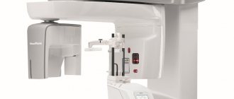
X-ray research methods
They are the most common in the diagnosis of rhinosinusitis, despite the fact that the cells of the ethmoidal labyrinth and the sphenoid sinus are not sufficiently accessible to it. Computed tomography is widely recommended by foreign standard diagnostics of sinusitis, but is a rather expensive method, so its use is impractical in everyday work to identify common forms of rhinosinusitis and monitor the effectiveness of conservative treatment. In addition, these diagnostic methods cannot be used in a certain group of patients (children under 3 years of age, pregnant women). There are also cases that some patients may refuse an X-ray examination on principle. In such a situation, the method of choice is ultrasound examination of the urinary tract.
Ultrasound of the nasal sinuses at ON CLINIC in Ryazan
Our doors are open every day - including on weekends and holidays. We employ experienced doctors of various specializations, whose high qualifications are confirmed by their diplomas and international certificates. Our diagnostic department is equipped with the best medical equipment - this contributes to the early detection of any pathologies and the timely prescription of their effective treatment.
We can perform ultrasound of the paranasal sinuses, as well as ultrasound of the abdominal organs, ultrasound of the prostate gland and other types of ultrasound examination on the day of your visit. Call us and make an appointment at any time convenient for you!
How does the procedure work?
No preliminary preparation for the study is required. Before the procedure, the patient is asked to remove all jewelry that is close to the face, as well as removable dental prostheses. This study is somewhat different from other types of ultrasound. Here the sensor is installed directly above the frontal or maxillary sinuses.
During the examination, the patient is asked to tilt his head to the sides and change his body position. If there is fluid, volume, formations, or foreign objects in the nasal sinuses, the direction and speed of the waves will change. The study allows you to determine the size of the identified formations.
Procedure
The sensor for physiotherapy is similar to that used for ultrasound diagnostics. The only difference will be that the conductor for ultrasound is not a gel, but an ointment based on hydrocortisone. The stages of the session are no different:
- disinfection of the skin area that will be affected;
- applying hydrocortisone ointment and distributing it evenly;
- exposure to ultrasound of a certain power within different frequencies.
This approach ensures rapid penetration of hydrocortisone into tissues. And alternating different frequencies gives good results in the long term, despite the short duration of the session. After completing the phonophoresis procedure with hydrocortisone, you should not remove the ointment from the body for another 2-3 hours. To obtain a positive effect, a course of 10–14 daily procedures is required. Please note that re-treatment is possible no earlier than after 3-5 months.
Mechanism of action
Transport of any drug through the skin is limited by its ability to penetrate the epidermis (top layer of skin) and dermis (deep layer). The easiest and surest route is through the sebaceous glands and hair follicles, so warming the skin before the procedure significantly increases the rate and percentage of drug transfer.
Warming up immediately after phonophoresis significantly increases the time of absorption of hydrocortisone by the body’s vascular system. Sensors and devices used in physical therapy have many modes of operation at different frequencies and intensities. When choosing a device, the doctor focuses on solving the main problem without damaging the skin. And only then, he sets the pace and frequency of the impact, taking into account the patient’s sensations and the characteristics of his body, excluding contraindications and pathologies.
What does an ultrasound of the nose show?
Paranasal sinuses are cavities inside the bones that are filled with air. They are located in the body of the upper jaw, which is attached to other bones of the face. The ultrasound method makes it possible to obtain the following information about these anatomical structures:
- whether there are lesions on the mucous membrane inside the sinuses; what is the condition of the nasal septum; structure and thickness of subcutaneous tissue; assess the parameters of nasal cartilage; condition of the vascular walls; assess the level of fluid inside the sinus cavity; examine the condition of the bone walls; determine the size and configuration of the tumor.
Ultrasound also allows you to find foreign objects that have entered the cavity of the paranasal sinus.