What is contrast x-ray - the history of the discovery of a diagnostic method
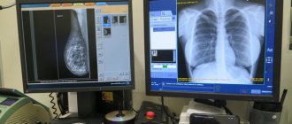
Contrast radiography is a set of X-ray examination techniques, the main feature of which is the use of contrast agents . Thus, the doctor receives images that have high diagnostic value.
The first contrast agent is the brainchild of French doctors - Jean-Athanase Sicard and Jacques Forestier . In 1921, lipiodol was used for injection into the cerebrospinal fluid. This helped to identify the exact location of intraspinal cysts and tumors. At the end of the 20s of the last century, American professor Moses Swick developed a new method for studying the urinary system, which involved the intravenous administration of a contrast agent. The specified iodine-containing drug was transported through the bloodstream to the kidneys, bladder and urinary tract, ensuring their visualization on x-rays. The modifications proposed by Swick were implemented by the German physician Arthur Binz , and presented in 1929 at the congress of the German Society of Urology.
In 1931, the German surgeon and urologist Werner Frosman tested his own method of cardiac catheterization. A similar experiment was successful and marked the beginning of angiocardiographic diagnostics.
Contrast agents for MRI and CT first saw the world in the 80s of the last century.
Based on the technology for introducing the specified substance, two types of contrast are distinguished:
- Invasive . Involves piercing the mucous membranes or skin. In this case, the necessary drug is administered by injection.
- Non-invasive. It is considered a non-traumatic technique. The coloring substance enters the body through the mouth or through an enema into the rectum.
A specific property of X-ray contrast agents is their ability to absorb X-rays that come from soft tissues.
These drugs are divided into two large groups:
- X-ray negative . Practiced when examining organs of the gastrointestinal tract and urinary system. Any gas can act as a contrast agent here.
- X-ray positive . They contain heavy chemical elements, which ensures better absorption of X-rays. Nowadays, the most popular are insoluble (barium sulfate) and iodine-containing coloring preparations.
Video: X-ray of the stomach with barium
The essence of contrast radiography - what does the study provide in diagnosis?
The method under consideration makes it possible to:
Examine the body's blood vessels to assess their general condition
In the case of using this examination technique, a catheter is inserted into the patient’s blood vessel, after which a contrast agent is immediately infused.
Often the coloring agent is administered 3 times. X-ray shooting is carried out very quickly.
- To study the structure of the cerebral arteries and the quality of blood flow in them. Timely diagnosis in the presence of alarming symptoms makes it possible to avoid serious disorders associated with cerebral circulation.
- Examine the cardiac arteries to prescribe adequate treatment for errors in the functioning of the heart.
- Examine the renal arteries for the presence of additional vessels, “plugs” in the vessels, and pathological neoplasms in the kidneys. In this case, the contrast agent is administered through a catheter, which is previously placed in the femoral artery.
- Check the quality of functioning of the veins of the lower extremities .
Examine the mammary ducts using contrast
Such a diagnostic procedure is called ductography, galactography or radiopaque mammography.
The coloring agent is administered through a catheter, which is placed into the mammary duct, which is located on the nipple.
Study the quality of work, the structure of the gastrointestinal tract organs
During the study, you can obtain information about the condition of the mucous membrane; about the shape and contours of the stomach, esophagus and small intestine; evaluate their tone and peristalsis.
The main ingredient in this examination is a suspension of barium sulfate. Depending on the type of contrast, a powder can be used, which is mixed with water and taken immediately before diagnosis, or a ready-made water suspension, which in consistency resembles thick sour cream.
The latter type of suspension is used when conducting double contrasting, and it retains its properties for 3-4 days.
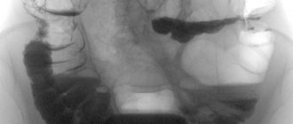
Examine the large intestine using irrigoscopy for the presence of scars, developmental anomalies, pathological neoplasms, fistulas
Using this method, it is also possible to identify cancerous and benign tumors in the liver, spleen, and organs of the female reproductive system; see interintestinal abscesses, bleeding in the gastrointestinal tract.
To perform this type of examination, a barium solution is injected rectally into the intestines. The patient may feel some discomfort in the form of abdominal distension and the urge to defecate - this is normal. First, the doctor takes targeted photographs, then a survey X-ray of the entire peritoneum.
After bowel movements, another x-ray is taken, thanks to which you can examine the mucous membrane of the large intestine and evaluate its ability to contract.
Examine the outflow of blood in ailments associated with erectile function, establish the localization of atrophied or sclerotic areas
Initially, caverject is injected into the sexual organ in order to ensure an erection, followed by iodine-containing preparations.
To study the structure of the female pelvic organs: fallopian tubes, body and cervix
This procedure is carried out in relation to the menstrual cycle.
The iodine-containing drug enters the uterine cavity through a special cannula.
Perform a review of the urinary system of a patient diagnosed with urological pathology
In this case, a contrast agent is injected into a vein in the elbow.
If prescribed by a doctor, separate examinations of the urethra, ureters, and prostate gland can be performed. In such cases, the dye is administered through the urethra.
In some cases, this procedure, due to its pain, is performed under general anesthesia.
I. Description of complications following the introduction of RCV
Complications of RCV administration can be divided into general, organ-specific and local. 1. General reactions General reactions to the administration of RCV are usually observed during intravascular administration and occur relatively rarely. The radiologist should always be aware of this possibility and take special measures to prevent and treat them. The frequency of adverse reactions has decreased significantly with the introduction of modern non-ionic contrast agents into practice. Most of the recorded adverse reactions are mild and do not threaten the life and health of the patient. The most dangerous reactions occur suddenly (usually at the end of the needle) or within the first 10 minutes after the drug enters the bloodstream. Common adverse reactions to RCV can be acute or late. 1.1. Acute reactions • Allergy-like reactions Allergy-like reactions are not associated with the phenomena of so-called iodism (intolerance to free iodine), since during circulation in the body, iodine atoms are practically not split off from the molecule, which is based on the benzene ring. The reaction develops as a direct response of the body to the chemical complex of the drug. The mechanism of development of such reactions is still being studied, but antibodies are not involved in it. There are no specific antidotes to prevent their occurrence. Clinical signs of acute allergic reactions to RCV are similar to those observed with the use of a wide variety of drugs. Relief of such reactions is carried out according to the same rules as for other allergy-like or allergic conditions. • Other reactions Some non-allergic reactions to contrast media are associated with some chemo- and osmotoxicity of RCV. They can be expressed by heart rhythm disturbances, decreased myocardial contractility, pulmonary edema, and convulsions. When using nonionic low- or iso-osmolar drugs, such reactions can be classified as casuistic, occurring only in patients with severe concomitant changes in the cardiovascular system. There are mild, moderate and severe degrees of reactions to RCV. Mild Mild acute reactions may include nausea, vomiting, urticarial skin rashes, sore throat, and minor swelling of the tongue or face. Typically, these symptoms do not require medical intervention. The listed manifestations most often occur when using ionic RCVs with increased osmolarity and are often associated with an increase in the volume of the administered contrast agent in excess of that recommended in the instructions. Pain during intravascular administration of a contrast agent is caused by high concentrations of PCV and is not considered an allergy-like reaction. Mild reactions usually do not require treatment. Patients with such reactions should be observed for 30 minutes after RCV administration, when the risk of serious complications has subsided. Moderate Reactions of moderate severity do not pose a threat to the patient’s life, but require medical attention. These reactions can be expressed by severe urticaria or erythema, bronchospasm, moderate swelling of the face and tongue, transient arterial hypotension with tachycardia, and vomiting. In such patients, the possibility of immediate initiation of relief measures should be considered. Severe Severe reactions can pose a threat to the patient's life. They usually develop rapidly and require immediate intervention. Their symptoms are altered consciousness, swelling of the larynx, severe respiratory depression, up to severe bronchospasm, and disruption of the cardiovascular system, as well as a sharp decrease in blood pressure. There have been known cases of death. 1.2. Late reactions Late reactions to intravenous administration of RCV most often manifest themselves as skin symptoms, are mild and occur in the period from 30-60 minutes to several days after the study. These reactions occur with a frequency of 0.5-10 percent. They usually manifest themselves as urticaria or a small papular rash of various sizes and locations and may be accompanied by itching. It is extremely rare that more severe late reactions are possible in the form of the development of toxic epidermal necrolysis and cutaneous vasculitis. Severe delayed noncutaneous reactions to contrast agents are extremely rare. It should be noted that severe hypotension and cardiopulmonary shock may not always be associated with the administration of RCV, but often develop due to an underlying disease associated with severe damage to internal organs. 2. Organ-specific reactions Nephrotoxicity It is known that RCV can impair renal function and, in rare cases, lead to the development of acute renal failure. To more accurately assess the risk of renal dysfunction, the term “contrast-induced nephropathy” (CIN) was introduced. The course of CIN is usually manifested by a transient asymptomatic increase in serum creatinine. Typically, creatinine begins to rise within 24 hours after intravascular contrast agent administration, peaks after 4 days, and often returns to baseline levels after 7 to 10 days. According to international agreement, CIN is a condition in which renal function decreases (serum creatinine increases by more than 25% or more than 44 mmol/L (0.5 mg/dL) within 3 days after intravascular administration of a contrast agent in the absence of other etiological reasons. Serum creatinine concentration is the most common and objective indicator of kidney function. However, as an accurate measure of glomerular filtration rate (GFR), it has its limitations. Serum creatinine depends significantly on gender, muscle mass, nutrition, age of the patient and the presence of blood so-called creatinine-like substances. Impaired renal function can occur even with normal creatinine levels. More accurate is the assessment of glomerular filtration rate (GFR) using special formulas (for example, Cockroft's formula). 3. Local reactions Extravasation of contrast agent Extravasation of contrast agent during intravascular administration occurs in 0.1% - 0.9% and depends on the qualifications of medical personnel, as well as the individual characteristics of the constitution and angioarchitecture of the patient. If complications occur, patients complain of swelling in the injection area, pain and discomfort in the presence of erythema and increased sensitivity. Severe but relatively rare complications are tissue necrosis and skin ulcers. The patient is observed until the condition improves. Consultation with a surgeon is indicated for patients with increasing swelling or pain, obvious disruption of tissue trophism and persistent increased sensitivity in the area of extravasation. The fact of RCV extravasation and the list of treatment measures taken must be recorded in the patient’s medical documents, and the attending physician must be notified about this. The patient should be given clear instructions about the need to immediately seek medical help if the condition worsens, the appearance of neurological symptoms (paresthesia) or signs of circulatory problems. Air embolism can be noted as a separate complication. Air embolism Air embolism is a very rare complication of intravenous contrast agents. Asymptomatic air embolism usually occurs when contrast agent is injected manually using a syringe. The risk of clinical manifestations of embolism appears only when more than three ml of air enters the vascular bed. Using an automatic syringe (injector) minimizes the occurrence of this complication.
Contraindications to radiography with contrast and possible complications
This type of diagnosis has many contraindications. Depending on the area being examined and the type of contrast agent, restrictions may vary.
General contraindications for radiography using iodine-containing preparations are:
- Hypersensitivity to drugs that contain iodine.
- Kidney, heart or liver failure.
- Acute inflammatory processes in the area where they plan to inject a contrast agent.
- Serious mental disorders.
- Thrombosis, which is accompanied by inflammation in the vascular walls.
- Disorders associated with blood clotting - in the case of examination of the genitourinary system.
When examining cardiac vessels, the general list of restrictions regarding contrast X-rays is supplemented by the following conditions:
- Chronic illnesses, ulcers, and diabetes in the acute stage.
- Uncontrolled attacks of high blood pressure.
- Endocarditis.
Possible negative consequences of coronary angiography:
- Irregularities in heart rhythm.
- Violation of the integrity of the heart arteries.
- Formation of a blood clot in a coronary vessel.
The introduction of a dye into the blood vessels is fraught with bleeding at the puncture site with the formation of a hematoma in the future.
After contrasting the liver and biliary tract, the patient may have complaints of:
- Nausea.
- Minor pain in the abdominal area.
- Flashes of heat to the head.
The use of barium preparations is prohibited if the doctor has doubts regarding the integrity of the gastrointestinal tract .
The penetration of these substances into the peritoneum can cause the development of peritonitis .
If radiography with contrast involves the use of gases, the patient should not have glaucoma or prostate adenoma .
Contrast x-ray of the genitourinary system is contraindicated in the following cases:
- Pregnancy.
- Ulcerative colitis with a pronounced symptomatic picture.
- Deviations of a somatic nature.
- Perforation of the intestinal wall.
II. Risk factors for adverse reactions to RCV administration
The presence of a number of diseases in the patient increases the likelihood of developing complications following the introduction of RCV. 1. Risk factors for the development of general reactions • Bronchial asthma • History of allergic reactions of moderate and severe degree • Heart diseases: – angina pectoris, chronic heart failure, – severe aortic stenosis, – primary pulmonary hypertension, – cardiomyopathy • Severe liver diseases • Sickle cell anemia • Pheochromocytoma • Severe hyperthyroidism • Other risk factors: – age (in young children and the elderly the risk of adverse reactions increases); – use of beta-blockers 2. Risk factors for the development of contrast-induced nephropathy (CIN) Risk factors for the development of CIN include nosological forms and conditions leading to impaired renal function, accompanied by an increase in the level of creatinine in the blood plasma: • diabetic nephropathy • dehydration • chronic heart disease failure • acute myocardial infarction • severe arterial hypertension • taking nephrotoxic drugs • acute or chronic renal failure • use of high-osmolar RCV in large doses • use of a contrast agent more than once within 72 hours.
Preparing for an x-ray with a contrast agent - recommendations for patients before and after the examination
Preparation for a contrast X-ray examination of the body’s blood vessels consists of the following activities:
- Providing the doctor with information about existing allergic reactions, as well as about taking certain medications.
- Complete avoidance of spicy, fatty, smoked foods, as well as flour products a few days before the procedure. The diet during this period should consist of cereals and light soups. You cannot have breakfast on the day of contrast.
- 14 days before the procedure you must refrain from drinking alcoholic beverages, and 1 day before - from smoking.
- 12 hours before cholegraphy (examination of the biliary tract), the patient is administered bilitrast: intravenously or orally. Before the examination, 2 enemas are given: in the evening and 2 hours before the diagnosis. In some cases, sedatives or sleeping pills may also be prescribed.
Contrast studies of the genitourinary system are often performed using general anesthesia. In this case, the patient should refuse to eat 8 hours before the procedure and minimize the amount of liquid consumed.
After contrasting the blood vessels is completed, the patient remains in the clinic for a minimum of 6 hours.
In addition, the following recommendations should be followed:
- You need to drink as much fluid as possible - it will help remove iodine-containing substances from the body as quickly as possible.
- For the first day, you should adhere to bed rest: under no circumstances should you perform actions that will physically or psychologically tire the body. This also applies to driving a car.
- The bandage applied to the area where the skin was pierced during insertion of the cannula should not be removed for 24 hours.
- In the first 12 hours after contrast X-ray of blood vessels, you should not take a shower or bath.
Before coronary angiography, the patient should undergo some tests:
- General and detailed blood test.
- Determination of blood group and Rh factor.
- Testing the body for the presence of hepatitis B and C.
- Electro- and echocardiogram.
- Blood test for AIDS and syphilis.
Before conducting contrast radiography of the stomach and duodenum for patients who do not have abnormalities in the functioning of the gastrointestinal tract, the only preparation point is to abstain from eating for at least 6 hours.
IV. Treatment for the development of reactions and side complications
If adverse reactions develop, you must immediately stop administering the drug.
4.1. Skin lesions Acute mild urticaria: skin itching, isolated urticarial rashes with normal blood pressure. Intramuscular or intravenous administration of a solution of chloropyramine 2% 2 ml or clemastine 2 ml. 4.2. Acute urticaria and Quincke's edema Itching and hyperemia of the skin, widespread urticarial rashes, generalized urticaria. Swelling of the lips, tongue, swelling of other localization with normal blood pressure values. 1. Intramuscular or intravenous administration of a solution of chloropyramine 2% 2 ml or clemastine 2 ml. 2. Intravenous or intramuscular administration of steroids (prednisolone 30-90 mg or dexamethasone 4-8 mg). 4.3. Damage to the respiratory system Cough, shortness of breath, wheezing, suffocation, attack of bronchial asthma 1. Salbutamol through a nebulizer 2.5-5 mg or a metered dose inhaler with a spacer, 2 breaths 1-3 times. 2. Intravenous or intramuscular administration of steroids (prednisolone 30-90 mg or dexamethasone 4-12 mg). 3. If necessary, additional intravenous administration of aminophylline solution 2.4% -5.0-10.0 ml with saline solution. 4.4. A sharp decrease in blood pressure 1. Immediately stop administering the drug. At the same time, call an ambulance team, resuscitators, and other medical workers from neighboring offices and begin carrying out treatment measures. 2. Urgently inject a 0.1% solution of adrenaline intramuscularly into the middle of the anterolateral surface of the thigh in a dose of 0.2-0.5 ml. If necessary, repeat administration after 5 minutes. 3. Immediately place the patient on his back, raise his lower limbs, and turn his head to the side. Do not raise the patient's head or place him in a sitting position! Advance the lower jaw, assess the airway, clear the airway; remove removable dentures 4. Provide oxygen for breathing. 5. Leave intravenous access or establish it, administer 0.9% sodium chloride solution. 6. If there is no effect and severe hypotension persists, adrenaline 0.1% should be diluted intravenously in a stream of 1 ml in 10 ml of saline until hemodynamics are stabilized. 7. Inject steroids intravenously or intramuscularly (prednisolone 90-120 mg or dexamethasone 8-16 mg) 8. For bronchospasm - inhalation of salbutamol through a nebulizer or metered dose inhaler with a spacer 200-500 mg. Intravenously, if necessary, an additional solution of aminophylline 2.4% -10.0 ml, diluted with saline solution. 9. When blood pressure stabilizes, introduce antihistamines - chloropyramine solution 2% 2 ml or clemastine 2 ml 10. If there is no effect of therapy, perform cardiopulmonary resuscitation until resuscitators arrive. 11.Monitoring blood pressure, pulse rate and respiration. If possible, connect a monitor.
Materials taken from this source, from the specialized medical resource radiomed.ru